Pregnancy Identifies Novel Targets of Elf5
Renee L. Rogers1*¤a, Isabelle Van Seuningen2, Jodee Gould1, Paul J. Hertzog1, Matthew J. Naylor3,4¤a, Melanie A. Pritchard1*¤b
1Centre for Functional Genomics and Human Disease, Monash Institute of Medical Research, Clayton, Victoria, Australia,2Inserm, U837, Centre de Recherche Jean-Pierre Aubert, Lille, France,3Cancer Research Program, Garvan Institute of Medical Research, Darlinghurst, New South Wales, Australia,4St. Vincent’s Hospital Clinical School, Faculty of Medicine, University of New South Wales, Sydney, New South Wales, Australia
Abstract
Background:Elf5, an epithelial specific Ets transcription factor, plays a crucial role in the pregnancy-associated development of the mouse mammary gland.Elf52/2embryos do not survive, however the Elf5+/2 mammary gland displays a severe pregnancy-associated developmental defect. While it is known that Elf5 is crucial for correct mammary development and lactation, the molecular mechanisms employed by Elf5 to exert its effects on the mammary gland are largely unknown.
Principal Findings:Transcript profiling was used to investigate the transcriptional changes that occur as a result of Elf5 haploinsufficiency in theElf5+/2 mouse model. We show that the development of the mouseElf5+/2mammary gland is delayed at a transcriptional and morphological level, due to the delayed increase in Elf5 protein in these glands. We also identify a number of potential Elf5 target genes, includingMucin 4, whose expression, is directly regulated by the binding of Elf5 to anEtsbinding site within its promoter.
Conclusion:We identify novel transcriptional targets of Elf5 and show that Muc4 is a direct target of Elf5, further elucidating the mechanisms through which Elf5 regulates proliferation and differentiation in the mammary gland.
Citation:Rogers RL, Van Seuningen I, Gould J, Hertzog PJ, Naylor MJ, et al. (2010) Transcript Profiling ofElf5+/2Mammary Glands during Pregnancy Identifies
Novel Targets of Elf5. PLoS ONE 5(10): e13150. doi:10.1371/journal.pone.0013150
Editor:Jeffrey A. Whitsett, Cincinnati Children’s Hospital Medical Center, United States of America
ReceivedJune 24, 2010;AcceptedAugust 25, 2010;PublishedOctober 7, 2010
Copyright:ß2010 Rogers et al. This is an open-access article distributed under the terms of the Creative Commons Attribution License, which permits unrestricted use, distribution, and reproduction in any medium, provided the original author and source are credited.
Funding:RLR was the recipient of an Australian Postgraduate Award and a Women in Scientific Excellence (WISE) award. The authors also wish to thank the Australian Research Council (MAP), National Health and Medical Research Council (MAP), National Breast Cancer Foundation of Australia (MN), Gardiner Foundation (RLR, MAP) and Ligue Nationale contre le Cancer (Equipe labellisee 2008, IVS) for financial support. The funders had no role in study design, data collection and analysis, decision to publish, or preparation of the manuscript.
Competing Interests:The authors have declared that no competing interests exist. * E-mail: [email protected] (MAP); [email protected] (RLR)
¤a Current address: Cancer Research Program, Garvan Institute of Medical Research, Darlinghurst, New South Wales, Australia ¤b Current address: Department of Biochemistry and Molecular Biology, Monash University, Clayton, Victoria, Australia
Introduction
The mammary gland is one of a few organs able to undergo repeated phases of growth, differentiation and regression. At the onset of puberty in the female, the increase in ovarian steroids induces elongation and side-branching of the rudimentary mammary ductal system which formed embryonically. Club-shaped terminal end buds (TEBs) develop at the ends of the developing ducts which consist of a layer of cap cells at the proceeding edge and multiple inner body cell layers. These TEBs are the proliferating edge of the ducts penetrating the mammary fat pad and regress once the development of the ductal tree is complete. Some differentiation of the ductal system occurs at this stage, resulting in a compact glandular structure. The gland then remains relatively inactive until a pregnancy occurs [reviewed in 1, 2].
Lobuloalveolar development commences with the onset of pregnancy and is associated with the formation of spherical alveoli along the ductal network that formed during puberty. By the end of pregnancy, the ductal tree is densely populated with alveoli.
Functional differentiation of the alveoli commences during late pregnancy and this process is complete by parturition to enable lactation. The mammary gland continues to produce milk until weaning, at which time the gland regresses in a process known as involution. This cyclical development of the mammary gland is controlled by various hormonal and genetic signals [reviewed in 1, 2]
Using a genetic knock-out mouse model, we identified a key role for the epithelial-specific Ets transcription factor, Elf5, in the development of the mammary gland during pregnancy [3]. The complete lack of Elf5resulted in a lack of the extraembryonic ectoderm which led to early embryonic death [4]. Mice carrying one functional copy of the Elf5 gene (Elf5+/2) were viable.
However, unlike their wildtype littermates,Elf5+/2females were
unable to support their offspring due to a pregnancy-associated mammary gland developmental defect, which prohibited these dams from lactating [3].Elf5expression was downregulated in the
Prlr+/2 mammary gland while Prlr expression remained
un-changed in theElf5+/2mammary gland [3], indicating that Elf5
later confirmed using a genetic complementation approach whereby ectopic expression of Elf5 in the Prlr-null mammary epithelium was sufficient to rescue the developmental defect observed in the Prlr-null mice [5]. While it is known that Elf5 is able to activate the promoter of the milk protein genewhey acidic protein(Wap) [6], the mechanisms employed by Elf5 to induce its effect on mammary epithelial cell proliferation and differentiation are largely unknown. Here, we have used transcript profiling to investigate the transcriptional changes that occur as a result ofElf5
haploinsufficiency as a first step in determining the mechanisms of Elf5 action and the targets of this transcription factor. We show that mammary gland development in theElf5+/2female mouse is
delayed during pregnancy due to the delayed increase in Elf5 protein expression in these glands. We identify a number of potential Elf5 target genes including Muc4, which we show is directly regulated by the binding of Elf5 to its promoter.
Methods
Mice
All work performed using mice as part of this study was approved by the appropriate Monash University Animal Ethics Committee and as such, all animal work followed the committee’s guidelines and procedures. Mice were housed in a windowless room with controlled temperature (22uC62uC), on a 12 hour light and dark cycle and were fedad libitum. Pregnancy was determined by the presence of a vaginal plug and the day of detection designated 0.5 days post coitum (dpc). Mammary glands were collected between 11am and 1pm to control for circadian Prl release. Mice targeted at theElf5locus were generated previously in our laboratory [3] and backcrossed for more than 10 generations onto a C57Bl/6 genetic background.
Microarray analysis
Microarray analysis of the Elf5+/2 mammary glands was
performed using a common reference experimental design. Abdominal mammary glands (minus the associated lymph nodes) of 2 mice were pooled (to ensure enough material was obtained from virgin samples) and total RNA extracted using a RNeasy maxi kit (Qiagen) according to the manufacturer’s instructions. RNA for use as the reference was extracted from eight embryonic (17.5dpc) C57Bl/6 mice using the same method. The RNA samples were sent to the Adelaide Microarray Facility for the remainder of the processing (see website for processing protocols: http://www. microarray.adelaide.edu.au). Five stages of mammary gland development (virgin, 8.5dpc, 10.5dpc, 14.5dpc and 16.5dpc), in two distinct genotypes (Elf5+/+ and Elf5+/2) were examined in
triplicate. A total of 30 array slides was used to examine 3 independent pooled RNA samples per genotype per timepoint. The slides chosen for this study were 22,000 element Compugen array chips which were spotted with single stranded oligonucleotides of 60 base pair length. Processed slides were scanned and the resulting images analysed using Digital Genome (Molecularware). Data analysis was performed using the Genespring software suite (Agilent). All data produced is MIAME compliant and has been deposited in the NCBI Gene Expression Omnibus database under series accession number GSE23373 (http://www.ncbi.nlm.nih. gov/geo/query/acc.cgi?acc = GSE23373).
RNA extraction and gene expression analysis
Total RNA was extracted from mammary glands using Trizol reagent (Invitrogen) according to the manufacturer’s instructions. RNA was treated with DNase (Promega) before being converted to cDNA using Superscript III (Invitrogen) and random primers
(Promega). Quantitative real-time RT-PCR was performed on samples using TaqMan Gene Expression Assays (ABI) specific for Elf5 and Muc4. Relative expression was determined using the DDCTmethod [reviewed in 7], with the expression of the genes of interest first being normalised to the level of 18S expression.
Protein extraction and Western blotting
Snap frozen tissue was homogenised in tissue lysis buffer (50 mM Hepes pH 7.5, 100 mM NaCl, 1 mM EDTA, 10% (v/v) glycerol, 0.5% (v/v) Nonidet-P40, Roche protease inhibitor cocktail tablet), and centrifuged to remove any insoluble material. Protein (50mg) was separated using SDS-PAGE. Fractionated proteins were transferred to polyvinylidene fluoride (PVDF) membrane (Immobilon-P, Millipore) and membranes blocked for 1 hour at room temperature with 5% skim milk/TBST, before incubating with primary antibody overnight at 4uC. All secondary antibodies were conjugated to horseradish peroxidase and obtained from Dako. Membranes were incubated with Super-Signal West Pico chemiluminescent substrate (Pierce), after which they were exposed to Hyperfilm ECL film (Amersham Pharmacia), to detect chemiluminescence. Antibodies used in this study: anti-Elf5 (N-20) (Santa Cruz, catalogue number SC-9645); anti-mouse milk specific proteins (Accurate Chemical and Scientific Corpo-ration, catalogue number YNRMMSP). Blotting for Muc4 was performed as described previously [8]
Recombinant ELF5 protein preparation
Recombinant ELF5 protein was made by cloning a truncated
ELF5cDNA into the BamHI/SacII sites of the pQE-30 (Qiagen) vector enabling the production of a truncated Elf5 (D33) protein fused to a (HIS)6tag. Primers used to amplify the truncated cDNA were as follows: hELF5D33BamHI_F: 59 CGTAGGATCCGCC-TTTGAGCATCAGACAG 39 and hELF5D33SacI_R: 59
CA-TGGAGCTCAGCTTGTCTTCCTGCCACCC 39. TALON
beads (Clontech) were used to purify the protein as per the manufacturer’s instructions.
Cell transfection and reporter assays
The human breast carcinoma cell line T47D (ATCC) was grown in 1x RPMI with 10% (v/v) FCS plus 1% (v/v) penicillin/ streptomycin and maintained in culture at 37uC, 5% CO2.T47D cells were plated in a 24 well plate at a density of 56104cells/well in complete media. These cells were left to settle at 37uC, 5% CO2 for 24 hours before being transiently transfected with a total of 500 ng of DNA using the FuGENE6 transfection reagent according to the manufacturer’s instructions (using a 1:3 DNA:FuGENE ratio). Cells were transfected with a MUC4
Isolation and extraction of nuclei
Snap frozen mammary glands, excised from 16.5dpc mice, were crushed in a mortar and pestle under liquid nitrogen, to a fine powder which was then mixed with ice-cold buffer A (10 mM Hepes pH 7.9, 1.5 mM MgCl2, 10 mM KCl, 500mM PMSF, 500mM DTT 2900ml per gland). The suspension was centri-fuged, and the pellet resuspended in 20ml of buffer A with the addition of Nonidet-P40 (to 0.15% v/v) and incubated on ice for 10 mins. The sample was centrifuged and the pellet resuspended in 15ml of ice-cold buffer C (20 mM Hepes pH 7.9, 420 mM NaCl, 1.5 mM MgCl2, 0.2 mM EDTA, 25% (v/v) glycerol, 500mM PMSF) before centrifugation and transfer of the supernatant to a fresh tube containing 40ml of ice-cold buffer D (10 mM Hepes pH 7.9, 50 mM KCl, 0.2 mM EDTA, 20% (v/v) glycerol, 500mM PMSF, 0.5 mM DTT).
Electrophoretic mobility shift assay (EMSA)
Short double stranded oligonucleotides were end-labelled with (c-32P)dATP (Perkin-Elmer) using T4 polynucleotide kinase (Promega), according to the manufacturer’s instructions. When examining ELF5 binding to theMUC4promoter, an oligonucle-otide containing theEtssite predicted at2216 was used as a probe 59CCACCAGGAAAGAAAACACCG 39. A mutant sequence where GGAA was changed to AAAA was also used. Nuclear extract (4mg) or recombinant ELF5 protein (200 ng), with 1x bandshift binding buffer (40% (v/v) glycerol, 10 mM EDTA, 50 mM DTT, 100 mM Tris pH 7.5, 1 M NaCl, 1 mg/ml BSA) and a 100-fold molar excess of unlabelled competitor oligonucle-otide were combined in a total volume of 20ml. After 30 mins at room temperature, 30,000 counts per minute (cpm) of radiola-belled oligonucleotide probe was added and incubation continued for an additional 30 mins. Reactions were electrophoresed on polyacrylamide gels which were dried under vacuum onto blotting paper and then exposed to X-ray film.
Results
Transcript profiling indicated that pregnancy-associated development of theElf5+/2mammary gland was delayed
The changes in gene expression that occur as a result of the loss of oneElf5allele were investigated by transcript profiling. Having previously observed that the morphological development of the
Elf5+/2 mammary gland appears stunted [3], we sought to
examine this potential developmental delay by transcriptional profiling. Profiles were generated for both theElf5+/+
andElf5+/2
mammary glands at 5 stages of mammary development (virgin, 8.5dpc, 10.5dpc, 14.5dpc and 16.5dpc). To examine the similarities between the global transcriptional profiles of each of the 10 experimental conditions (five stages of mammary gland development in 2 genotypes), a dendrogram (Figure 1a) was generated within Genespring using an average linking clustering algorithm and a Spearman correlation similarity measure.
The virgin wildtype mammary gland was placed on its own node of the dendrogram while all the other conditions were clustered on a separate arm. The Elf5+/2 virgin and 8.5dpc
samples were placed together on a separate node suggesting that they were most similar to one another, and more similar to the other pregnant conditions rather than to the Elf5+/+
virgin condition. Notably, theElf5+/2 10.5dpc and 14.5dpc conditions
were clustered with the wildtype 8.5dpc sample, between the wildtype virgin and wildtype 10.5dpc samples, indicating that the expression profile of the 10.5dpc and 14.5dpc heterozygous gland was most similar to an earlier timepoint in the wildtype gland. Not surprisingly, the 14.5dpc and 16.5dpc wildtype conditions were most similar to one another. TheElf5+/216.5dpc condition was
placed on a separate node, but adjacent to the wildtype 14.5dpc and 16.5dpc conditions, suggesting that the gene expression profile of theElf5+/216.5dpc mammary gland had ‘caught up’ with the
wildtype to some extent, although obvious gene expression differences remained.
Figure 1. Comparing the Transcriptional profiles ofElf5+/+
andElf5+/2mammary glands. A.A condition dendrogram was generated within
the Genespring software suite using an average linking clustering algorithm and a Spearman correlation similarity measure. Individual genes are represented by horizontal bars, which are coloured according to their expression level relative to the virgin Elf5+/+(V
+/+) sample.B.Principal components analysis was performed in Genespring on all 10 conditions examined by microarray. Conditions are plotted on a 3-dimensional scatter plot according to their variance from the first 3 components. The conditions were normalised to the V+/+condition.
A principal components analysis (PCA) was also performed on the dataset to examine the relationships between the transcrip-tional profiles of the experimental conditions (Figure 1b). A three-dimensional scatter plot mapping the experimental conditions by the first three principal components showed that the Elf5+/+
conditions are separated based upon whether they originated from a pregnant animal or not i.e. the virgin Elf5+/+ condition was placed alone, whereas the 4 conditions representing pregnant
Elf5+/+ animals were clustered together. The 4 pregnant conditions showed increasingly greater variance from the first component in accordance with the stage of pregnancy. The separation of the wildtype pregnant conditions according to the stage of pregnancy also occurred in the direction of the third component. Here, the later the stage of pregnancy they represented the closer they were placed to 1 on the axis of the third component.
Supporting the condition dendrogram, the virgin Elf5+/2
sample was positioned closer to the pregnant samples of both genotypes than to the wildtype virgin sample. TheElf5+/214.5dpc
condition was placed closest to the 8.5dpc and 10.5dpc wildtype conditions suggesting that it had greatest similarity to the wildtype mammary gland at earlier stages of pregnancy. The 16.5dpc
Elf5+/2 condition was positioned closest to the 14.5dpc wildtype
sample indicating that the expression profile of theElf5+/2gland is
becoming more similar to a more developed gland after 14.5dpc. It should be noted however that morphologically and functionally the Elf5+/2 gland remains underdeveloped compared to the
wildtype gland [3].
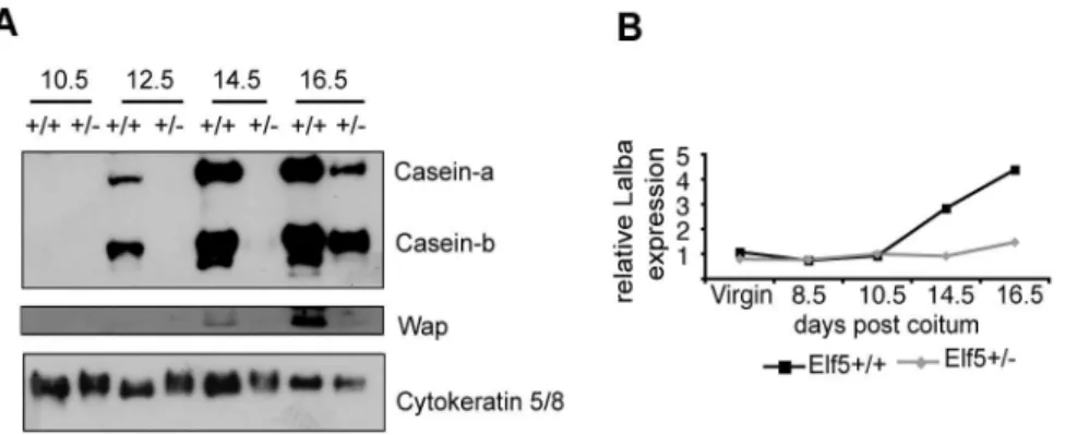
Expression of milk proteins is delayed in theElf5+/2 mammary gland
Milk protein expression in the mammary gland can be used as a measure of differentiation. Expression of a-casein and b-casein proteins was first evident at 12.5dpc in the Elf5+/+ mammary gland, while Wap appeared at 14.5dpc (Figure 2a). In contrast, in theElf5+/2 mammary gland, expression ofa- andb-caseins was
not apparent until 16.5dpc. This result supports the notion that at 14.5dpc theElf5+/2mammary gland is most similar to a very early
stage wildtype gland, while at 16.5dpc theElf5+/2gland is more
like a gland that has entered the differentiation program. Indeed, the milk protein expression profile of theElf5+/216.5dpc gland is
similar to the wildtype 12.5dpc. gland.
Chronologically, the milk protein genea-lactalbuminis the last milk protein gene to be expressed during pregnancy-associated mammary gland development [11]. The expression ofa-lactalbumin
has been used to determine whether a mammary gland has progressed from the first stage of lactogenesis known as the secretory initiation phase to the second stage, the secretory activation phase [12]. To determine whether the Elf5+/2
mammary gland had proceeded through secretory initiation, we examined the level of a-lactalbumin expression in the Elf5+/2 mammary gland in our microarray experiment (Figure 2b). Unlike the wildtype gland, at 14.5dpc there was no significant increase in a-lactalbuminexpression in theElf5+/2gland and furthermore, the static expression level of a-lactalbumin in the Elf5+/2 gland suggested that the Elf5+/2 gland had not proceeded through
secretory initiation by 16.5dpc.
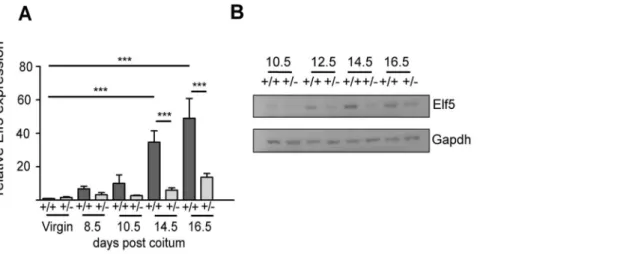
Pregnancy associated increase of Elf5 expression is delayed in theElf5+/2 mammary gland
To examine the correlation betweenElf5expression levels and the gene expression profiles observed we measuredElf5 mRNA and protein inElf5+/+
and Elf5+/2 mammary glands at various
stages of pregnancy (Figure 3). In theElf5+/+
mammary glandElf5
mRNA expression increased during the course of pregnancy, reaching a 49 fold induction in expression by 16.5dpc compared to its expression in the wildtype virgin gland (Figure 3a). Others have shown a similar Elf5 expression pattern during pregnancy-associated mammary gland development in C57Bl/6 x 129SVPas mice [5]. Expression ofElf5remained relatively low in theElf5+/2
mammary glands throughout early pregnancy but began to increase at the later timepoints (Figure 3a). In theElf5+/2gland
at 14.5dpc,Elf5expression increased by 5.94 fold with respect to the virgin wildtype gland, compared with 34.72 fold in theElf5+/+
gland (p,0.001). Likewise, at 16.5dpc Elf5 expression in the
Elf5+/2 mammary gland had only increased by 13.69 fold (with
respect to the virgin wildtype condition), whereas in the pregnant wildtype mammary gland at this stage, Elf5 expression was increased by 49.08 fold (p,0.001). In the wildtype mammary gland, Elf5 protein was detected at all time points examined and its expression increased throughout pregnancy as previously de-scribed (Figure 3b) [3,5]. Similar to its mRNA expression pattern, Elf5 protein was detected in allElf5+/2mammary gland samples,
however the levels of Elf5 expression remained comparable to that in the 10.5dpc Elf5+/2 mammary gland until 16.5dpc where it
increased slightly (Figure 3b).
Identification of potential Elf5 target genes
To identify potential Elf5 targets, gene expression changes due to Elf5 heterozygosity were examined at each individual timepoint.
Figure 2. Differentiation of theElf5+/2mammary gland during pregnancy is delayed. A.Western blot analysis of the milk proteinsa
-casein,b-casein and Wap in the mammary glands ofElf5+/+
andElf5+/2mice.B.Lactalbumin (Lalba) expression in the mammary glands ofElf5+/+
and
Elf5+/2mice measured by microarray analysis. Expression is shown relative to the virginElf5+/+sample.
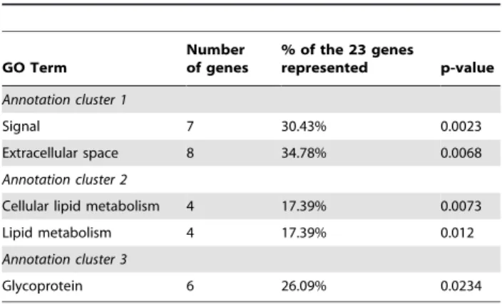
Lists of the gene expression changes and the gene ontology analysis of these changes can be found in supplementary Tables S1, S2, S3, S4, S5, S6, S7, S8, S9, S10, S11, S12, S13, S14 with accompanying descriptions in supplementary files S1 and S2. We also used clustering algorithms to organise our data. This type of analysis assembles genes with common patterns of expression into groups, called clusters. Our rationale for choosing clustering was that genes grouped according to their similar expression patterns were likely to be co-regulated. By clustering our data set and identifying the cluster containing Wap, a known Elf5 target gene [6], we anticipated identifying other Elf5 targets.
Employing two commonly used algorithms, K-means and self organising maps (SOMs), we searched for the genes that clustered with Wap. Independent K-means clustering was performed 10 times, the first time with a K value of 5, after which the K value was increased by 5 each time until 50 clusters was reached. Of the 10 K-means analyses performed, K = 15 produced the highest explained variability (70.435%) and for this reason was used in the remainder of our analysis. We also generated multiple SOMs (464, 465, 466, 565, 566 and 666), with the 666 SOM generating the highest explained variability (79.419%). We identified the clusters from each of the 15 cluster K-means and the 666 SOM that containedWapand compared the two lists to identify 23 common genes (Figure 4). The expression of all 23 of these genes was downregulated at all timepoints in the pregnant
Elf5+/2gland compared to the pregnant wildtype gland.
Next we used the DAVID functional annotation clustering tool (http://david.abcc.ncifcrf.gov/home.jsp) [13] to determine wheth-er any gene ontology classification was ovwheth-er-represented in the 23 genes that consistently clustered with Wap (Table 1). The gene ontology (GO) terms signal, extracellular space, cellular lipid metabolism, lipid metabolism and glycoprotein were all identified as being significantly enriched in the list with each having a p-value less than 0.05. The GO terms signal and extracellular space defined under annotation cluster 1, represented 34.78% of the 23 genes and included Csng, Lamp1, Col9a1, Wap, Btn1a1, Glycam1,
Aplp and Pp11r. The terms cellular lipid metabolism and lipid metabolism defined under annotation cluster 2, represented 17.39% of the genes and includedCte1,Lrat,Fabp3,Alox12e. The term glycoprotein was also significantly enriched (annotation cluster 3) and represented the genes Lamp1, Col9a1, Btn1a1,
Glycam1,AplpandIghg.
Figure 3. Elf5 expression profile during pregnancy associated mammary gland development. A.Elf5 mRNA expression in in the mammary glands ofElf5+/+
andElf5+/2mice measured by qRT-PCR. Expression is shown relative to the average expression ofElf5in theElf5+/+
virgin samples. ***p,0.001. n = 3.B.Western blot analysis of Elf5 protein expression in the mammary glands ofElf5+/+andElf5+/2mice during pregnancy.
doi:10.1371/journal.pone.0013150.g003
Figure 4. Genes that clustered with the known Elf5 target gene Wap.Expression profiles of the 23 genes that consistently clustered withWapusing 2 clustering algorithms, in the mammary glands of
Elf5+/+
andElf5+/2mice. Gene expression is shown relative to expression
in the virginElf5+/+condition.
Identification ofEtsbinding sites in the upstream regions of potential Elf5 target genes
We hypothesised that the promoter of a gene whose expression is directly regulated by Elf5 would contain anEtsbinding site – and more specifically, anEtssite with a specific flanking sequence reported to be preferred by Elf5 [59(A/C)GGAA(G/A)(T/G)(A/ G)NNC 39] [14]. The predicted promoter regions (1000 bp upstream of the transcriptional start site) of 20 of the 23 genes clustered with Wap were obtained using EZretrieve. (http:// siriusb/umdnj.edu:18080/EZRetrieve/index.jsp) and Promoser (http://biowulf.bu.edu/zlab/PromoSer). The upstream sequences for the genesAcbd7,Ighgand Gm566were not identified by these programs. Putative promoter sequences were searched using the Transcriptional Element Search System (TESS), which is
accessible via the web-based Baylor College of Medicine (BCM) Search Launcher (http://searchlauncher.bcm.tmc.edu/). Of the 20 promoter sequences searched, 5 (Wap,Muc4,Col9a1,Pp11rand
Lamp1) contained at least one putativeEtsbinding site. We chose to investigateMuc4as a potential direct transcriptional target of Elf5 since Muc4has a role in the pregnant mammary gland in rodents and in humans and dysregulated expression ofMUC4has been associated with breast cancer [15]. Moreover, MUC4 is transcriptionally regulated by another Ets factor, PEA3 [9]. In addition, the predictedEtssites at nucleotide positions2216 and
21613 (where numbering is relative to the initiating ATG) of the humanMUC4promoter are almost identical to the preferredElf5
binding site, with only the 39 nucleotide differing from the predictedElf5preferred sequence.
Muc4expression is decreased in theElf5+/2 mammary gland
The Muc4 expression pattern observed in the microarray analysis (Figure 5a) was confirmed by quantitative real time RT-PCR on RNA samples distinct from those used in the microarray experiments (Figure 5b). While there was no significant difference in Muc4 expression between the two genotypes in the virgin, 8.5dpc or 10.5dpc glands, a 10 fold reduction inMuc4expression was observed at 14.5dpc in the Elf5+/2 gland compared to the
wildtype (55.16 fold in the wildtype compared with 5.26 fold in
Elf5+/2; p,0.001). A significant 2.3 fold decrease in Muc4
expression was observed in theElf5+/2gland at 16.5dpc compared
with the 16.5dpc wildtype gland (43.65 fold in the wildtype gland compared with 18.76 fold inElf5+/2; p,0.05). Correspondingly,
while Muc4 protein increased dramatically at 18dpc and was sustained at 1dpp in theElf5+/+
mammary gland, Muc4 protein was undetectable in theElf5+/2 mammary glands at these times
(Figure 5c).
Table 1.Functional annotation clustering of the 23 genes
that clustered with Wap.
GO Term
Number of genes
% of the 23 genes
represented p-value
Annotation cluster 1
Signal 7 30.43% 0.0023
Extracellular space 8 34.78% 0.0068
Annotation cluster 2
Cellular lipid metabolism 4 17.39% 0.0073 Lipid metabolism 4 17.39% 0.012
Annotation cluster 3
Glycoprotein 6 26.09% 0.0234
doi:10.1371/journal.pone.0013150.t001
Figure 5. ExaminingMuc4as a potential Elf5 target gene. A.Muc4mRNA expression in the mammary glands ofElf5+/+
andElf5+/2mice
measured by microarray analysis. Expression is shown relative to the virginElf5+/+condition.B.Muc4expression in the mammary glands ofElf5+/+and
Elf5+/2mice measured by qRT-PCR. Expression is shown relative to the average expression ofMuc4in the virginElf5+/+samples. ***p
,0.001; *p,0.05. n = 3.C. Western blot examining Muc4 protein expression inElf5+/+
andElf5+/2mammary glands.
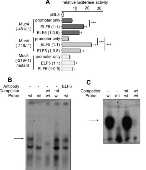
ELF5 can activate theMUC4promoter via theEtsbinding site at2216
The full-length proximal hMUC4promoter (2461/21) and a truncated proximal promoter (2219/21) were used to drive a
luciferase reporter gene [10]. The proximal promoter contains a predictedELF5-preferredETS binding site at position 2216, in addition to a predictedETSbinding site at2349 [14]. The human breast carcinoma cell line T47D was used since it expressesMUC4
[16] and therefore contains all the factors required for MUC4
expression. Cells were transfected with the promoter constructs alone, or co-transfected with an ELF5 expression plasmid [6]. The full-length proximalMUC4promoter triggered a 7.49 fold increase in luciferase activity over the empty pGL3 vector (Figure 6a).
Co-expression of ELF5 in these cells at a 1:1 molar ratio (ELF5:MUC4 promoter) resulted in a significant increase in promoter activity (16.06 fold over the promoterless vector; p,0.001). Luciferase expression driven by the truncatedMUC4
proximal promoter increased 4.69 fold above the baseline of the promoterless vector and significant increases in luciferase activity were observed when ELF5 was over-expressed in these cells at a 0.5:1 or 1:1 (ELF5:promoter) molar ratio (p,0.001) indicating that ELF5 was acting on theMUC4promoter.
To confirm that ELF5 was activating theMUC4promoter via the preferredELF5binding site at nucleotide position2216, the promoter-luciferase reporter experiments were repeated with a
MUC4 2219/21 promoter-luciferase construct in which the Ets
Figure 6. ELF5 directly regulates MUC4 promoter activity. A. The effect of ELF5 expression onMUC4promoter activity was tested usingMUC4
promoter-luciferasereporter gene assays with different regions of theMUC4promoter. Cells were transfected with either the promoter construct alone or with the promoter construct plus an ELF5 expression vector at molar ratios of 1:1 or 1:0.5 (promoter-luciferasevector:ELF5 expression vector). Luciferase expression is shown relative to the expression of the pGL3 promoterless vector. *** p,0.001; **p,0.01. n = 3.B.EMSA was used to determine whether Elf5 could bind the2216Etssite within theMUC4promoter. Binding was observed (arrow) in lane 1 when a nuclear extract from a 16.5dpcElf5+/+
mammary gland was incubated with a radioactively labelled oligonucleotide encompassing the2216Etssite from theMUC4
promoter (wt). The binding observed was not evident when theEtssite in the probe was mutated (mt) (lane 2), indicating that the upper band represents specific binding to theEtssequence. Binding to the wt probe was competed off with a 100-fold molar excess of unlabelled wt oligo (lane 3) but not with mt oligo (lane 4). Binding to the wt probe was not shifted with the addition of anti-ELF5 antibody (lane 5).C.The EMSA from (A) was performed again using aD33 ELF5 recombinant protein in place of the nuclear extract. The truncated recombinant ELF5 protein bound the labelled wt oligo (lane 1), but not the mt oligo (lane 2). Binding to the wt oligo could not be competed off with the mt oligo (lane 3), but was successfully competed off using an excess of unlabelled wt oligo (lane 4).
binding site at2216 had been mutated (GGAA was changed to AAAA) [9]. This2216/21 mutant construct showed promoter activity equivalent to that of the wildtype 2216/21 region, indicating that basal MUC4 expression in T47D cells was not dependant on the Etsbinding site at position 2216. However, over-expression of ELF5 failed to induce activity of the mutated
MUC4promoter (Figure 6a), indicating that ELF5 was acting on theEtsbinding site at nucleotide position2216.
ELF5 binds theEtsbinding site at2216 in the MUC4 promoter
To confirm that ELF5 regulatedMUC4expression by directly binding its target sequence, we performed electrophoretic mobility shift assays (EMSA). Two bands were evident upon incubation of the wildtype probe with the d16.5 pregnant mammary gland nuclear extract (Figure 6b, lane 1) indicating binding to the probe. When a probe containing the mutatedEtsbinding site was used only the lower band was evident (lane 2) suggesting that the higher band represented specific binding. Binding to the wildtype probe could be competed off by the addition of a 100-fold molar excess of a non-radiolabelled wildtype probe to the nuclear extract (lane 3) while the addition of an excess of mutated non-radiolabelled probe had no effect on binding activity (lane 4) confirming that binding to the2216Etssite was specific. To determine whether it was Elf5 in the nuclear extract binding to the site we performed a supershift assay by adding an anti-Elf5 antibody to the nuclear extract/probe mix. The addition of this antibody had no effect on the mobility of the probe/protein complex (lane 5). However, it is possible that this anti-Elf5 antibody does not function in a supershift assay.
Since we could not determine the identity of the protein in the nuclear extract binding the MUC4 promoter, we performed an EMSA using recombinant ELF5 protein. Full length recombinant ELF5 protein does not bind DNA efficiently due to the presence of a negative regulatory domain at the amino end of the protein [17], therefore a truncated ELF5 protein lacking 33 amino acids from the amino terminus was produced. The D33 ELF5 protein was able to bind the wildtype probe (Figure 6c, lane 1) but was unable to bind the mutated probe (lane 2), indicating that the binding to the wildtype probe was specific. Incubation with a 100-fold molar excess of the unlabelled mutated probe did not alter binding to the wildtype probe (lane 3) but addition of unlabelled wildtype probe competed off all binding (lane 4).
Discussion
Elf5 plays a major role in pregnancy-associated mammary gland development. Re-expression of Elf5 in the epithelium of the Prlr2/2 mammary gland restored mammary gland development during pregnancy [5], revealing thatElf5acts downstream of the Prlr, presumably as a Stat5 target gene [1,2]. However for the most part, the mechanism underpinning Elf5 function remains unclear. We used transcript profiling to examine the transcrip-tional changes induced by the loss of oneElf5allele, as a first step to defining this mechanism.
Transcript profiling revealed a delay in the development of theElf5+/2mammary gland during pregnancy
It has been proposed that Elf5 plays a major role in the co-ordination of proliferation, differentiation and apoptosis in the mammary epithelial cell compartment [18]. Our results, and the phenotype of the Elf5-over-expressing mouse [19], support the notion that Elf5 is essential for mammary epithelial cell differentiation. We have shown that the global gene expression
profile of the mammary gland shifts upon robust expression of Elf5 protein and coincides with the first expression of milk proteins. We observed delayed synthesis of milk proteins in theElf5+/2gland
where increased expression of Elf5 protein was delayed until 16.5dpc, suggesting that these mammary glands are not entering the differentiation phase of the developmental program until this time.
With the exception ofWap[6], the direct transcriptional targets of Elf5 in the mammary gland are unknown. Clustering analysis allowed us to generate a list of 23 genes with expression profiles similar to Wap, with six already known to play roles in the mammary gland (Csng[20];Glycam1[21];Muc4[22];Fabp3[23];
Btn1a1[24] andCx26[25]). Functional annotation of the 23 gene cluster revealed a significant enrichment for genes associated with signalling and the extracellular space, lipid metabolism and glycoproteins.
Of the 23 genes identified through clustering analysis, only 5 contained at least one Ets binding site in the promoter region analysed. A number of possible explanations exist. Firstly, the remaining genes may not be direct targets of Elf5 but may be regulated by factors which are themselves dependent on Elf5. Secondly, it is possible that Elf5 forms a complex with one or more other proteins and that this complex binds the promoter regions of target genes via the binding sites of the non-Ets proteins within the complex. Thirdly, the possibility exists that Ets binding sites involved in the regulation of these genes are located within a region outside of the 1000 base pairs searched in our study.
MUC4gene expression is directly regulated by ELF5 Muc4 is a glycoprotein located within the membrane of secretory epithelial cells [26] and its expression is dysregulated in breast cancer [15]. Here we have shown thatMuc4expression is significantly down-regulated in the Elf5+/2 mammary gland
compared to the wildtype gland at 14.5dpc and 16.5dpc. In the T47D breast cancer cell line, exogenous addition of ELF5 was able to induce expression from theMUC4promoter. Furthermore, we have shown that ELF5 is able to directly bind theMUC4promoter via a preferred ELF5 binding site.
Interestingly, the binding site used by ELF5 to regulateMUC4
expression is the same site used by another Ets factor, PEA3, to regulate MUC4expression in the pancreas [9], in HC11 mouse mammary epithelial cells and in MAT-B1 and MAT-C1 rat mammary tumour cells [27], suggesting some functional redun-dancy. However,Elf5 and Pea3are expressed at different stages of post-natal mammary gland development and the expression pattern of Muc4 in the pregnant mammary gland most closely resembles that of Elf5. The expression of bothElf5and Muc4is induced at mid pregnancy, peaking early in lactation, and diminishing during involution [reviewed in 3,5,26], whereas Pea3 protein expression is highest during puberty and during early pregnancy [reviewed in 28]. It therefore seems most likely thatMuc4expression in the pregnant mammary gland is regulated by Elf5 and not Pea3.
In rat mammary epithelial cells, Muc4 acts as a ligand for the ErbB2 receptor [29]. It has been suggested that a Muc4/ErbB2 complex is involved in the maintenance and survival of alveolar epithelial cells during pregnancy and that the downregulation of
Muc4andErbB2 expression during involution plays a role in the initiation of apoptosis [30]. Although Muc4 protein is absent in
Elf5+/2 mammary glands during pregnancy, we have previously
shown that there is no increase in apoptosis inElf5+/2mammary
glands [3].
autophosphorylation on the tyrosine residue at position 1248 [31]. In polarised epithelial cells, ErbB2 resides on the basolateral surface of the cell along with other ErbB receptors. The association of Muc4 with ErbB2 leads to the translocation of ErbB2 from the basolateral to the apical surface of the cell [31]. This repositioning effectively separates ErbB2 from its usual heterodimerisation partner - ErbB3. ErbB2/ErbB3 dimers are strong inducers of cellular proliferation [32], therefore the separation of these proteins prevents downstream proliferation signals from these receptors. The separation of ErbB2 from ErbB3 induced by Muc4 is thought to trigger the ‘switch’ from the proliferative phase to the differentiation phase in mammary epithelial cells [29,31].
The absence of Muc4 protein in the Elf5+/2 gland would
abrogate the formation of the Muc4/ErbB2 complex and the subsequent phosophorlation of ErbB2. Therefore it is likely that the switch from proliferation to differentiation is not induced in
Elf5+/2mammary glands. However we did not observe excessive
proliferation in theElf5+/2mammary glands. Rather, these glands
contained fewer alveolar structures than the wildtype gland. Therefore there must be other mechanisms involved in the cessation of mammary epithelial cell proliferation. Our data is consistent with Elf5 acting as a controller of a switch to differentiation, since robust expression of Elf5 protein in the
Elf5+/2 mammary gland resulted in a marked shift in the
transcriptional profile of the gland and the first expression of milk proteins – definitive markers of differentiation. We propose that Elf5 controls this switch partly via its regulation ofMuc4.
It is not unprecedented for an Ets family member to be involved in the control of the cellular cues that drive proliferation versus differentiation. Pea3 is a positive regulator of Muc4 expression in pancreatic cancer cells, whilst also acting as a negative regulator of ErbB2 [9]. Via its control of the balance between relative expression levels of Muc4 and ErbB2, Pea3 is thought to promote pancreatic cancer cell differentiation [9]. There is the possibility that the delicate balance of proliferation and differentiation may be a mechanism often driven by Ets factors.
Our hypothesis that Elf5 regulates the switch from mammary epithelial cell proliferation to differentiation is supported by the report of a mouse in which ectopic over-expression of Elf5 in the mammary gland led to a reduction in epithelial proliferation and forced differentiation of the mammary epithelium [19]. This resulted in the expression of numerous milk proteins in non-pregnant mammary glands [19]. This phenotype may have occurred via a direct upregulation of Muc4 by Elf5 and the subsequent termination of proliferative signals generated by ErbB2 and ErbB3 dimers on the basolateral surface of the epithelial cells. The novel findings of our study give insight into a newly defined role for Elf5 in the pregnant mammary gland. We hypothesise that via the direct regulation ofMuc4expression, Elf5 co-ordinates the switch between mammary epithelial cell proliferation and differentiation during pregnancy, making it a master regulator of cellular processes in this organ.
Supporting Information
File S1 Genes with significantly different expression in the
Elf5+/2 mammary gland compared with the Elf5+/+
mammary gland. Tables S1-S10.
Found at: doi:10.1371/journal.pone.0013150.s001 (0.02 MB DOC)
File S2 Functional Annotation Clustering - Tables S11-S14.
Found at: doi:10.1371/journal.pone.0013150.s002 (0.03 MB DOC)
Table S1 Genes upregulated inElf5+/2virgin mammary gland
compared toElf5+/+
virgin mammary gland.
Found at: doi:10.1371/journal.pone.0013150.s003 (0.03 MB DOC)
Table S2 Genes downregulated in Elf5+/2 virgin mammary
gland compared toElf5+/+
virgin mammary gland.
Found at: doi:10.1371/journal.pone.0013150.s004 (0.03 MB DOC)
Table S3 Genes upregulated in Elf5+/2 mammary gland
compared toElf5+/+
mammary gland at 8.5dpc.
Found at: doi:10.1371/journal.pone.0013150.s005 (0.03 MB DOC)
Table S4 Genes downregulated in Elf5+/2 mammary gland
compared toElf5+/+
mammary gland at 8.5dpc.
Found at: doi:10.1371/journal.pone.0013150.s006 (0.03 MB DOC)
Table S5 Genes upregulated in Elf5+/2 mammary gland
compared toElf5+/+
mammary gland at 10.5dpc.
Found at: doi:10.1371/journal.pone.0013150.s007 (0.03 MB DOC)
Table S6 Genes downregulated in Elf5+/2 mammary gland
compared toElf5+/+
mammary gland at 10.5dpc.
Found at: doi:10.1371/journal.pone.0013150.s008 (0.03 MB DOC)
Table S7 Genes upregulated in Elf5+/2 mammary gland
compared toElf5+/+
mammary gland at 14.5dpc.
Found at: doi:10.1371/journal.pone.0013150.s009 (0.03 MB DOC)
Table S8 Genes downregulated in Elf5+/2 mammary gland
compared toElf5+/+
mammary gland at 14.5dpc.
Found at: doi:10.1371/journal.pone.0013150.s010 (0.04 MB DOC)
Table S9 Genes upregulated in Elf5+/2 mammary gland
compared toElf5+/+mammary gland at 16.5dpc
Found at: doi:10.1371/journal.pone.0013150.s011 (0.04 MB DOC)
Table S10 Genes downregulated in Elf5+/2 mammary gland
compared toElf5+/+
mammary gland at 16.5dpc.
Found at: doi:10.1371/journal.pone.0013150.s012 (0.07 MB DOC)
Table S11 Functional annotation clustering of genes
dysregu-lated in theElf5+/2virgin mammary gland.
Found at: doi:10.1371/journal.pone.0013150.s013 (0.03 MB DOC)
Table S12 Functional annotation clustering of genes
downreg-ulated in theElf5+/2mammary gland at 14.5dpc.
Found at: doi:10.1371/journal.pone.0013150.s014 (0.04 MB DOC)
Table S13 Functional annotation clustering of genes
downreg-ulated in theElf5+/2mammary gland at 16.5dpc.
Found at: doi:10.1371/journal.pone.0013150.s015 (0.03 MB DOC)
Table S14 Functional annotation clustering of genes
upregu-lated in theElf5+/2mammary gland at 16.5dpc.
Acknowledgments
The authors wish to thank Ashley Connolly of the Adelaide Microarray Facility, Ashley Mansell and Shamith Samarajiwa of Monash Institute of Medical Research, Nicolas Jonckheere of Inserm and C. Marcelo Sergio of the Garvan Institute for technical advice and assistance.
Author Contributions
Conceived and designed the experiments: RLR MP. Performed the experiments: RLR. Analyzed the data: RLR MP. Contributed reagents/ materials/analysis tools: IVS JG PJH MJN MP. Wrote the paper: RLR MP. Assisted with the editing of the final paper and provided financial assistance: MJN PJH MP.
References
1. Hennighausen L, Robinson G (2005) Information networks in the mammary gland. Nature Reviews Molecular Cell Biology 6: 715–725.
2. Oakes SR, Rogers RL, Naylor MJ, Ormandy CJ (2008) Prolactin regulation of mamamry gland development. J Mammary Gland Biol Neoplasia 13: 13–28. 3. Zhou J, Chehab R, Tkalcevic J, Naylor MJ, Harris J, et al. (2005) Elf5 is essential
for early embryogenesis and mammary gland development during pregnancy and lactation. Embo J 24: 635–644.
4. Donnison M, Beaton A, Davey HW, Broadhurst R, L’Huillier P, et al. (2005) Loss of the extraembryonic ectoderm in Elf5 mutants leads to defects in embryonic patterning. Development 132: 2299–2308.
5. Harris J, Stanford PM, Sutherland K, Oakes SR, Naylor MJ, et al. (2006) Socs2 and Elf5 mediate prolactin-induced mammary gland development. Mol Endocrinol 20: 1177–1187.
6. Thomas RS, Ng AN, Zhou J, Tymms MJ, Doppler W, et al. (2000) The Elf group of Ets-related transcription factors. ELF3 and ELF5. Adv Exp Med Biol 480: 123–128.
7. Yuan JS, Reed A, Chen F, Stewart CNJ (2006) Statistical analysis of real-time PCR data. BMC Bioinformatics 7: 85.
8. Jonckheere N, Vincent A, Perrais M, Ducourouble MP, Male AK, et al. (2007) The human mucin MUC4 is transcriptionally regulated by caudal-related homeobox, hepatocyte nuclear factors, forkhead box A, and GATA endodermal transcription factors in epithelial cancer cells. J Biol Chem 282: 22638–22650. 9. Fauquette V, Perrais M, Cerulis S, Jonckheere N, Ducourouble MP, et al. (2005) The antagonistic regulation of human MUC4 and ErbB-2 genes by the Ets protein PEA3 in pancreatic cancer cells: implications for the proliferation/ differentiation balance in the cells. Biochem J 386: 35–45.
10. Perrais M, Pigny P, Ducourouble MP, Petitprez D, Porchet N, et al. (2001) Characterization of human mucin gene MUC4 promoter: importance of growth factors and proinflammatory cytokines for its regulation in pancreatic cancer cells. J Biol Chem 276: 30923–30933.
11. Robinson GW, McKnight RA, Smith GH, Hennighausen L (1995) Mammary epithelial cells undergo secretory differentiation in cycling virgins but require pregnancy for the establishment of terminal differentiation. Development 121: 2079–2090.
12. Naylor MJ, Oakes SR, Gardiner-Garden M, Harris J, Blazek K, et al. (2005) Transcriptional changes underlying the secretory activation phase of mammary gland development. Mol Endocrinol 19: 1868–1883.
13. Dennis G, Sherman BT, Hosack DA, Yang J, Gao W, et al. (2003) DAVID: Database for Annotation, Visualization, and Integrated Discovery. Genome Biol 4: 3–9.
14. Yaniw D, Hu J (2005) Epithelium-specific ets transcription factor 2 upregulates cytokeratin 18 expression in pulmonary epithelial cells through an interaction with cytokeratin 18 intron 1. Cell Res 15: 423–429.
15. Rakha EA, Boyce RW, Abd El-Rehim D, Kurien T, Green AR, et al. (2005) Expression of mucins (MUC1, MUC2, MUC3, MUC4, MUC5AC and MUC6) and their prognostic significance in human breast cancer. Mod Pathol 18: 1295–1304.
16. Yuan Z-L, Guan YJ, Wang L, Wei W, Kane AB, et al. (2004) Central Role of the Threonine Residue within the p+1 Loop of Receptor Tyrosine Kinase in STAT3 Constitutive Phosphorylation in Metastatic Cancer Cells. Mol Cell Biol 24: 9390–9400.
17. Oettgen P, Kas K, Dube A, Gu X, Grall F, et al. (1999) Characterization of ESE-2, a novel ESE-1-related Ets transcription factor that is restricted to
glandular epithelium and differentiated keratinocytes. J Biol Chem 274: 29439–29452.
18. Oakes SR, Hilton HN, Ormandy CJ (2006) The alveolar switch: coordinating the proliferative cues and cell fate decisions that drive the formation of lobuloalveoli from ductal epithelium. Breast Cancer Res 8: 207.
19. Oakes SR, Naylor MJ, Asselin-Labat ML, Blazek KD, Gardiner-Garden M, et al. (2008) The Ets transcription factor Elf5 specifies mammary alveolar cell fate. Genes Dev 22: 581–586.
20. Yu-Lee LY, Rosen JM (1983) The rat casein multigene family. I. Fine structure of the gamma-casein gene. J Biol Chem 258: 10794–10804.
21. Groenen MA, Dijkhof RJ, van der Poel JJ (1995) Characterization of a GlyCAM1-like gene (glycosylation-dependent cell adhesion molecule 1) which is highly and specifically expressed in the lactating bovine mammary gland. Gene 158: 189–195.
22. Carraway KLr, Rossi EA, Komatsu M, Price-Schiavi SA, Huang D, et al. (1999) An intramembrane modulator of the ErbB2 receptor tyrosine kinase that potentiates neuregulin signalling. J Biol Chem 274: 5263–5266.
23. Yang Y, Spitzer E, Kenney N, Zschiesche W, Li M, Kromminga A, et al. (1994) Members of the fatty acid binding protein family are differentiation factors for the mammary gland. 127 4.
24. Ogg SL, Weldon AK, Dobbie L, Smith AJ, Mather IH (2004) Expression of butyrophilin (Btn1a1) in lactating mammary gland is essential for the regulated secretion of milk-lipid droplets. Proc Natl Acad Sci U S A 101: 10084–10089. 25. Locke D, Perusinghe N, Newman T, Jayatilake H, Evans WH, et al. (2000)
Developmental expression and assembly of connexins into homomeric and heteromeric gap junction hemichannels in the mouse mammary gland. J Cell Physiol 183: 228–237.
26. Carraway KL, Price-Schiavi SA, Komatsu M, Jepson S, Perez A, et al. (2001) Muc4/Sialomucin Complex in Mammary Gland and Breast Cancer. Journal of Mammary Gland Biology and Neoplasia 6: 323–337.
27. Perez A, Barcos R, Fernandez I, Price-Schiavi SA, Carraway KL (2003) PEA3 transactivates the Muc4/Sialomucin complex promoter in mammary epithelial and tumour cells. J Biol Chem 278: 36942–36952.
28. Shepherd T, Hassell JA (2001) Role of Ets transcription factors in mammary gland development and oncogenesis. J Mammary Gland Biol Neoplasia 6: 129–140.
29. Jepson S, Komatsu M, Haq B, Arango ME, Huang D, et al. (2002) Muc4/ Sialomucin complex, the intramembrane ErbB2 ligand, induces specific phosphorylation of ErbB2and enhances expression of p27kip, but does not activate mitogen-activated kinase or protein kinase B/Akt pathways. Oncogene 21: 7524–7532.
30. Price-Schiavi SA, Andrechek E, Idris N, Li P, Rong M, et al. (2005) Expression, location, and interactions of ErbB2 and its intramembrane ligand Muc4 (sialomucin complex) in rat mammary gland during pregnancy. J Cell Physiol 203: 44–53.