ZDB-Number: 2668735-5
IC Journal No: 8192
Volume 2 Issue 1
Online Available at www.phytojournal.com
Journal of Pharmacognosy and Phytochemistry
Vol. 2 No. 1 2013 www.phytojournal.com Page | 209
Cnidarian from the Coast of Goa – Identified to the
Species Level
J. Mythili Krishna 1* and A. Gophane 2
1. Department of Pharmacognosy, Goa College of Pharmacy, Goa 403 001, India [E-mail: [email protected]]
2. Department of Zoology, Willingdon College, Sangli, Maharashtra-416415 India
Cnidaria represents a phylum of marine organisms consisting of colony forming aquatic (mostly marine) animals which have a rich potential as a source of drugs and therapeutic agents. One such organism which has gained importance in the contemporary times is zoanthid. There is a growing interest in ascertaining their taxonomic diversity using a combination of molecular, morphological and histological techniques. The unusual life style, symbiotic photosysthesis, ability to form colonies with polyps and the marine niche which they occupy make them a very interesting organism for research. There are many reports of toxicological properties as well as characterization of the methanolic extract of Zooanthids from Indian ocean. However, there is very little work on systematics of these organisms. We report isolation and identification of this soft coral from the intertidal rocky shore of Anjuna (Goa) following standard protocols of morphological, histological and molecular analyses.
Keyword: Cnidaria, Zoanthid, Molecular Techniques and Histological Techniques.
1. Introduction
Marine organisms are an integral part of pharmaceutical search for new discoveries to combat the increasing ailments affecting the human race. This inquisitiveness ostensibly started as early as 1872, when the first organized exploration of the oceans was launched by Sir C.
Wyville Thomson as Challenger Expedition
(1872-1876 AD) and was ensued by the John
Murray - Mabahiss Expedition (1933-1934 AD)[1]. The first step for any novel research is a clear taxonomic identification of the organism under study.
Presently, it has been approximated that the range of marine species on earth is from 1.4 to 1.6 million, wherein a total of only 2,12,000 (+/-
29%) species only are exactly known. This huge
divergence between the two estimates has been mainly because of insufficient information on the biodiversity of smaller organisms whereas
abundant data is available on marine mammals[2].
One such relatively small organisms of
significance are zoanthids (soft corals). These have become recent interests in the field of scientific inquisition for two reasons. The first is the challenge they offer in their taxonomic discernment: due to the morphological variability within a species and paucity of research to define dependable canon for segregation into genera and
species[3,4]. The second is the presence of certain
biochemical substances which are of
Vol. 2 No. 1 2013 www.phytojournal.com Page | 210
Palythoa sp[5]; norzoanthamine group of alkaloids which have shown promise in the treatment of
osteoporosis, from the zoanthid Zoanthus sp.[6]
and the latest, important contribution to the group of biotechnological revolutionary tools, Zs Green (zFP506) and Zs Yellow (zFP538), the respective coloured fluorescent proteins, from the zoanthid
Zoanthus sp[7]. The first facet has been taken up globally, although verily none from India.
The subcontinent of India has an 8,000 Km long coastline. Due to its sub-tropical climatic conditions, possesses limited number of coral reef fields, in contrast to other regions of the globe. Out of these, many are present on the east and west coast, with only few patches found in the
region of Ratnagiri, Malwan and Goa[8]. Malwan
is the nearest coast to Goa with a distance of 62 Km or 38 miles approximately. There is very little detailed literature available with respect to zoanthids from the Malwan coast; only one report by Dr. Parulekar, 1981, mentioning the presence
of a zoanthid Epizoanthus elongatum (Verill),
Epizoanthidae[9] and none from the coast of Goa.
Some records are available about the biochemical aspects of the zoanthids of India, albeit, the work has been limited to methanolic extracts of the same[10,11,12]: Hariamide – a novel sulphated
sphingolipid by CDRI[13], Lucknow, and
2-deoxyecdysterone – a novel oxytoxic agent by the
NIO[14], Goa (fig. 1 and 2).
1.1 Zoanthids:
Fundamentally, zoanthids are hexacrorallian organisms (soft corals having hexamerous symmetry). They belong to the order Zoantharia/Zoanthidea/Zoanthinaria. This order is categorized in the class Anthozoa, which in turn is placed in the phylum/kingdom called cnidaria/coelenterate. Basically, the order is characterized by colonies of soft bodied polyps forming mats or solitary in nature and carrying two rows of tentacles[15] (fig. 3 and 4). Overall, zoanthids are composed of three layers, Ectoderm (outer layer), Endoderm (inner layer) and mesoglea (the middle layer) (fig. 5).
Besides the external morphological features, the internal elements include three major criteria for
identification: ‘Actinopharynx’, ‘Siphonoglyph’ and ‘Mesenteries’. Actinopharynx also called as stomadeum or gullet is a colon or a tube-like structure lined by endodermic cells, and protrudes itself into the gastrovascualr cavity or coelenteron of the organism. Siphonoglyph otherwise known as sulcus is the highly glandular region of the actinopharynx having a thick covering of cilia. The sulcus ascertains the dichotomic alignment inside a polyp. This symmetrical alignment is further delineated due to the existence of ‘Mesenteries’. ‘Mesenteries’ also referred to as ‘septa’ are radially-arranged layers of tissues, present ‘all’ or ‘portion’ of the distance between the body wall to the central tube or actinopharynx. These are arranged in cycles of six (hexamerous symmetry). The distribution of mesentries and the type (complete or incomplete) of the fifth pair of mesentry with respect to the dorsal injunction decides the further division of the order into the sub-orders: Brachycnemina (incomplete fifth pair) and macrocnemina (complete fifth pair). These sub-orders are further segregated into families – Sphenopidae and Zoanthidae (Brachycnemina); Epizoanthidae,
Abysszoanthidae and Neozoanthidae
(Macrocnemina)[15].
1.2 Taxonomic Pandemonium:
The perplexity of zoanthid taxonomy is attributed to both the morphological as well as histological aspects. Morphologically the flexibility and
malleability exhibited hinders the precise
subjective evaluation. Many morphological identification dictums have been hypothesized in the yester years grading from color, sphincter muscle anatomy, tentacle number etc. Hence the characterization of original specimen/species varied in accordance with the authors describing them. Also poor conservation or maim or mutilation of the specimen lead to lack of
handiness or accessibility of reference
samples[16]. A fair understanding of the situation
can be obtained by analyzing the literature available on zoanthids. Systematic study on
Zoanthus erythrochloros and Z. gnophodes was
carried out in the year 1957 by Pax and Muller[17].
Vol. 2 No. 1 2013 www.phytojournal.com Page | 211
zoanthid, Z. pacificus was matriculated by Walsh
and Bowers[18]. Subsequently, after about 30
years 2001, similar results of morphological study have been documented by Uchida and Soyama regarding the above three species along
with a new one Z. sansibaricus[19]. A more,
appropriate range of the values was recorded, for
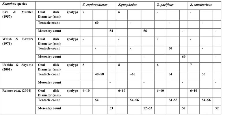
each feature of the zoanthid referred above, by Reimer et.al in 2004[20]. This range ensures the flexibility and plasticity of growth conditions which has not been reflected in the previous instances and henceforth forms a better standard for comparison. The summary of the above data is presented in Table 1.
Table 1: Summary of Zoanthus species morphological characteristics from previous literature.17-20
Zoanthus species Z. erythrochloros Z.gnophodes Z. pacificus Z. sansibaricus
Pax & Mueller (1957)
Oral disk (polyp) Diameter (mm)
7 6 - -
Tentacle count 60 - - -
Mesentry count 54 56 - -
Walsh & Bowers (1971)
Oral disk (polyp) Diameter (mm)
- - 7 -
Tentacle count - - 60 -
Mesentry count - - 60 -
Uchida & Soyama (2001)
Oral disk (polyp) Diameter (mm)
8 8 6 7
Tentacle count 48~58 ~60 54 56
Mesentry count - - - -
Reimer et.al. (2004) Oral disk (polyp) Diameter (mm)
6~10 6~10 6~10 6~10
Tentacle count 54 54~56 54~58 54~56
Mesentry count 53 52~53 52 52
Histological study of zoanthid internal anatomy is another labrynthine aspect to deal with. Presence of indurate substances like sand encrustations, bone pieces etc. makes the sectioning difficult as there is a risk of damaging the soft tissues if excess pressure is applied. Thus accurate understanding of the internal morphology can be obtained only after ensuring the complete elimination of the hindrances. Previously a method involving the use of HF was commonly used to deliquesce the sand particles from the
encrusted zoanthids – termed as desilification[21].
This was the most frequent method applied, despite the hazardous nature of HF. Yet the results of such a treatment were not satisfactory with regard to the quality. It was then realized that the fluoride ion of HF, indomitably bonds to
the free calcium ion, produced insoluble CaF2. This infers that the zoanthids which contain calcium in the form of bone pieces etc., ingrained in the mesoglea, require another supplementary
technique of decalcification preceding
desilification. A protocol to this affect has now been formulated, standardized and documented
by Reimer et.al[22]. This approach was
Vol. 2 No. 1 2013 www.phytojournal.com Page | 212 Identification studies have recently proceeded
productively due to the designing of DNA barcoding. Instances wherein morphological data when by itself proved inadequate for analysis, application of genetic information to realize the
output have proved to be eventful[23]. DNA
barcoding encompasses several domains,
diversifying from biodiversity estimation,
traceability of commercialized organisms to the understanding of the connection between various life stages[24- 26].
The primary innovative attribute of barcoding is the selection of one or a few standardized markers which help in interspecific segregation of the species. In general partial cytochrome oxidase subunit I (COI) sequences or the large mitochondrial ribosomal subunit (mt 16S rDNA)
gene is used as barcode. Sinniger F. et.al., have
done comprehensive study of the applicability of the two markers in identifying zoanthids and have reported that either both or one of the markers
can be used to testify zoanthids[27].
In the present study, a systematic approach has been applied for a zoanthid from the Indian coast to identify it till the species level, using standardized protocols, involving morphological, histological and molecular techniques, which
would then form a basis for further
phytochemical study.
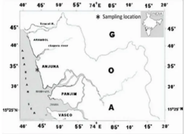
2. Materials and Methods 2.1 Sampling
Zoanthid samples, in the form of mat of organisms were collected from the intertidal rocky shore of Anjuna beach, along the coast of Goa (fig. 1 and fig. 2) and preserved in absolute alcohol. As samples were being collected photographs were taken to assist in identification and recording of morphological (external) diagnostic character data (oral disk/polyp diameter and tentacle count).
2.2 Decalcification and Hydrofluoric acid Desilification Protocol:
Initially from the mat of the zoanthids collected, small portions were cut and transferred to 75% ethanol followed by 10% formalin seawater for
24 hours. The polyps were then decalcified by chelation with a mixture of 20% citric acid and 50% formic acid subsequently diluted to 50% strength with distilled water. Decalcification was stopped when bubbles no longer emitted from the polyps (~6hrs). Colonies were then rinsed overnight in distilled water, with water being changed multiple times. Polyps were then treated with 15% HF with pH of <1 for 48 hrs. During desilification treatment all safety protocols were followed (triple gloves, safety goggles, conducted in fume hood, etc.). After desilification, polyps were rinsed overnight with multiple changes of distilled water until pH was approximately 7.0
and then stored in 70% ethanol until
sectioning[22].
Fig 1: Rocky intertidal zone of Anjuna beach, Goa
Vol. 2 No. 1 2013 www.phytojournal.com Page | 213
2.3 Histology:
The specimens were dehydrated through an ethanol-xylene series. Some specimens in 100%
ethanol were placed in vacuo for approximately
30min to remove air bubbles in the coelenteron. From the mat sample, individual specimens were separated. Then they were embedded in paraffin and blocks were prepared. Serial sections of 5-10µm thick were prepared with a rotary
microtome and stained with Delafields’
hematoxylin and eosin. The slides so prepared were examined with a light microscope (Olympus IX51 Inverted Microscope). The following morphological characters and conditions were examined; mesentry condition, number and form (in particular fifth mesentry from dorsal directive complete or incomplete; presence or absence of sand and debris in mesoglea; overall condition of the tissue and in particular ectoderm and endoderm.
2.4 DNA Extraction and PCR Amplification and Sequencing:
DNA was extracted from ethanol-preserved
samples using Chloroform: Phenol:
Isoamylalcohol extraction protocol. Small pieces of the polyps were cut (~200mg), dried and subjected to digestion using a lysis buffer (100mM TrisHCl pH 8, 100mM Na2EDTA pH 8, 1.5M NaCl, 1% CTAB) for 1 hr at 60°C in a water bath. The lysed tissues were subjected to centrifugation at maximum RPM of 12,000 for 15 minutes. To the above supernatant 500µl of Chloroform : Phenol : Isoamylalcohol (25:24:1) mixture was added and subjected to rotor mixing for 30 minutes, followed by centrifugation at maximum RPM of 12,000 for 10 minutes. The above step was repeated again for maximum extraction of residual proteins. To the supernatant 500µl of Chloroform: Isoamylalcohol (24:1) mixture was added and centrifuged at maximum RPM of 12,000 for 10 minutes. To the above supernatant equal volume of 100% ice cold ethanol was added (to precipitate DNA extracted) and centrifuged at maximum RPM of 12,000 for 10 minutes. All the supernatant liquid was removed carefully without disturbing the pellet obtained. The pellet so obtained was incubated
for 5-7 minutes with 70% ethanol and then centrifuged to wash it clear of any residual contamination. The pellet was then dried and
eluted into 50µl of pure water[28, 29].
Mitochondrial 16s Ribosomal DNA (mt 16s rDNA) was amplified using zoanthid - specific
primers 16Sant0a
5’-GAAGTAGGCTTGGAGCCAGCCA-3’
(Forward) and 16SbmoH
5’-CGAACAGCCAACCCTTGG-3’ (Reverse), with the following thermal cycle conditions: an initial denaturing step at 95°C for 2 minutes, followed by 35 cycles of 1 minute denature at 95°C, 30 seconds annealing at 52°C and 90 seconds extension at 72°C, followed by 7 minutes
extension at 72°C[30].
The amplified PCR products were checked by 1.2% agarose gel electrophoresis. Cycle sequencing was accomplished in both directions using the forward and reverse primers separately. Reagents and reaction conditions were as specified in the ABI Prism Big Dye v.3.1 Terminator cycle sequencing ready reaction kit (PE Applied Biosystems, Foster City, CA, USA). Reaction products were analysed on an Applied Biosystems 3130xL genetic analyser (Division of Perkin Elmer, Foster City, CA, USA). The sequences were analysed by Sequencing Analysis Software v5.3.1 (ABI Software, Division of Perkin Elmer, Foster City, CA, USA).
3. Result and Discussion
Vol. 2 No. 1 2013 www.phytojournal.com Page | 214
between the zoanthids under study and Zoanthus
sansibaricus[30(a)]. Also the oral disc diameter,
tentacle count and mesentry count of the present polyp under investigation matched well with the
recently documented data on Zoanthus
sansibaricus from the coast of Japan[31].
Slides resulting after the treatment and sectioning are shown below (fig. 5). Cross-sections did not
have any sand or debris remaining. Holes resembling lacunae were seen in the mesoglea, and little portions of ectodermal tissue were found damaged. There were complete and
incomplete mesentries and the 5thmesentry from
the dorsal directive were incomplete indicating a
Vol. 2 No. 1 2013 www.phytojournal.com Page | 215 3.2 16S mtrDNA Gene Sequence:
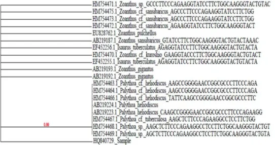
The amplified gene of 16S mtrDNA was found to be approximately 1200 bp from the agarose gel picture (fig. 6). This could be further sequenced partially. The partial sequence of 16S mtrDNA was obtained and the nucleotide sequence was
deposited in Gen Bank (accession number HQ840729). The partial nucleotide sequence (~556bp) when BLAST in NCBI was found to be >99% similar (fig. 7) to 16S mt rDNA of
Zoanthus sansibaricus(AB219187.1). The match is as follows:
Fig. 7: Phylogenetic analysis of the sample based on 16S mtr DNA gene
4. Discussion
4.1 Morphological Analysis:
Species of the genus Zoanthus are colonial and
new polyps arise from the horizontal main body of the organism. Zoanthid polyps are found in all oceans where they form colonies of separate individuals that are either tightly packed or sparse, depending on the species. They have a wide variety of colours, shapes and sizes. From the photographs (fig. 3 and fig. 4) of the zoanthid sample under study and by subjective evaluation, they have been found to possess the following characters: a green colour oral disc with pink ring around it, which is typical of zoanthids, as they are reported to possess such bright fluorescent colours; the oral disc could be seen only when they open up; overall the colour was that which could be camouflaged in the rocks; the polyps were tough and hard to be separated; the colonies were present as mats of polyps covering the rocky intertidal region; and overall they were looking like a blob of jelly when freshly washed by the
waves and are appearing similar to the picture of Zoanthus sansibaricus present in the compendium of Zoanthids from New Caledonia. The measurements of the oral disc diameter and polyp height were found to be in the range of 8~10mm and 6~8mm respectively. The count of tentacle number and mesentry number were found to be in the range of 52~54 and 52~56. Till now four different species of Zoanthus having similar range of morphological characters, have been
reported Z. sansibaricus, Z. gnophodes, Z.
pacificus, and Z. erythrochloros. The morphological characters of the above zoanthids reported being, oral disc diameter (6~10 mm), tentacle count (54~58) and mesentery count (52~53), despite showing small variation between
individual polyps[20]. Hence from the
Vol. 2 No. 1 2013 www.phytojournal.com Page | 216 4.2 Histological Analysis:
Cross-section after the treatment did not have any sand or debris remaining and had only holes equivalent to lacunae in the mesoglea. Mesentry form and shape were chiefly well retained with only minority of mesentries being cut. Admitting in certain zones the ectoderm was afflicted, the endoderm was overall found to be satisfactory. Such a treatment helps in clearly identifying the arrangement of mesentries which is found to be brachycneminic in the present study sample.
4.3 Molecular analysis:
In Zoantharia, mitochondrial and nuclear
molecular markers have been used to clarify the phylogeny of the order as well as to describe new
zoanthid species[33-35]. A comparative study
between partial sequences of two mitochondrial genes, cytochrome c oxidase subunit I (COI) and the large mitochondrial ribosomal subunit (mt 16S rDNA gene) as potential barcodes to identify zoanthid species revealed that 16S sequences present distinct advantages over COI. 16S
sequences are slightly more variable in zoanthids than COI and possess insertions and deletions (indels), which are a source of additional
taxonomic information [27]. The partial sequence
obtained from the BLAST to find sequence alignment revealed a high percentage (>99%) of
identity to Zoanthus sansibaricus. The next
closest match in the nucleotide BLAST was
Zoanthus gigantus (AB219193.1 &
AB219192.1), Zoanthus kuroshio (HM754470.1)
and Zoanthus pulchellus (EU828762.1) which differed in the morphological features.
5. Conclusion
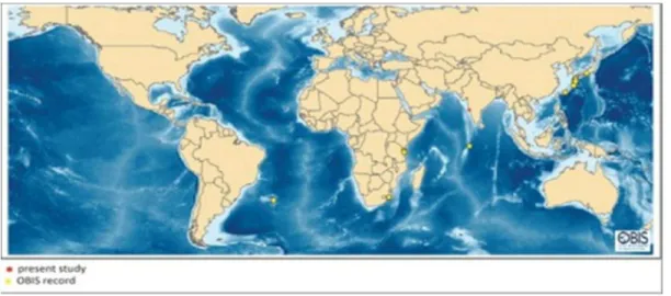
Zoanthus sansibaricus is one of the abundant and widespread Zoanthid. While generally found in intertidal areas, some colonies have been found as deep as 40m and on subtidal reef walls. According to OBIS database global distribution of Z. sansibaricus (fig. 7) indicates its record mainly from the coast of Japan and along the tropics [36].
Fig. 7: Global distribution of Zoanthus sansibaricus (Zoanthidae)
The published scientific reports made so far from India on zoanthids are few and merely deal with the genus level identification, perhaps due to its complicated morphological characteristics. This situation has been rescued by the advent of molecular studies and recent documentation of
data world over, though this is the first report of molecular and morphological data from India. All the results obtained concluded that the species
under identification is Zoanthus sansibaricus
Vol. 2 No. 1 2013 www.phytojournal.com Page | 217 6. Acknowledgement
The authors are grateful to Dr. Fredric Sinniger, Department of Zoology and Animal Biology, Molecular Systematic Group, University of Geneva, Switzerland and Dr. James Reimer. D., Department of Chemistry, Biology and Marine Science, Faculty of Science, Okinawa, Japan University of the Ryukyus, for their expert advice. The authors wish to thank, Director, NIO, for providing necessary facilities for the work. We also wish to thank Dr. Deepti Deobagkar, Department of Zoology, University of Pune, Pune, Dr. Loka Bharathi and Dr. Ramaiah, Biological Oceanography Division, NIO, Goa, for their persuasion and Mr. R. M. Meena for the support of molecular studies. A
special thanks and profound gratitude to Dr. Baban Ingole and Dr. C. Ravindran,
Biological Oceanography Division, NIO, Goa, for encouraging and providing all the support for the entire work.
7. References
1. Challenger Expedition.
http://www.britannica.com 14 May, 2012. 2. Bouchet, P. The magnitude of marine
biodiversity. In: Carlos M. Duarte, editor. The exploration of marine diversity: Scientific and Technological Challenges. Fundacion BBVA; Paris, 2006. 31-62.
3. Burnett, W. J., Benzic, J. A. H., Beardmore, J. A., and Ryland, J. S. Zoanthids (Anthozoa, Hexacorallia) from the Great Barrier Reef and Torres Straits, Australia: Systematics, evolution and a key to species. Coral Reefs 1997; 16: 55-68.
4. Ryland, J. S., Lancaster, J. E. A review of
Zoanthid nematocyst types and their
population structure. Hydrobiologia 2004; 530/531: 179-187.
5. Moore, R. E., and Schener, P. J. Palytoxin: a new marine toxin from a coelenterate. Science 1971; 172: 495-498.
6. Fukuzawa, S., Hayashi, Y., Ucmura, D., Nagatsu, A., Yamada, K., and Ijuin, Y. The siolation and
structure of five new alkaloids,
norzoanthamine, oxyzoanthamine,
norzoanthamine, cyclozoanthamine, and
epizoanthamine. Heterocyclecs
Communications 1995; 1: 207-214.
7. Stewart, C. N. Jr. Go with the glow: fluorescent proteins to light transgenic organisms. Trends Biotechnology 2006; 24: 155-162.
8. Muley, E. V., Alfred, J. R. B., Venkataraman, K. and Wafar, M. V. M. Status of Coral Reefs of India. 9 ICRS, Bali, Indonesia, October, 2009. 9. Parulekar, A. H. Marine Fauna of Malvan,
Central West Coast of India. Mahasagar- Bulletin of the NIO. 1981; 14 Supple 1:33-34. 10. Rao, C. B., Anjaneyulu, A. S. R., Sarma, N. S.,
Venkateswarulu, Y., Rosser, R. M., Faulkner, D. J., Chen, M. H. M., and Cardy, J. Zoanthamine: anovel alkaloid from a marine Zoanthid. Journal of American Chemical Society 1984; 106: 7983-7984.
11. Rao, C. B., Anjaneyulu, A. S. R., Sarma, N. S., and Venkateswarulu, Y. Alkaloids from marine Zoanthid. Journal of Organic Chemistry 1985; 50: 3757-3760.
12. Rao, C. B., Rao, D. V., Raju, V. S. N., Sullivan, B. W., and Faulkner, D. J. Two new alkaloids from Indian species of Zoanthus. Heterocycles 1989; 28: 103.
13. Babu, U. V., Bhandari, S. P. S., and Garg, H, S. Hariamide, a novel sulphated sphingolipid from a Zoanthus sp. of the Indian coast. Journal of Natural Products 1997; 60: 1307-1309.
14. Parameswaran, P. S., Naik, C. G., Gonsalves, C., and Achuthankutty, C. T. Isolation of 2-deoxyecdysterone, a novel oxytocic agent, from a marine Zoanthus sp. Journal of Indian Institute of Science. 2001; 81: 169-173.
15. Daly,M., Brugler,M.R., Cartwright,P., Collins, A. G., Dawson, M. N., Fautin, D. G., et.al. The Phylum Cnidaria: A review of phylogenetic patterns and diversity 300 years after Linnaeus. In: Zhang, Z. Q., and Shear, W. A., editors.
Linneaus Tercentenary: Progress in
Invertebrate Taxonomy, Zootaxa 2007; 1668: 127-182.
16. Burnett, W. J., Benzic, J. A. H., Beardmore, J. A., and Ryland, J. S. Zoanthids (Anthozoa, Hexacorallia) from the Great Barrier Reef and Torres Straits, Australia: systematics, evolution and a key to species. Coral Reefs 1997; 16: 55-68.
17. Pax, F. And Mueller, I. Memoires du Museum National d’Histoire Naturell 16 Sirie A. Zoologie 1957; 1-40.
18. Walsh, G. E. And Bowers, R. L. A review of Hawaiian Zoanthids with descriptions of three new species. Zoological Journal of Linnean Society. 1971; 50: 161-180.
Vol. 2 No. 1 2013 www.phytojournal.com Page | 218 20. Reimer, J. D., Ono, S., Fujiwara, Y., Takishita, K.,
and Tsukhara, J. Reconsidering Zoanthus spp. Diversity: Molecular evidence of conspecificity within four previously presumed species. Zoological Science. 2004; 21: 517-525.
21. Ryland, J. S., and Babcock, R. C. Annual cycle of gametogenesis and spawing in a tropical zoanthid. Hydrobiologia 1991; 216: 117-123. 22. Reimer, J. D., Nakachi, S., Hirose, M., Hirose, E.,
and Hashiguchi, S. Uning hydrofluoric acid for morphological investigations of zoanthids (cnidarian: Anthozoa): A critical assessment of
methodology and necessity. Marine
Biotechnology. 2010; 12: 605-617.
23. Hebert, P. D. N., Cywinska, A., Ball, S. L., and deWaard, J. R., Biological identifications through DNA barcodes. Proceedings of Royal Society of London B. 2003; 270: 313-322. 24. Bucklin, A., Frost, B. W., Bragdfor-Grieve, J.,
Allen, L. D., and Copley, N. J. Molecular systematic and phylogenetic assessment of 34 calanoid copepod species of Calanidae and Clausocalanidae. Marine Biology. 2003; 142: 333-343.
25. Ward, R. D., Zimlak, T. S., Innes, B. H., Last, R. R., and Hebert, P. D. DNA barcoding Australia’s fish species. Philosophical Transactions Royal Society of London B. 2005; 360: 1847-1857. 26. Hebert, P. D. N., Renton, E. H., Burns, J. M.,
Janzen, D. H., and Hallwachs, W. Ten species in one: DNA barcoding reveals cryptic species in the Neotropical skipper butterfly Astraples fulgerator. Proceedings of National Academy Science USA. 2004a; 101: 14812-14817.
27. Sinniger, F., Reimer, J. D., and Pawlowski, J. Potential of DNA sequences to identify zoanthids (Cnidaria: Zoantharia). Zoological Science. 2008; 25: 1253-1260.
28. Rogers, S. O., and Bendich, A. J. Extraction of DNA from milligram amounts of fresh, herbarium and mummified plant tissues. Plant Molecular Biology 1985; 5: 69-76.
29. Sacchi, C. N. Single-step method of RNA isolation by acid guanidium thiocyanate phenol chloroform extractions. Analytical Biochemistry 1987; 162: 156-9.
30. Sinniger, F., Reimer, J. D., and Pawlowski, J. The parazoanthidae (Hexacorallia: Zoantharia) DNA taxonomy: description of two new genera. Marine Biodiversity. 2010; 40: 57-70.
31. 30 (a). Zoantharia of New Caledonia. http://ird.nc/biodec/synthese_generale.html. 32. Reimer, J. D. Key to field identification of
shallow water brachycnemic zoanthids(Order Zoantharia: Suborder Brachycnemina) present in Okinawa. Galaxea. Journal of Coral Reef Studies. 2010; 12: 23-29.
33. Sinniger, F., Montoya-Burgos, J. I., Chevoldonne, P., and Pawlowski. J. Phylogeny of the order zoantharia (Anthozoa, Hexacorallia) based on the mitochondrial ribosomal genes. Marine Biology 2005; 147: 1121-1128.
34. Reimer, J. D., Ono. S., Takishita, K., Tsukahara, J.,
and Maruyama, T. Molecular evidence
suggesting species in the zoanthid genera
Palythoa and Protopalythoa (Anthozoa: Hexacorallia) are congeneric. Zoological Science. 2006; 23: 87-94.
35. Reimer, J. D., Nonaka, M., Sinniger, F., and Iwase,
F., Morphological and molecular
characterization of a new genus and new
species of parazoanthid (Anthozoa:
Hexacorallia: Zoantharia) associated with Japanese red coral. Coral Reef 2008; 27: 935-949.
36. Reimer, J. D., Ono, S., Iwama, A., Tsukahara, J., Takishita, K., and Maruyama, T., Morphological and molecular revision of Zoanthus (Anthozoa: Hexacorallia) from southwestern Japan with description of two new species. Zoological Science. 2006a; 23:261-275.