Metabolic Characteristics of a
Glucose-Utilizing
Shewanella oneidensis
Strain Grown
under Electrode-Respiring Conditions
Gen Nakagawa, Atsushi Kouzuma*, Atsumi Hirose, Takuya Kasai, Gen Yoshida, Kazuya Watanabe
School of Life Sciences, Tokyo University of Pharmacy and Life Sciences, Tokyo, Japan
Abstract
In bioelectrochemical systems, the electrode potential is an important parameter affecting the electron flow between electrodes and microbes and microbial metabolic activities. Here, we investigated the metabolic characteristics of a glucose-utilizing strain of engineered Shewanella oneidensisunder electrode-respiring conditions in electrochemical reactors for gaining insight into how metabolic pathways in electrochemically active bacteria are affected by the electrode potential. When an electrochemical reactor was operated with its working electrode poised at +0.4 V (vs. an Ag/AgCl reference electrode), the engineeredS. oneidensisstrain, carrying a plasmid encoding a sugar permease and glucose kinase of Escherichia coli, generated current by oxidizing glucose to acetate and produced D-lactate as an intermediate metabolite. However, D-lactate accumulation was not observed when the engineered strain was grown with a working electrode poised at 0 V. We also found that transcription of genes involved in pyruvate and D-lactate metabolisms was upregulated at a high electrode potential compared with their transcription at a low electrode potential. These results suggest that the carbon catabolic pathway ofS.oneidensiscan be modified by con-trolling the potential of a working electrode in an electrochemical bioreactor.
Introduction
The environmental redox state is an important factor determining the growth and metabolic activities of microorganisms because it affects the availability of electron acceptors and cellular energy conservation processes, such as respiration and fermentation [1]. In the respiratory chain, the difference in redox potentials between electron donors and acceptors determines the amount of energy conserved during electron transfer reactions. The intracellular redox balance, which influences fermentative products, is also associated with the environmental redox state [2]. In biotechnological processes, the amount of electron acceptors (e.g., oxygen) is used for controlling the production rate and target compound yields [3].
Recent studies have suggested that bioelectrochemical systems (BES) are useful for control-ling microbial metabolic activities [4]. BES are biotechnological systems that utilize the
OPEN ACCESS
Citation:Nakagawa G, Kouzuma A, Hirose A, Kasai T, Yoshida G, Watanabe K (2015) Metabolic Characteristics of a Glucose-UtilizingShewanella oneidensisStrain Grown under Electrode-Respiring Conditions. PLoS ONE 10(9): e0138813. doi:10.1371/journal.pone.0138813
Editor:Patrick C. Cirino, University of Houston, UNITED STATES
Received:February 12, 2015
Accepted:September 3, 2015
Published:September 22, 2015
Copyright:© 2015 Nakagawa et al. This is an open access article distributed under the terms of the Creative Commons Attribution License, which permits unrestricted use, distribution, and reproduction in any medium, provided the original author and source are credited.
Data Availability Statement:All relevant data are within the paper and its Supporting Information files.
Funding:This work was mainly supported by JSPS KAKENHI Grant Numbers 24880030 and 26850056 (http://www.jsps.go.jp/english/index.html) and partly supported by the Noguchi Institute (http://www. noguchi.or.jp/index.php?lang = en). The funders had no role in study design, data collection and analysis, decision to publish, or preparation of the manuscript.
association between electrodes and electrochemically active bacteria (EAB). BES have attracted considerable attention because of their wide applicability for valuable biotechnological pro-cesses, including electricity generation (i.e., microbial fuel cells; MFC) [5] and the production of fuels and chemicals (i.e., microbial electrosynthesis) [6]. In BES, the redox state of electron acceptors (or donors) and rate of electron flow during metabolism can be altered by controlling the electrode potential, allowing modification of the intracellular redox balance in electrode-associated microbes [6,7]. Studies have also demonstrated that differences in electrode poten-tial and/or catalytic current in BES influence the gene expression profiles of EAB [8–10]. To cite an example, Matsuda et al. [10] have reported that the expression levels of the genes associ-ated with the TCA cycle inShewanella loihicaPV-4 were markedly altered when cells were cul-tivated in the presence of electrodes poised at different potentials. However, limited
information is available on how EAB control metabolism and alter their intracellular metabolic products in response to changes in the electrode potential.
Shewanella oneidensisMR-1 is one of the most extensively studied EAB, owing to its anno-tated genome sequence [11], ease of genetic manipulation [4], and capability to transfer elec-trons to extracellular electrodes without an exogenously added mediator [12]. Further, recent studies have demonstrated the potential applicability of this strain for microbial electrosynth-esis systems [13]. Because MR-1 has also been well characterized in terms of its central carbon utilization pathways [14–19], it is considered suitable as a model bacterium for studying the metabolic characteristics of EAB during electrochemical cultivation in BES. However, studies have also revealed that MR-1 and many otherShewanellastrains preferably utilize low-molecu-lar-weight organic acids, including lactate and pyruvate, as carbon and energy sources. They do not prefer five- and six-carbon carbohydrates, including glucose, although these sugars are widely used as substrates for MFC and bioproduction processes. Therefore, metabolic engi-neering of MR-1 to confer the ability to utilize glucose may be valuable not only for expanding the applicability of this strain for biotechnological applications but also for understanding how EAB metabolize carbohydrates under potential-controlled conditions.
Spontaneous and engineered glucose-utilizingS oneidensisstrains have been obtained in previous studies. Howard et al. [20] have reported that MR-1 acquired the ability to utilize glu-cose after an initial exposure to gluglu-cose. More recently, Choi et al. [21] successfully constructed glucose-utilizingShewanellamutants by introducing the glucose facilitator (glf) and glucoki-nase (glk) genes fromZymomonas mobilis. However, although they also demonstrated that engineered MR-1 was able to generate current using glucose as the electron donor in an MFC reactor [21], the metabolic profiles of sugar-utilizingShewanellastrains during electrode respi-ration have not been investigated. In the present study, we constructed an engineeredS. onei-densisstrain by introducing glycolytic genes fromEscherichia coli. We then characterized the glucose utilization profiles of this strain under electrode-respiring conditions in BES, with a particular focus on differences in D/L-lactate production from glucose and the expression lev-els of genes involved in pyruvate and lactate metabolism.
Materials and Methods
Bacterial strains and growth conditions
S.oneidensisMR-1 was obtained from the American Type Culture Collection (ATCC).E.coli
strains [22] were routinely cultured in Luria–Bertani (LB) medium at 37°C. TheE.colimating strain (WM6026) required supplementation of the medium with 100μg/ml
2,6-diaminopime-lic acid (DAP) for growth.Shewanella oneidensisstrains were cultured at 30°C in LB or in lac-tate minimal medium (LMM) [23] containing 15 mM lactate as the carbon source and
mineral solution. Glucose minimal medium (GMM), which contained 10 mM or 15 mM glu-cose instead of the lactate in LMM, was used for cultivation with gluglu-cose as the carbon and energy source. For aerobic cultivation,S.oneidensisstrains were introduced into 300-ml baffled Erlenmeyer flasks containing 100 ml LMM or GMM, and were cultivated with shaking on a rotary shaker at 180 rpm. For anaerobic cultivation,S.oneidensisstrains were introduced into 100-ml bottles containing 80 ml GMM supplemented with 5 mM or 40 mM fumarate. The bot-tles containing the anaerobic cultures were capped with Teflon-coated butyl rubber septa, sealed with aluminum crimp seals, and purged with pure nitrogen gas. The optical densities at 600 nm (OD600) of the cultures were measured using a DU800 spectrophotometer (Beckman). When necessary, 15μg/ml gentamicin (Gm) was added to the culture media. Agar plates
con-tained 1.6% Bacto agar (Difco).
Mutant construction
Theglk(ECK2384) andgalP(ECK2938) genes were amplified from the genomic DNA of anE.
coliK-12 derivative, strain DH5α, using primer sets glk-F-KpnI and glk-R-XhoI, and
galP-F-X-hoI and galP-R-PstI, respectively (seeS1 Tablein the Supporting Information). The PCR prod-ucts obtained were digested by the restriction enzymes corresponding to the sites incorporated in the primers (XhoI and either KpnI or PstI) and then ligated into KpnI-PstI-digested pBBR1MCS-5 [24]. The resultant plasmid, pBBR-glk-galP, was introduced intoS.oneidensis
cells by filter mating withE.coliWM6026.
In-frame disruption of thedld-IIandldhAgene in strain MR-1 was performed using a two-step homologous recombination method with suicide plasmid pSMV-10, as described previ-ously [22,23,25]. Briefly, a 1.6-kb fusion product, consisting of upstream and downstream sequences of thedld-IIorldhAgene joined by an 18-bp linker sequence, was constructed by PCR andin-vitroextension using the primers listed inS1 Table. The amplified fusion product was ligated into the SpeI site of pSMV-10, generating pSMV-dld-II or pSMV-ldhA, which was then introduced into MR-1 by filter mating withE.coliWM6026. Transconjugants (single-crossover clones) were selected on LB plates containing 50μg/ml kanamycin (Km) and were
further cultivated for 20 h in LB medium lacking antibiotics. The cultures were then spread onto LB plates containing 10% (w/v) sucrose to isolate Km-sensitive double-crossover mutants. Disruption of the target gene in the obtained strains was confirmed by PCR. One representative mutant strain in which thedld-IIorldhAgene was disrupted in-frame was selected and desig-natedΔdld-IIorΔldhA, respectively. To construct adld-II/ldhAdouble-knockout mutant
(Δdld-IIΔldhA), pSMV-ldhA was introduced into theΔdld-IIcells, and the double-crossover
mutants were screened as described above.
Operation of electrochemical cells
A small double-chambered EC (36 ml total capacity) was used to monitor and compare cur-rents generated byS.oneidensisstrains. This EC was equipped with a graphite felt working electrode (3.0 cm2; poised at +0.4 Vvs. Ag/AgCl) and an Ag/AgCl reference electrode (HX-R5, Hokuto Denko) in the anode chamber and a platinum wire counter electrode (5 cm,φ0.3 mm;
(3.0 cm2). Coulombic efficiency was calculated by dividing the total number of electrons trans-ferred to the working electrode by the theoretical maximum number of electrons produced by complete substrate oxidation to CO2(24 e–/mol for glucose and 12 e–/mol for lactate).
A large double-chambered EC (360 ml total capacity) was used for metabolite and transcrip-tional analyses of MR-1(pBBR-glk-galP). This EC was equipped with a graphite felt working electrode (8 cm2) and an Ag/AgCl reference electrode in the anode chamber and a platinum wire counter electrode (10 cm) in the cathode chamber. A Nafion 117 proton exchange mem-brane (28 cm2) was used to separate the anode and cathode chambers. The anode chamber was filled with 150 ml of GMM supplemented with 2 mM glucose and 170 mM NaCl, and then inoculated with bacterial cells at an initial OD600of 0.01. The cathode chamber was filled with 150 ml of the same medium without glucose. A current density (μA/cm2) was calculated based
on the projected area of working electrode (4.0 cm2). Reproducibility was assessed using at least three independent measurements, and typical data are shown here. For metabolite analy-ses, the working anode electrode was poised at 0 V or +0.4 V (vs. Ag/AgCl) using a VPM3 potentiostat. For transcriptional analyses, the working electrode was poised at 0 V, and then after the electric current became stable, the electrode potential was changed to +0.3 V or–0.3 V. After further cultivation in the ECs for 2 h, the bacterial cells attached to the working elec-trode were collected and subjected to RNA extraction.
Metabolite analyses
After the cells were removed by filtration through a membrane filter unit (0.20μm pore size,
DISMIC-25HP; Advantec), the amounts of acetate, formate, and some other organic acids in the EC supernatant were measured using a previously described high-performance liquid chro-matography (HPLC; Agilent 1100 series) method [22]. Glucose and D/L-lactate in the filtered supernatant were measured with enzymatic assays that were performed using an F-kit (J. K. international) according to the manufacturer’s instructions.
RNA extraction
Total RNA was extracted using Trizol reagent (Invitrogen) according to the manufacturer’s instructions and subsequently purified using an RNeasy Mini Kit and an RNase-Free DNase Set (Qiagen). The quality of extracted RNA was evaluated using an Agilent 2100 Bioanalyzer with RNA 6000 Pico reagents and RNA Pico Chips (Agilent Technologies) according to the manufacturer’s instructions.
Quantitative RT-PCR
Quantitative RT-PCR (qRT-PCR) was performed using a LightCycler 1.5 instrument (Roche) according to a previously described method [26–28]. The PCR mixture (20μl) contained 15 ng
total RNA, 1.3μl of 50 mM Mn(OAc)2solution, 7.5μl LightCycler RNA Master SYBR Green I
(Roche), and 0.15μM of the primers listed inS1 Tablein the Supporting Information. To
gen-erate standard curves, DNA fragments from the target genesdld-II,lldF,ldhA,aceF,pykA,eda,
performed in triplicate at a minimum, and the data were statistically analyzed by the Student’s t-test. APvalue of 0.01 was considered statistically significant.
Results and Discussion
Construction of glucose-utilizing mutant
The annotated genomic sequence data ofS.oneidensisMR-1 (Genbank accession no. AE014299) suggest that this strain is able to metabolize glucose-6-phosphate and its down-stream glycolytic metabolites through the Entner–Doudoroff (ED) and pentose phosphate (PP) pathways. However, this strain is unable to take up and phosphorylate glucose because a frameshift exists in the glucose/galactose transporter gene,gluP(SO_2214) [16,29], and no glu-cokinase gene (glk) can be found in the genome. Although MR-1 has a complete set of genes encoding the phosphoenolpyruvate (PEP):sugar phosphotransferase system for glucose (PTSGlc;ptsHI-crrandptsG), it is known that this system does not support the growth of bacte-ria on glucose through the ED and PP pathways. This is because these glycolytic pathways can-not produce a sufficient amount of PEP for the phosphotransferase reaction of the PTS [16]. However, previous studies have reported that the introduction ofglkand a glucose/galactose-proton symporter gene (galP) restored the ability of PTSGlc-inactivatedE.colimutants to utilize glucose by allowing glucose uptake and phosphorylation without the consumption of PEP [30–
32]. In the present study, we constructed plasmid pBBR-glk-galP, which contains theglkand
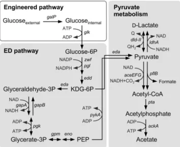
galPgenes derived fromE.coliK-12, and introduced it into MR-1 to confer the ability to utilize glucose on this strain (Fig 1).
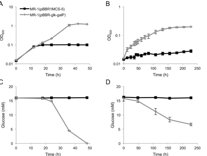
When MR-1(pBBR-glk-galP) cells were cultivated in GMM under aerobic or anaerobic, fumarate-reducing conditions, cell growth accompanied by glucose consumption was observed (Fig 2A to 2D), demonstrating that introduction of theseE.coliglycolytic genes allowed MR-1 to acquire the ability to grow on glucose. However, when the cells were incubated under anaer-obic condition in the absence of any electron acceptor, substantial cell growth was not observed within the first 5 days of incubation (data not shown). This indicates that the engineered strain cannot acquire sufficient energy for growth under these fermentation conditions. A similar
Fig 1. The glycolytic pathway in the engineeredS.oneidensisMR-1.The engineered pathway constructed in this study is shown in a white box. Intrinsic catabolic pathways shown in shaded boxes are depicted based on findings reported in the literature [15,17–19,35].
result has also been observed for another engineered strain ofS.oneidensis[21], which exhib-ited only very low biomass production even when cells were cultivated for 18 days under glu-cose-fermenting conditions. The poor growth of the MR-1 derivatives during glucose fermentation is likely related to the low ATP yields that result from glycolysis through the ED pathway [33].
Current generation by MR-1(pBBR-
glk
-
galP
)
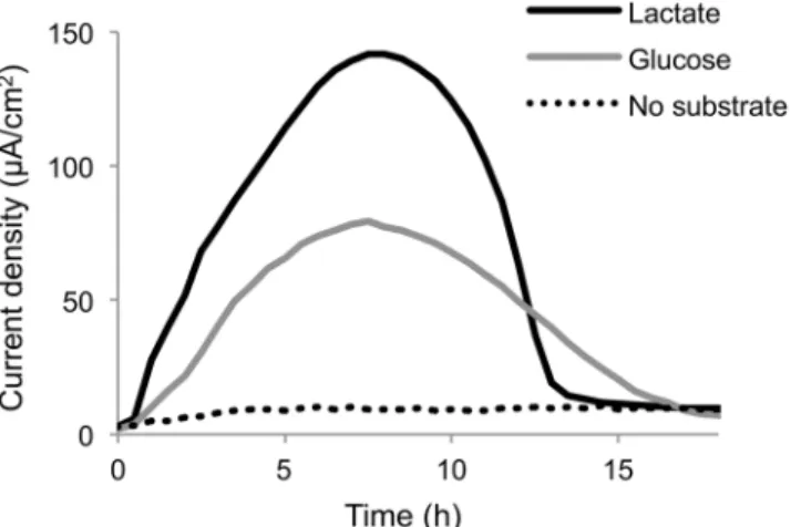
Current generation from glucose by MR-1(pBBR-glk-galP) was analyzed and compared with that from lactate using a small double-chambered EC equipped with a working electrode poised at +0.4 V (vs. Ag/AgCl) (Fig 3). The results demonstrate that the engineered strain is able to generate current using glucose as the electron donor. However, the maximum current density obtained from glucose (77.3μA/cm2) was 55% lower than that obtained from lactate
(140μA/cm2), suggesting that the rate of glucose metabolism in the engineered MR-1 was
lower than the rate of lactate metabolism. This difference in the current densities may reflect a difference in the growth rates of this strain in GMM and LMM (0.014 h–1and 0.35 h–1, respec-tively, under fumarate-reducing conditions). Coulombic efficiencies in glucose- and lactate-supplemented ECs were calculated to be 10.3% and 19.3%, respectively, indicating that many Fig 2. Growth (A, B) and glucose consumption (C, D) ofS.oneidensisderivatives under aerobic (A, C) and fumarate-reducing (B, D) conditions.S.
oneidensiscells harboring pBBR1MCS-5 (control vector) or pBBR-glk-galPwere grown in GMM-containing 15 mM glucose as the electron acceptor. Anaerobic cultures were supplemented with 40 mM fumarate as the electron donor. Error bars represent standard deviations calculated from triplicate measurements.
of the supplemented substrates were not completely oxidized in these ECs. Previous studies have reported thatShewanellastrains exhibits low coulombic efficiencies in lactate-supple-mented MFC because they partially oxidize lactate and produce acetate as the major metabolite [22,34]. Similarly, in the present study, acetate was detected at concentrations of 2.3 mM and 2.9 mM in the supernatant of the glucose- and lactate-supplemented ECs, respectively, when the substrates were completely consumed (18 h after commencing the incubation). The molar yields of acetate from glucose and lactate were 58% and 73%, respectively. These results indi-cate that current generation from glucose by the engineered MR-1 mainly occurs through par-tial substrate oxidation of glucose to acetate, as is the case for current generation from lactate.
Metabolic responses to different electrode potentials
To investigate the influence of electrode potentials on the metabolic activity of engineeredS.
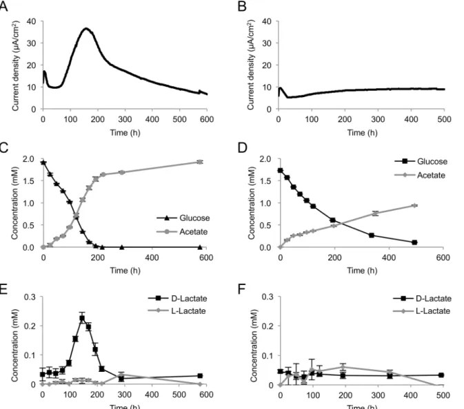
oneidensis, current generation and metabolite production from glucose were monitored using an EC equipped with a working electrode poised at +0.4 V (vs. Ag/AgCl) (high potential, HP) or 0 V (low potential, LP) (Fig 4A to 4F). In this experiment, we used a large double-chambered EC for stable sampling of supernatants from the anode chamber. Higher electric current and glucose-consumption rate were observed under the HP condition (Fig 4A and 4C) than were observed under the LP condition (Fig 4B and 4D). Although Matsuda et al. [10] have reported thatS.loihicaPV-4 generated decreased current when cells were grown in an EC equipped with a tin-doped, In2O3(ITO)-coated glass working electrode poised at a high potential, such decreases in current at higher potentials were not observed in the present study. It is likely that differences in electrode materials and configuration of the EC affect the current-generation profiles ofShewanellastrains under poised potential conditions.
Acetate was detected in ECs operated under the HP and LP conditions, along with current generation and glucose consumption (Fig 4C and 4D). Interestingly, a significant amount of D-lactate was detected, along with an increase in current density, under the HP condition (Fig 4E). This demonstrates that the engineered MR-1 was able to ferment glucose and produce D-lactate as an intermediate metabolite under electrode respiration. D-D-lactate was completely consumed within 300 h (Fig 4E), indicating that this metabolite was oxidized to acetate for cur-rent generation. However, under the LP condition, substantial quantities of D-lactate were not Fig 3. Current generation from glucose or lactate by MR-1 (pBBR-glk-galP).Cells were introduced into ECs supplemented with a minimal medium containing lactate or glucose as the electron donor and grown in the presence of a working electrode poised at +0.4 V (vs. Ag/AgCl). Results represent means of at least two parallel but independent experiments.
detected (Fig 4F). These results suggest that the difference in the electrode potential influences the glycolytic flux to D-lactate in the engineeredS.oneidensis. L-lactate did not remarkably accumulate under HP or LP conditions (Fig 4E and 4F). Formate and other organic acids, including succinate, fumarate, propionate, and maleate, were not detected in this experiment (data not shown), while Choi et al. [21] have reported that a glucose-utilizingS.oneidensis
strain produced formate in addition to acetate and lactate (the chirality was not identified in that experiment) during glucose metabolism in the presence or absence of Fe(III) oxide. This difference is likely due to the rapid oxidation of formate under poised electrode conditions.
Identification of D-lactate-production pathways
Based on the annotated genomic sequence data,S.oneidensisMR-1 is predicted to have two D-lactate dehydrogenase (D-LDH) genes, i.e.,dld-II(SO_1521) andldhA(SO_0968). Previous studies have reported that, while Dld-II functions as the respiratory LDH required for D-Fig 4. Current (A, B) and metabolite (C, D, E, F) production from glucose by MR-1(pBBR-glk-galP) in ECs operated with HP (A, C, E) or LP (B, D, F) electrode.Cells were cultivated in ECs supplemented with 2 mM glucose as the electron donor. The error bars represent the standard deviations calculated from triplicate measurements.
lactate oxidation to pyruvate in MR-1 [15], this enzyme is also involved in pyruvate reduction to D-lactate during pyruvate fermentation [35]. LdhA belongs to a family of fermentative NADH-dependent D-LDHs [36], although the function has not yet been characterized in MR-1. To identify the gene(s) involved in D-lactate production in the engineered MR-1, we con-structed two single-knockout mutants for these D-LDH genes (Δdld-IIandΔldhA) and a
dou-ble-knockout mutant (Δdld-IIΔldhA), and examined their ability to produce D-lactate from
glucose. In this experiment, wild-type MR-1 (WT),Δdld-II,ΔldhA, andΔdld-IIΔldhAcells
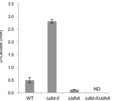
transformed with pBBR-glk-galPwere grown in GMM under electron acceptor (fumarate)-limited conditions, and D-lactate accumulation in culture supernatants was determined and compared (Fig 5). The results revealed that the production of D-lactate byΔldhAcells
(0.122 ± 0.002 mM) was substantially lower than that by WT cells (0.493 ± 0.107 mM). Although Pinchuk et al. [35] reported that the deletion ofldhAin MR-1 did not impair lactate production during pyruvate fermentation, the above results indicate that LdhA functions as a major fermentative D-LDH during sugar utilization. However,ΔldhAcells retained the ability
to produce a small amount of D-lactate, whileΔdld-IIΔldhAcells did not produce this
metabo-lite at a detectable level (Fig 5). This observation indicates that Dld-II is partially involved in D-lactate production from glucose. We also found thatΔdld-IIaccumulated a higher
concentra-tion of D-lactate compared to WT (Fig 5), supporting that Dld-II is mainly involved in the oxi-dation of D-lactate to pyruvate. Taken together, these results indicate that, in the engineered glycolytic pathway, a substantial portion of pyruvate produced during glucose fermentation is converted to D-lactate by LdhA, and partly by Dld-II, although the product is largely recon-verted to pyruvate by Dld-II (Fig 1).
Expression of lactate and pyruvate metabolism genes
To explore the reason for the electrode potential-dependent accumulation of D-lactate, we investigated the transcriptional levels of the genes involved in lactate and pyruvate metabolism Fig 5. D-lactate production from glucose by D-LDH-deficientS.oneidensisderivatives.Cells were anaerobically grown in GMM containing 10 mM glucose and 5 mM fumarate until the electron acceptor was completely consumed (for 24 h). Error bars represent standard deviations calculated from triplicate measurements. ND, not detected (below detection limits; 0.02 mM).
in cells grown at different electrode potentials. In this experiment, MR-1(pBBR-glk-galP) cells were cultivated at a poised electrode potential of 0 V until the electric current became stable (for 9 h), and then the potential was altered to +0.3 V or–0.3 V. This experiment confirmed that electric current was steeply increased or decreased by the shift in the electrode potential (Fig 6A). To investigate the influence of shifts in electrical potential on gene expression without substantial changes in metabolite concentrations, cells attached to the working electrodes were collected 2 h after the potential shifts. Total RNA extracted from these cells was analyzed using qRT-PCR. Expression levels of D- and L-LDH genes (dld-II,ldhA, andlldF) are shown inFig 6B. Interestingly, the expression levels ofdld-IIwere significantly increased with the increase in the electrode potential, while those ofldhAandlldF, which encodes a component of the respi-ratory L-LDH complex (LldEFG) [15], were not significantly affected by the potential shift (P<0.01;Fig 6B). Potential-dependent expression was also observed for three genes involved
in pyruvate metabolism, i.e.,pykA,eda, andaceF(Fig 6C). ThepykA(SO_2491) andeda
(SO_2486) genes are annotated respectively as pyruvate kinase that catalyzes the conversion of PEP to pyruvate and 2-keto-3-deoxygluconate 6-phosphate (KDG-6P) aldolase that catalyzes the conversion of KDG-6P to pyruvate and glyceraldehyde 3-phosphate (seeFig 1). TheaceF
(SO_0425) gene is reported to encode a component of the pyruvate dehydrogenase (PDH) Fig 6. Current generation (A) and expression levels of the genes involved in lactate (B) and other carbon (C) metabolism under potential-controlled conditions.MR-1(pBBR-glk-galP) cells were cultivated in ECs containing media supplemented with 2 mM glucose as the electron donor. The arrow indicates the time point at which the electrode potential was shifted. Gene expression levels were analyzed by quantitative RT-PCR analysis. Error bars represent standard deviations calculated from at least three measurements. Astarisks indicate statistically significant differences (P<0.01).
complex (AceEFG) involved in the conversion of pyruvate to acetyl-CoA and CO2[35]. How-ever, the expression of the pyruvate formate-lyase (pflB; SO_2912) and phosphotransacetylase (pta; SO_2916) genes was not significantly affected by the potential shift (P<0.01;Fig 6C).
These results suggest that the activity of key enzymes involved in pyruvate and D-lactate metabolism (i.e., PykA, PDH, and Dld-II; seeFig 1) is increased under HP conditions, although the mechanisms underlying these transcriptional changes and the accumulation of D-lactate are currently unclear. Since pyruvate oxidation to acetly-CoA by PDH involves the production of NADH, it is conceivable that the activation of PDH under HP conditions results in an increase in intracellular NADH, thereby enhancing the production of D-lactate catalyzed by LdhA. It is also possible that the activation of Dld-II contributes to a transient accumulation of D-lactate, as this enzyme catalyzes the bidirectional conversion between D-lactate and pyruvate (Fig 5). Further studies are underway to elucidate the complex carbon and electron fluxes for the D-lactate production under potential-controlled conditions.
Although reasons for the potential-dependent expression of thedld-II,pykA,eda, and PDH genes are currently unknown, it is conceivable that changes in intracellular redox states may influence the expression of these genes via redox-sensing regulators, such as PAS-domain con-taining proteins [1,37], as HP electrodes can act as efficient electron acceptors and promote the oxidation of intracellular molecules. Because the cultivation of MR-1 in the presence of D-lac-tate did not result in a significant increase in the expression of thedld-IIandldhAgenes (S1 Fig), it is not likely that the accumulation of D-lactate under HP conditions induces the sion of these D-LDH genes. In contrast, previous studies have demonstrated that the expres-sion of the L-LDH genes (lldEFG) requires LlpR (L-lactate-positive-regulator; SO_3460) [15,38], although the molecular mechanism underlying the positive regulation oflldEFGby this regulator remains unclear. As it has been reported that the transcription of L-LDH genes (lldRDP) inE.coliis regulated by the LldR regulator in a L-lactate-dependent manner [39], it is possible that the expression of thelldEFGgenes in MR-1 is also regulated by the presence or absence of L-lactate. However, our data suggest that it is not linked to changes in extracellular or intracellular redox status.
Conclusions
Supporting Information
S1 Fig. qRT-PCR analyses ofdld-IIandldhAin MR-1 cells grown with D-lactate and pyru-vate.MR-1 cells were cultivated in LMM supplemented with 15 mM D-lactate as the carbon and energy source or in a pyruvate minimal medium containing 15 mM pyruvate (in substitu-tion for lactate in LMM) up to the early stasubstitu-tionary growth phase. Results are expressed as rela-tive values to mRNA levels in cells grown on pyruvate. Error bars represent standard deviation calculated from at least three measurements.
(PDF)
S1 Table. Primers used in this study. (PDF)
Author Contributions
Conceived and designed the experiments: AK KW. Performed the experiments: GN AK AH TK GY. Analyzed the data: GN AK AH TK. Contributed reagents/materials/analysis tools: AK KW. Wrote the paper: AK KW.
References
1. Green J, Paget MS. Bacterial redox sensors. Nat Rev Microbiol. 2004; 2: 954–966. PMID:15550941 2. Iuchi S, Weiner L. Cellular and molecular physiology ofEscherichia coliin the adaptation to aerobic
environments. J Biochem. 1996; 120: 1055–1063. PMID:9010748
3. Liu CG, Xue C, Lin YH, Bai FW. Redox potential control and applications in microaerobic and anaerobic fermentations. Biotechnology Advances. Elsevier Inc.; 2013. p. 257–65.
4. Sydow A, Krieg T, Mayer F, Schrader J, Holtmann D. Electroactive bacteria-molecular mechanisms and genetic tools. Appl Microbiol Biotechnol. 2014; 98: 8481–8495. doi:10.1007/s00253-014-6005-z PMID:25139447
5. Watanabe K. Recent developments in microbial fuel cell technologies for sustainable bioenergy. J Biosci Bioeng. 2008; 106: 528–536. doi:10.1263/jbb.106.528PMID:19134546
6. Rabaey K, Rozendal RA. Microbial electrosynthesis—revisiting the electrical route for microbial produc-tion. Nat Rev Microbiol. 2010; 8: 706–716. doi:10.1038/nrmicro2422PMID:20844557
7. Flynn JM, Ross DE, Hunt KA, Bond DR, Gralnick JA. Enabling unbalanced fermentations by using engineered electrode-interfaced bacteria. mBio. 2010; 1: 1–8.
8. Matsuda S, Liu H, Kato S, Hashimoto K, Nakanishi S (2011) Negative faradaic resistance in extracellu-lar electron transfer by anode-respiringGeobacter sulfurreducenscells. Environ Sci Technol 45: 10163–10169. doi:10.1021/es200834bPMID:22047596
9. Ishii S, Suzuki S, Norden-Krichmar TM, Tenney A, Chain PSG, Scholz MB, et al. A novel metatranscrip-tomic approach to identify gene expression dynamics during extracellular electron transfer. Nat Com-mun. 2013; 4: 1601. doi:10.1038/ncomms2615PMID:23511466
10. Matsuda S, Liu H, Kouzuma A, Watanabe K, Hashimoto K, Nakanishi S. Electrochemical gating of tri-carboxylic acid cycle in electricity-producing bacterial cells ofShewanella. PLoS One. 2013; 8: e72901. doi:10.1371/journal.pone.0072901PMID:23977370
11. Heidelberg JF, Paulsen IT, Nelson KE, Gaidos EJ, Nelson WC, Read TD, et al. Genome sequence of the dissimilatory metal ion-reducing bacteriumShewanella oneidensis. Nat Biotechnol. 2002; 20: 1118–1123. PMID:12368813
12. Kim BH, Kim HJ, Hyun MS, Park DH. Direct electrode reaction of Fe(III)-reducing bacterium, Shewa-nella putrefaciens. J Microbiol Biotechnol. 1999; 9: 127–131.
13. Ross DE, Flynn JM, Baron DB, Gralnick JA, Bond DR. Towards electrosynthesis inShewanella: ener-getics of reversing the Mtr pathway for reductive metabolism. PLoS One. 2011; 6: e16649. doi:10. 1371/journal.pone.0016649PMID:21311751
14. Yang C, Rodionov DA, Li X, Laikova ON, Gelfand MS, Zagnitko OP, et al. Comparative genomics and experimental characterization of N-acetylglucosamine utilization pathway ofShewanella oneidensis. J Biol Chem. 2006; 281: 29872–29885. PMID:16857666
15. Pinchuk GE, Rodionov DA, Yang C, Li X, Osterman AL, Dervyn E, et al. Genomic reconstruction of
utilization. Proc Natl Acad Sci U S A. 2009; 106: 2874–2879. doi:10.1073/pnas.0806798106PMID:
19196979
16. Rodionov DA, Yang C, Li X, Rodionova IA, Wang Y, Obraztsova AY, et al. Genomic encyclopedia of sugar utilization pathways in theShewanellagenus. BMC Genomics. 2010; 11: 494. doi: 10.1186/1471-2164-11-494PMID:20836887
17. Scott JH, Nealson KH A biochemical study of the intermediary carbon metabolism ofShewanella putre-faciens. J Bacteriol 1994; 176: 3408–3411. PMID:8195102
18. Tang YJ, Meadows AL, Kirby J, Keasling JD Anaerobic central metabolic pathways inShewanella onei-densisMR-1 reinterpreted in the light of isotopic metabolite labeling. J Bacteriol 2007; 189: 894–901. PMID:17114268
19. Tang YJ, Hwang JS, Wemmer DE, Keasling JDShewanella oneidensisMR-1 fluxome under various oxygen conditions. 2007;Appl Environ Microbiol 73: 718–729. PMID:17098921
20. Howard EC, Hamdan LJ, Lizewski SE, Ringeisen BR. High frequency of glucose utilizing mutants in
Shewanella oneidensisMR-1. FEMS Microbiol Lett. 2011; 327: 9–14. doi:10.1111/j.1574-6968.2011.
02450.xPMID:22092702
21. Choi D, Lee SB, Kim S, Min B, Choi I-G, Chang IS. Metabolically engineered glucose-utilizing Shewa-nellastrains under anaerobic conditions. Bioresour Technol. 2014; 154: 59–66. doi:10.1016/j.biortech.
2013.12.025PMID:24384311
22. Newton GJ, Mori S, Nakamura R, Hashimoto K, Watanabe K. Analyses of current-generating mecha-nisms ofShewanella loihicaPV-4 andShewanella oneidensisMR-1 in microbial fuel cells. Appl Environ Microbiol. 2009; 75: 7674–7681. doi:10.1128/AEM.01142-09PMID:19837834
23. Kouzuma A, Meng X-Y, Kimura N, Hashimoto K, Watanabe K. Disruption of the putative cell surface polysaccharide biosynthesis gene SO3177 inShewanella oneidensisMR-1 enhances adhesion to elec-trodes and current generation in microbial fuel cells. Appl Environ Microbiol. 2010; 76: 4151–4157. doi:
10.1128/AEM.00117-10PMID:20453127
24. Kovach ME, Elzer PH, Hill DS, Robertson GT, Farris MA, Roop RM, et al. Four new derivatives of the broad-host-range cloning vector pBBR1MCS, carrying different antibiotic-resistance cassettes. Gene. 1995; 166: 175–176. PMID:8529885
25. Saltikov CW, Newman DK. Genetic identification of a respiratory arsenate reductase. Proc Natl Acad Sci U S A 2003; 100: 10983–10988. PMID:12939408
26. Kouzuma A, Hashimoto K, Watanabe K. Influences of aerobic respiration on current generation by She-wanella oneidensisMR-1 in single-chamber microbial fuel cells. Biosci Biotechnol Biochem. 2012; 76: 270–275. PMID:22313754
27. Kouzuma A, Hashimoto K, Watanabe K. Roles of siderophore in manganese-oxide reduction by She-wanella oneidensisMR-1. FEMS Microbiol Lett. 2012; 326: 91–98. doi:10.1111/j.1574-6968.2011.
02444.xPMID:22092340
28. Kouzuma A, Oba H, Tajima N, Hashimoto K, Watanabe K. Electrochemical selection and characteriza-tion of a high current-generatingShewanella oneidensismutant with altered cell-surface morphology and biofilm-related gene expression. BMC Microbiol. 2014; 14: 190. doi:10.1186/1471-2180-14-190
PMID:25028134
29. Romine MF, Carlson TS, Norbeck AD, McCue LA, Lipton MS. Identification of mobile elements and pseudogenes in theShewanella oneidensisMR-1 genome. Appl Environ Microbiol. 2008; 74: 3257– 3265. doi:10.1128/AEM.02720-07PMID:18378659
30. Flores N, Xiao J, Berry A, Bolivar F, Valle F. Pathway engineering for the production of aromatic com-pounds inEscherichia coli. Nat Biotechnol. 1996; 14: 620–623. PMID:9630954
31. Flores S, Gosset G, Flores N, de Graaf AA, Bolívar F. Analysis of carbon metabolism inEscherichia coli
strains with an inactive phosphotransferase system by13C labeling and NMR spectroscopy. Metab
Eng. 2002; 4: 124–137. PMID:12009792
32. Hernández-Montalvo V, Martínez A, Hernández-Chavez G, Bolivar F, Valle F, Gosset G. Expression of
galPandglkin aEscherichia coliPTS mutant restores glucose transport and increases glycolytic flux to fermentation products. Biotechnol Bioeng. 2003; 83: 687–694. PMID:12889033
33. Conway T. The Entner-Doudoroff pathway: history, physiology and molecular biology. FEMS Microbiol Rev. 1992; 9: 1–27. PMID:1389313
34. Lanthier M, Gregory KB, Lovley DR. Growth with high planktonic biomass inShewanella oneidensis
fuel cells. FEMS Microbiol Lett. 2008; 278: 29–35. PMID:17995953
35. Pinchuk GE, Geydebrekht OV, Hill EA, Reed JL, Konopka AE, Beliaev AS, et al. Pyruvate and lactate metabolism byShewanella oneidensisMR-1 under fermentation, oxygen limitation, and fumarate respi-ration conditions. Appl Environ Microbiol 2011; 77: 8234–8240. doi:10.1128/AEM.05382-11PMID:
36. Bunch PK, Mat-Jan F, Lee N, Clark DP. TheldhAgene encoding the fermentative lactate dehydroge-nase ofEscherichia coli. Microbiology. 1997; 143: 187–195. PMID:9025293
37. Taylor BL, Zhulin IB. PAS domains: internal sensors of oxygen, redox potential, and light. Microbiol Mol Biol Rev. 1999; 63: 479–506. PMID:10357859
38. Brutinel ED, Gralnick JA. Preferential utilization of D-lactate byShewanella oneidensis. Appl Environ Microbiol. 2012; 78:8474–8476. doi:10.1128/AEM.02183-12PMID:23001660
39. Aguilera L, Campos E, Giménez R, Badía J, Aguilar J, Baldoma L. Dual role of LldR in regulation of the