A Novel Phthalimide Derivative, TC11, Has
Preclinical Effects on High-Risk Myeloma Cells
and Osteoclasts
Maiko Matsushita1, Yoshie Ozaki1, Yuka Hasegawa1, Fukiko Terada1, Noriko Tabata2, Hirokazu Shiheido2, Hiroshi Yanagawa2, Tsukasa Oikawa3, Koichi Matsuo3, Wenlin Du4, Taketo Yamada4, Masashi Hozumi1, Daiju Ichikawa1, Yutaka Hattori1*
1Clinical Physiology and Therapeutics, Faculty of Pharmacy, Keio University, Tokyo, Japan,2Department of Biosciences and Informatics, Faculty of Science and Technology, Keio University, Yokohama, Japan, 3Cell and Tissue Biology, School of Medicine, Keio University, Tokyo, Japan,4Department of Pathology, School of Medicine, Keio University, Tokyo, Japan
Abstract
Despite the recent advances in the treatment of multiple myeloma (MM), MM patients with high-risk cytogenetic changes such as t(4;14) translocation or deletion of chromosome 17 still have extremely poor prognoses. With the goal of helping these high-risk MM patients, we previously developed a novel phthalimide derivative, TC11. Here we report the further characterization of TC11 including anti-myeloma effectsin vitroandin vivo, a
pharmacoki-netic study in mice, and anti-osteoclastogenic activity. Intraperitoneal injections of TC11 sig-nificantly delayed the growth of subcutaneous tumors in human myeloma-bearing SCID mice. Immunohistochemical analyses showed that TC11 induced apoptosis of MM cellsin vivo. In the pharmacokinetic analyses, the Cmaxwas 2.1μM at 1 h after the injection of
TC11, with 1.2 h as the half-life. TC11 significantly inhibited the differentiation and function of tartrate-resistant acid phosphatase (TRAP)-positive multinucleated osteoclasts in mouse osteoclast cultures using M-CSF and RANKL. We also revealed that TC11 induced the apo-ptosis of myeloma cells accompanied byα-tubulin fragmentation. In addition, TC11 and
lenalidomide, another phthalimide derivative, directly bound to nucleophosmin 1 (NPM1), whose role in MM is unknown. Thus, through multiple molecular interactions, TC11 is a po-tentially effective drug for high-risk MM patients with bone lesions. The present results sug-gest the possibility of the further development of novel thalidomide derivatives by drug designing.
Introduction
Multiple myeloma (MM) is a neoplasm of plasma cells that is accompanied by various clinical manifestations including lytic bone lesions, hypercalcemia, renal dysfunction,
immunodefi-ciency, and anemia [1,2]. Despite recent advances in the use of newly developed drugs
OPEN ACCESS
Citation:Matsushita M, Ozaki Y, Hasegawa Y, Ter-ada F, Tabata N, Shiheido H, et al. (2015) A Novel Phthalimide Derivative, TC11, Has Preclinical Effects on High-Risk Myeloma Cells and Osteoclasts. PLoS ONE 10(1): e0116135. doi:10.1371/journal. pone.0116135
Academic Editor:Dominique Heymann, Faculté de médecine de Nantes, FRANCE
Received:April 18, 2014
Accepted:November 24, 2014
Published:January 24, 2015
Copyright:© 2015 Matsushita et al. This is an open access article distributed under the terms of the
Creative Commons Attribution License, which permits unrestricted use, distribution, and reproduction in any medium, provided the original author and source are credited.
Data Availability Statement:All relevant data are within the paper.
including immune-modulatory drugs (IMiDs) such as thalidomide, lenalidomide, and pomali-domide and proteasome inhibitors such as bortezomib, carfilzomib, and MLN9708, MM is still an incurable disease [3–7]. In particular, MM patients harboring 17p deletion, t(14;16), t(14;20), or t(4;14) are classified as a high-risk group and have shown significantly shorter sur-vival [8–10]. For example, it is reported that even lenalidomide plus dexamethasone or
borte-zomib could not substantially improve the survival of refractory patients with del 17p [11,12].
With the goal of helping prolong the survival of these high-risk MM patients, we screened 29 synthetic phthalimide derivatives and found a novel compound,
2-(2,6-diisopropylphenyl)-5-amino-1H-isoindole-1,3-dione (TC11), which induced the apoptosis of KMS34 cells with t
(4;14) and del17p13 [13].
Bone lytic lesions, which are seen in 80%–90% of MM patients, have clinical manifestations that include pain, pathologic fractures, spinal cord compression, and hypercalcemia, thus pro-viding a negative impact on the quality of life of MM patients [1]. In the clinical setting, bis-phosphonate, radiotherapy, and surgery are used to treat bone disease. Bisphosphonate can significantly reduce skeletal-related events in MM patients [14], but side effects such as renal
impairment and osteonecrosis of the jaw are seen in some patients [15,16]. The development
of novel agents to effectively treat bone lesions without severe side effects is thus necessary.
In the present study, we evaluate the anti-myeloma effects of TC11in vitroandin vivo, and
we investigated the effects of TC11 on the differentiation of osteoclasts to determine whether this new drug could be effective for treating high-risk MM patients with bone lesions. We also examined nucleophosmin-1 (NPM1) as a molecule that binds directly to phthalimide, and the
results raised the possibility thatα-tubulin is involved in the anti-myeloma effect of TC11.
Materials and Methods
Cell lines
Human myeloma cell lines KMS11, KMS 26, KMS28, and KMS34 were established by Dr. T.
Otsuki (Kawasaki Medical School, Kurashiki, Japan) from Japanese patients [17,18] and these
cell lines were kindly provided by him. MUM24 was established from a patient with thalido-mide-resistant MM [19]. These cell lines were maintained in RPMI1640 medium (Sigma-Al-drich, St. Louis, MO) containing 10% fetal bovine serum (FBS). The human macrophage cell line RAW264.7 was purchased from American Type Culture Collection (Rockville, MD) and cultured in DMEM medium (Sigma-Aldrich) containing 10% FBS (Hyclone Laboratories, Logan, UT).
Reagents
We synthesizedTC11 [2-(2,6-diisopropylphenyl)-5-amino-1H-isoindole-1,3-dione] from
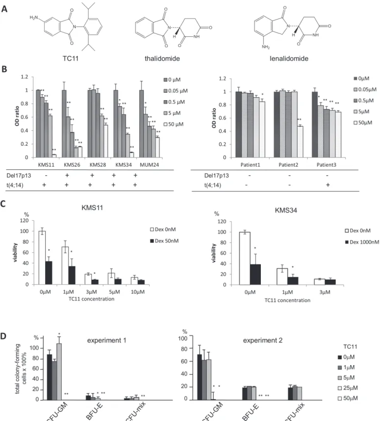
phthalic acid anhydride with a nitro group and amines, followed by catalytic hydrogenation under a nitrogen atmosphere. The benzene ring moiety of TC11 is different from those of tha-lidomide and lenatha-lidomide (Fig. 1A). Dexamethasone sodium phosphate (MSD K.K., Tokyo)
was used forin vitroproliferation assay.
Patients
’
samples
Bone marrow samples were obtained from patients with MM treated in Keio University Hospi-tal. Mononuclear cells were isolated by Ficoll density gradient centrifugation and cryopreserved in a liquid nitrogen tank until further use. All samples contained more than 30% myeloma cells. This study was approved by the ethics committee of the Keio University School of
analysis, decision to publish, or preparartion of the manuscript.
Medicine (99–7 and 15–21) and Faculty of Pharmacy (G110107–1). Written informed consent was obtained from all patients.
Cell viability assay
MM cells (2×10⁴cells per well) were seeded in 96-well plates and incubated with various
con-centrations of TC11 (0–50 µM) at 37°C for 48 h. The number of viable cells was assessed by MTT dye absorbance (Roche Diagnostics, Indianapolis, IN) according to the manufacturer’s instructions.
Colony-forming cell assay
To evaluate the hematological toxicity of TC11, 4×10⁴cells/mL of bone marrow cells from
13-wk-old male ICR mice were cultured in methylcellulose medium (Stem Cell Technologies, Vancouver, BC) containing FBS, 2-mercaptoethanol, 20 ng/mL mouse stem cell factor
(mSCF), 20 ng/mL mouse interleukin 3 (mIL-3), 10 ng/mL mouse interleukin-6 (mIL-6), and 1 U/mL human erythropoietin (hEPO) (kindly provided by Kyowa Hakko Kirin Co., Tokyo) in the presence or absence of TC11. On day 14, various types of colony-forming cells were counted.
In vivo
tumor growth assay
All of the animal experiments were approved by the Ethics Committee for Animal Experiments
at Keio University Faculty of Pharmacy (Approval no.09118-(0), 09118-(1)). Thein vivo
tumor-inhibitory activity assay was performed as described with several modifications [18].
Briefly, 3×10⁷KMS34 or KMS11 cells were subcutaneously inoculated into 5-wk-old male
ICR/SCID mice (Clea Japan, Tokyo) and plasmacytoma developed in 4–7 wks. In addition, twenty mg/kg of TC11 dissolved in 10% DMSO (Sigma-Aldrich)-1% Tween80 at the concen-tration of 2.5 mg/mL or only 10% DMSO-1% Tween80 as a control was injected intraperitone-ally twice every 3 days for 15 days (n = 7). The tumor volume was calculated according to the
following formula as described [18]: width × length2× 0.52.
Histopathologic examination
The histopathologic analysis was performed as described with several modifications [18].
When the subcutaneous tumors reached 50 mm3, the intraperitoneal injections of TC11 was
started. After 14 days of observation, the mice were sacrificed and the isolated tumors were fixed with 10% formalin and embedded in paraffin. Sliced sections were stained with hematox-ylin and eosin (H. E.). Anti-human cleaved PARP (Asp214) polyclonal antibody (Cell Signaling Technology Japan, Tokyo), anti-cleaved caspase-3 (Asp175) polyclonal antibody (Cell Signal-ing Technology Japan) and anti-human Ki-67 monoclonal antibody (clone MIB-1) (Dako Japan, Tokyo) were used for immunohistochemistry.
Pharmacokinetics study
To evaluate the pharmacokinetics of TC11, we obtained peripheral blood with a heparinized needle from the tail veins of 5-wk-old male ICR mice at 0.5, 1, 1.5, 4, 8, 12, and 24 h after an in-jection of a low dose (20 mg/kg) or a high dose (100 mg/kg) of TC11. Blood samples were
centrifuged immediately at 3400gfor 15 min at 4°C. The plasma fraction was transferred to a
polypropylene tube and stored at−80°C until the assay. The plasma samples were thawed and
diluted with 10% ethanol in phosphate-buffered saline (PBS). A stock solution of TC11 was prepared in ethanol at 1 mg/mL. A series of standard solutions at designated concentrations were prepared by diluting the stock solution with ethanol. All of the samples were analyzed by high-pressure liquid chromatography (HPLC; a Jasco HPLC system, Jasco, Tokyo). The C18 column (Sep-Pak; Waters Associates, Milford, MA) was used. The mobile phases were acetoni-trile and 25 mM ammonium acetate (60:40).
Osteoclast differentiation assay
We prepared murine osteoclasts from bone marrow cells as described [20]. In brief, cells
ob-tained from the bone marrow of 5-wk-old male ICR mice were cultured inα-MEM containing
10% FBS with macrophage-colony stimulating factor (M-CSF; R&D Systems, Minneapolis, MN) (10 ng/mL). After 3 days of culture, we removed the floating cells and used the attached cells including bone marrow-derived macrophages (BMMs) as osteoclast precursors. To gener-ate osteoclasts, BMMs were further cultured with M-CSF (10 ng/mL) and receptor activator of
nuclear factorκB ligand (RANKL; R&D Systems) (10 ng/mL). After an additional 3–6 days of
culture, the cells were fixed and stained for tartrate-resistant acid phosphatase (TRAP) as de-scribed [20]. TRAP-positive multinucleated cells containing more than three nuclei were
con-sidered TRAP+multinuclear osteoclasts (TRAP+MNCs).
Pit formation assay
RAW 264.7 cells were incubated for 5–8 days with RANKL (10 ng/mL). After maturation into osteoclasts, the cells were seeded on BioCoat Osteologic multi-test slides (BD Falcon, BD Bio-sciences, San Jose, CA). Various concentrations of TC11, thalidomide (Wako, Osaka, Japan), bortezomib (Toronto Research Chemicals Inc., ON, Canada), and osteoprotegerin (OPG; R&D Systems) were added every 2 days for 7 days. Finally Von Kossa stain was conducted to visual-ize resorption pits. The resorption pits were observed by fluorescence microscopy (BZ-9000, Keyence, Tokyo). The pit area was quantified using Image J software (NIH).
Immunocytochemistry
KMS34 cells were treated with 5 µM TC11 for 4 h and attached to a slide using CYTOSPIN4 (Thermo Fisher Scientific, Rockford, IL), then fixed with 2% paraformaldehyde and permeabi-lized with 0.5% Triton X followed by blocking with 1% bovine serum albumin (BSA) in PBS.
The sample was stained with antibody againstα-tubulin (Sigma-Aldrich) followed by
FITC-conjugated anti-mouse IgG (Takara Bio, Shiga, Japan), and then observed by fluorescence mi-croscopy (BZ-9000).
Surface plasmon resonance (SPR) analysis
We determined the binding kinetics by a surface plasmon resonance (SPR) analysis with the Biacore 3000 system (GE Healthcare, Buckinghamshire, UK). All experiments were performed at 25°C using TBS buffer (25 mM Tris-HCl, pH 7.4, 137 mM NaCl, 3 mM KCl). Recombinant
NPM1 was expressed inE. coliand purified. The NPM1 obtained (1.84 µM, 100 µL) was
dissociation were 60 and 300 s, respectively. The binding data were analyzed with 1:1 binding with the mass transfer model in the BIA evaluation software ver. 4.1 (Biacore).
Statistical analysis
The significance of differences was determined using Student’st-test. The level of significance
was specified as p<0.05.
Results
TC11 inhibited the growth of MM cells with chromosomal abnormalities
in vitro
We examined whether TC11 could inhibit the growth of four different MM cell lines harboring del 17p and/or t(4;14) in addition to previously reported KMS34 cells [13]. We also analyzed TC11’s effect on the bone marrow cells obtained from three MM patients. In an MTT assay, TC11 inhibited the proliferation of the KMS11, KMS26, KMS28, and MUM24 cells as well as the proliferation of all of the bone marrow cells from the MM patients, in a dose-dependent manner (Fig. 1B). TC11 was effective both for high risk and standard risk myeloma cells. As
shown inFig. 1C, dexamethasone (Dex) potentiated TC11 anti-myeloma effect of TC11 in
KMS 11 as well as KMS34 cells.
Suppression of the growth of normal hematopoietic cells by TC11
To evaluate the effect of TC11 on normal hematopoiesis, we conducted a colony formation assay using bone marrow cells from ICR mice (Fig. 1D). 1–5 µM of TC11 (IC50 values of most MM cell lines were 3–5 µM) did not suppress the production of the total colonies of normal he-matopoietic cells. 25 µM or higher concentrations of TC11 almost completely inhibited the col-ony formation. TC11 did not inhibit the production of colcol-ony-forming unit-granulocyte macrophage (CFU-GM), even though the number of burst-forming unit-erythroid (BFU-E)
was decreased by the addition of a 5 µM of TC11 (p<0.05) (Fig. 1D).
TC11 inhibited the growth of MM cells in the xenograft mouse model
through the induction of apoptosis
in vivo
We evaluated the anti-myeloma effect of TC11in vivoin the KMS34-bearing xenograft model.
KMS34 tumor xenografts (~50 mm3) were treated with intraperitoneal injections of 20 mg/kg
TC11 twice every 3 days for 2 wks, followed by a time-course analysis of the tumor volumes for
15 days.Fig. 2Ashows that the tumor growth was significantly suppressed by the TC11
injec-tions compared to the control (p<0.01). The average weight of the tumors of the TC11-treated
mice on day 14 was 153.94 mg, whereas that of the control mice was 314.14 mg (p<0.01)
(Fig. 2C). In addition, neither weight loss nor organ damage in lung, kidney or liver was ob-served macroscopically after the administration of TC11 (data not shown). Higher dose of TC11 (100 mg/kg) was injected to the xenograft model derived from another myeloma cell
line, KMS11. As shown inFig. 2D, TC11 significantly delayed the tumor growth. In one
xeno-graft model, the tumor had almost disappeared after injection of TC11. During treatment of higher dose of TC11, significant body weight loss was not observed in TC11-treated mice.
all active phases of the cell cycle but is absent in resting cells, was less weakly stained in
TC11-treated tumors (Fig. 2E). These results suggested that TC11 exhibited anti-tumor activityin
vivothrough the inhibition of cell proliferation and the induction of apoptosis of myeloma
cells.
Pharmacokinetics of TC11 in the mouse model
For the pharmacokinetic study, we examined the plasma concentrations of TC11 in mice after a single injection of 20 mg/kg or 100 mg/kg of TC11 using HPLC (Fig. 2F). The maximum
con-centration (Cmax) was 2.1 µM, which was observed at 1 hr after the injection (Tmax). The
plas-ma concentration of TC11 gradually decreased to 0.25 µM at 4 h and to 0.04 µM at 24 h. The elimination half-life (T1/2) was estimated to be 1.2 h. When a higher dose of TC11 (100 mg/
kg) was injected, the Cmaxwas 18.1 µM and the Tmaxwas 1.5 h; the plasma concentration was
0.45 µM at 8 h and 0.16 µM at 24 h. The T1/2 was estimated to be 2.6 h.
TC11 inhibited the differentiation of osteoclasts and bone resorption
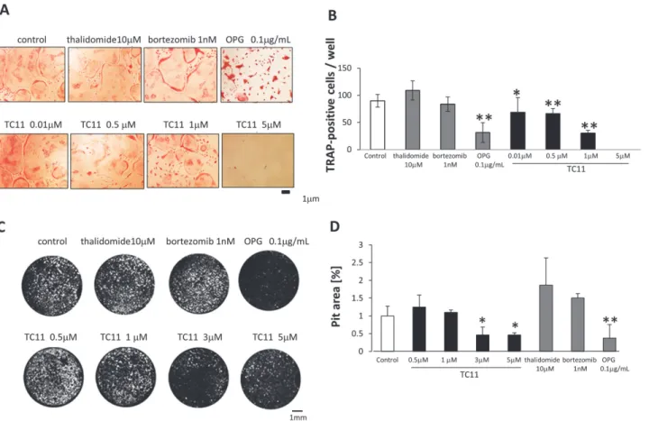
The hyperactivity of osteoclasts is considered one of the causes of lytic bone lesions, suggesting that the control of osteoclast activity could be a key to treating bone lesions in MM patients. We therefore examined the effects of TC11 on the differentiation and function of osteoclasts using mouse bone marrow primary cell culture. We analyzed M-CSF and RANKL-treated oste-oclasts by TRAP staining after treatment with various concentrations of TC11 (0.01µM–5 µM) (Fig. 3A). The number of TRAP-positive multinuclear cells was significantly lower in the cells treated with more than 0.5 µM of TC11 (Fig. 3B). In contrast, 10 µM of thalidomide or 1 nM of bortezomib did not affect the number of TRAP-positive cells.
We next examined TC11’s effect on the bone resorption activity of TC11 in a pit formation assay (Fig. 3C). After the treatment of RAW264.7 cells with TC11 (3 µM, 5 µM), the bone re-sorption area was reduced in a dose-dependent manner (Fig. 3D). Treatment with thalidomide (10 µM) or bortezomib (1 nM) did not change the resorption area compared to the control.
TC11 induced the fragmentation of
α
-tubulin
Because we previously found that TC11 binds toα-tubulin, we examined the tubulin formation
in KMS34 cells after TC11 treatment. The immunohistochemical analysis showed that
TC11-treated cells exhibited elevated levels ofα-tubulin fragmentation (Fig. 4A). The percentage of
cells with tubulin fragmentation was 6.5% in the cells treated with TC11, which is significantly higher compared to the 0.8% in the untreated cells (Fig. 4B).
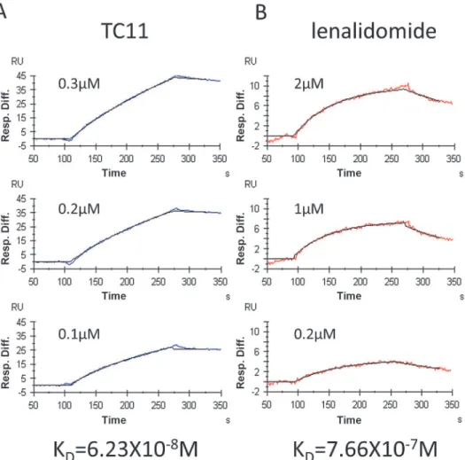
Binding between NPM and TC11 or lenalidomide
We previously identified NPM1 as one of the TC11-binding proteins, by mRNA display. In our previous observation, TC11 bound to the NH2-terminal 183-amino-acid (aa) region of monomeric NPM1. In the present study, we assessed the binding capacity of both TC11 and lenalidomide to the full length of NPM1. The SPR analysis showed that not only TC11 but also
lenalidomide could bind to NPM1 (Fig. 5A,B). The KDvalues of TC11 and lenalidomide were
6.23×10⁻⁸M and 7.66×10⁻⁷M, respectively.
subcutaneous plasmacytomas was examined. (E) Pathological examination of KMS11-derived tumors was shown. Immunohistochemical staining of the KMS11-derived tumors were also shown. (F) Pharmacokinetics of TC11. Plasma concentrations of TC11 in mice after a single injection of 20 mg/kg or 100 mg/kg of TC11 determined by HPLC.
Discussion
MM remains difficult to treat and cure, and MM patients with high-risk cytogenetic changes in particular have shown very poor prognosis when treated with existing drugs. To overcome this unmet clinical need, in previous work we screened phthalimide derivatives and identified TC11 as a novel candidate drug for MM. TC11 is different from other IMiDs in chemical struc-ture, lacking glutarimide moiety. That is, TC11 is a phthalimide derivative containing a ben-zene ring instead of a glutarimide structure (Fig. 1A). In the present study, we observed that TC11 inhibited the growth of MM cell lines, even though some of the myeloma cells harbored high-risk chromosomal abnormalities such as del17, or t(4;14). In addition, growth inhibition of bone marrow cells obtained from the relapsed MM patients was also examined. TC11 more or less inhibited proliferation of myeloma cells obtained from all the patients. Patient #3 showed t(4;14), and patient #2 had extramedullary disease in the liver. Even though these two patients were high-risk cases, TC11 regulated proliferation of the tumor cells (Fig. 1B). More-over, the growth of total colony-forming cells was not suppressed by the treatment with 1–5
Figure 3. TC11 inhibited both the maturation and function of bone marrow-derived osteoclasts.Mice bone marrow cells were cultured with
M-CSF (10 ng/mL) for 3 days, then further cultured with M-CSF (10 ng/mL) and RANKL (10 ng/mL). After an additional 3–6 days of culture with the indicated concentration of TC11, thalidomide, bortezomib, or DMSO, the cells were fixed and stained for TRAP as described in theMaterials and Methodssection. TRAP-positive multinucleated cells containing more than three nuclei were considered TRAP+multinuclear osteoclasts. (A) The number and size of TRAP+ multinuclear osteoclasts were decreased by treatment with 0.5–1µM of TC11. (B) The number of TRAP+multinuclear osteoclasts in each well of a 96-well plate was counted. Bars: means±SD.*p<0.05,**p<0.01 (Student’st-test). (C) RAW 264.7 cells were incubated for 5–8 days with RANKL (10 ng/mL) and
seeded on multi-test slides. The indicated concentrations of TC11, thalidomide, bortezomib, or OPG were added every 2 days for 7 days, followed by Von Kossa staining. The resorption pits were observed by fluorescence microscopy and the number of the pits was quantified using Image J software. (B) Bone resorption is indicated by white spots (pit). Bone resorption was suppressed by treatment with 3–5µM of TC11. (D) The pit area was measured by ImageJ software. The pit area of the DMSO-treated wells was used as the control ( = 1). Data shown are representative of three independent experiments. Bars: means±SD.*p<0.05,**p<0.01 (Student’st-test)
µM of TC11 even though TC11 significantly inhibited the growth of all MM cell lines in such concentrations, suggesting low hematological toxicity. It is speculated that myeloma cells may proliferate more dependently on TC11-associated molecules such as NPM1. Further study is needed to discriminate the molecular function of TC11 on myeloma cells from that on normal hematopoietic cells.
We also confirmed anin vivoanti-myeloma effect of TC11 by using intraperitoneal
injec-tions of TC11 to KMS34 and KMS11-bearing ICR/SCID mice. The tumor growth in these mice was significantly delayed by the administration of 20 mg/kg or higher dose of TC11. The histo-pathological examination revealed apoptosis of MM cells, which is consistent with our previous
study showing that TC11 induces apoptosis through the activation of caspase 3, 8 and 9in vitro
[13]. In addition, no systemic side effects including weight loss occurred.
Our pharmacokinetics study indicated that TC11 was completely eliminated from the plas-ma at 4 h after the injection of a low dose (20 mg/kg) of TC11 and at 8 h after the injection of a high dose (100 mg/kg) of TC11. These data suggest that TC11 would not accumulate in pa-tients by the administration of TC11 twice at a 3-day interval. To obtain stronger anti-tumor effects, it might be necessary to develop a new drug-delivery system or change the schedule of drug administration.
Figure 4. TC11 altered theα-tubulin formation of myeloma cells.KMS34 was treated with 5µM TC11 for 4 hours and attached to a slide by cytospin. The sample was fixed and stained with antibody againstα-tubulin followed by FITC-conjugated anti-mouse IgG (green). Nucleus was stained with DAPI (blue). A,
Representative mitotic cells with or without TC11 treatment. B, The percentage of cells with fragmentedα-tubulin in 2500 cells was calculated independently
in 9 areas.**P<0.01 (Studentt-test). A and B indicate representative data from 3 experiments.
Lytic bone disease in MM is thought to be associated with both enhanced activity of osteo-clasts and impaired function of osteoblasts. It was reported that bortezomib could cause an in-crease in bone formation by activating osteoblastic function, which leads to the improvement of osteolytic lesions [21]. Here, we found that TC11 inhibited both the maturation and function of osteoclasts. Osteoclasts are differentiated from hematopoietic stem cells through stimulation with M-CSF and RANKL. Under normal conditions, the balance between bone formation and bone resorption is well-controlled.
However, MM cells produce macrophage inflammatory protein-1 (MIP-1), which
stimu-lates osteoclasts to express receptor activator of nuclear factor-κB (RANK). RANK interacts
with RANKL on stromal cells or osteoblasts. This interaction leads to the activation of nuclear
factor-kappa B (NF-κB) and nuclear factor of activated T cell c1 (NFATc1) via an activation of
AP-1 [22]. In our M-CSF and RANKL-stimulated mouse bone marrow model, we observed that the differentiation of osteoclasts was significantly suppressed by at least 0.5 µM TC11. We also found that the function of osteoclasts was reduced by 3 µM TC11. These anti-osteoclastic effects might be characteristic of TC11, because these effects were not seen in the cells treated with thalidomide nor bortezomib. We also found that this effect was not due simply to the in-duction of apoptosis of the bone marrow cells, but rather occurred by a direct inhibition of
Figure 5. NPM1 interacts with TC11 and lenalidomide.Recombinant NPM1 was expressed inE. coliand
purified. The NPM1 obtained was immobilized onto the sensor chip NTA followed by an SPR analysis. Panels A and B show representative biosensorgrams of TC11 and lenalidomide, respectively.
osteoclast maturation, since apoptosis of osteoclasts was not observed after treatment with 3 µM TC11 (data not shown). It would be worthwhile to investigate the effects of TC11 on the expression of osteoclast-related genes such as AP-1.
To clarify the mechanism of TC11’s anti-myeloma effect, we investigated the effects of TC11 on NPM1, since we have found that NPM1 is one of the binding targets of TC11 by an mRNA display (i.e., the IVV method) [13]. In the present study, the results obtained by the
SPR analysis indicated that TC11 binds to the full length of NPM1, with theKDvalue of
6.23 × 10⁻⁸M. However, the pharmacological significance of the binding of TC11 to NPM1
has not been elucidated.
NPM1 is a nuclear phosphoprotein which plays a variety of roles in cancer cells by affecting DNA repair, centrosome duplication, and molecular chaperoning [23–25]. NPM1 is also im-portant in hematopoiesis by controlling the cell cycle of hematopoietic progenitor cells [26]. In addition, mutation of NPM1 is found in one-third of acute myelogenous leukemia (AML) pa-tients, which changes the localization of NPM1 from the nucleus to the cytoplasm [27–29].
Little is known about the role of NPM1 in MM patients. To our knowledge, there is only one report on NPM and MM, showing an overexpression of NPM1 in hyperdiploid MM cells [30]. All of the MM cell lines we used in the present study expressed NPM1 proteins, but NPM1 gene mutation was not observed in these MM cell lines, and TC11 did not cause an ac-cumulation of NPM1 in the cytoplasm of KMS34 as seen in leukemic cells (data not shown).
Another possibility explaining the role of NPM1 in TC11’s effects is that TC11 blocks the oligomerization of NPM1, since the binding site of TC11 contains the oligomerization domain of NPM1. NPM1 normally exists in oligomer form [31]. Therefore, TC11 might affect myelo-ma cells by blocking the oligomerization of NPM1 and inhibiting its various functions. Qi et al. showed that the inhibition of the oligomerization of NPM1 by a small-molecule inhibitor, NSC348884, induces the apoptosis of cancer cells through the activation of p53 [32]. Balusu et al. reported that NSC348884 could induce the apoptosis of AML cell lines [33].
It is also possible that TC11 impairs centrosomal disruption by binding to NPM1, because NPM1 has been reported to be indispensable to normal centrosomal duplication [34]. The NPM1 gene is located on chromosome 5q35, which is occasionally deleted in myelodysplastic
syndrome (5q−MDS) [35]. Considering our observation that lenalidomide could also bind to
NPM1, the contribution of these interactions to the anti-tumor effects should be further investigated.
We then analyzed the effects of TC11 onα-tubulin’s structure, since we identifiedα-tubulin
as another TC11-binding protein using the IVV method in our previous work [13]. Alpha-tubulin is a component of microtubules that is important for cell division and cellular trans-port. Drugs that alter the formation of microtubules, such as vinca alkaloids, have been used in cancer therapy. However, the target sites and effects of these drugs on microtubules are
differ-ent [36,37]. In the case of TC11, we observed abnormalα-tubulin fragmentation in
TC11-treated KMS34 cells (Fig. 4), which suggests that the apoptosis induced by TC11 might be trig-gered by an abnormal formation of microtubules. In the previous report, knockdown of NPM1 gene induced centrosomal disruption followed by activation of caspase-9 and significant reduc-tion in cell viability [12]. Therefore, it is speculated that the principal acreduc-tions of TC11 on mye-loma cells are centrosomal disruption and abnormal microtubule assembly by binding to
NPM1 andα-tubulin, respectively. Consequently, TC11-treated myeloma cells fall into mitotic
Cereblon (CRBN: cerebral protein with ion protease), a component of the E3 ubiquitin
li-gase complex, has been identified as a target of thalidomide-mediated teratogenicity [38,39]. It
was shown that not only thalidomide but also lenalidomide and pomalidomide bind to CRBN, attenuating the proliferation of myeloma cells through several pathways such as the down-regulation of autoubiquitination of CRBN [40–41]. However, our experiments using the IVV method failed to screen CRBN as a TC11-binding protein. The difference might be explained by the structure of TC11, because CRBN was reported to bind to a glutarimide moiety of lenali-domide or pomalilenali-domide, which was not included in TC11. On the other hand, NPM1 may bind to the phthalimide moiety which is a common component of TC11 and other IMiDs, since our data showed both TC11 and lenalidomide could bind to NPM1.
Thus, TC11 exerts its anti-myeloma effect via molecular interactions which do not involve CRBN. In addition, TC11 does not form racemate and is expected to lack teratogenicity. The results of our present study suggest that new phthalimide derivatives other than thalidomide, lenalidomide and pomalidomide could be developed by drug designing for the treatment of MM.
In conclusion, we have demonstrated that TC11, a novel phthalimide derivative, has anti-tumor activity against MM cells with high-risk genetic abnormality including del 17p and t
(4;14),in vitroandin vivo.This novel compound also down-regulates the differentiation and
function of osteoclasts. Our data provide a strong preclinical rationale for TC11 as a safe and effective drug for the treatment of high-risk MM patients with bone disease. The actions of this
drug relating toα-tubulin and NPM1 remain to be further investigated.
Acknowledgments
We thank Dr. Takemi Otsuki for gift of multiple myeloma cell lines.
Author Contributions
Conceived and designed the experiments: HY TO KM TY Y. Hattori. Performed the experi-ments: YO Y. Hasegawa FT NT HY TO WD TY MH Y. Hattori. Analyzed the data: MM YO Y. Hasegawa FT NT HS HY TO KM WD TY DI Y. Hattori. Wrote the paper: MM HY TO KM DI Y. Hattori.
References
1. Rajkumar SV (2013) Multiple myeloma: 2013 update on diagnosis, risk-stratification, and management. Am J Hematol. 88(3): 226–235. doi:10.1002/ajh.23390
2. Bird JM, Owen RG, D'Sa S, Snowden JA, Pratt G, et al. (2011) Guidelines for the diagnosis and man-agement of multiple myeloma 2011. Br J Haematol. 154(1): 32–75. doi:10.1111/j.1365-2141.2011. 08573.xPMID:21569004
3. Terpos E, Kanellias N, Christoulas D, Kastritis E, Dimopoulos MA (2013) Pomalidomide: a novel drug to treat relapsed and refractory multiple myeloma. Onco Targets Ther. 6: 531–538. doi:10.2147/OTT. S34498PMID:23690693
4. McCurdy AR, Lacy MQ (2013) Pomalidomide and its clinical potential for relapsed or refractory multiple myeloma: an update for the hematologist. Ther Adv Hematol. 4(3): 211–216. doi:10.1177/
2040620713480155PMID:23730498
5. Siegel DS (2012) Relapsed/Refractory multiple myeloma: defining refractory disease and identifying strategies to overcome resistance. Semin Hematol. 49 Suppl 1: S3–15. doi:10.1053/j.seminhematol. 2012.05.005PMID:22727390
6. Lacy MQ (2011) New immunomodulatory drugs in myeloma. Curr Hematol Malig Rep. 6(2): 120–125. doi:10.1007/s11899-011-0077-yPMID:21327565
8. Bergsagel PL, Mateos MV, Gutierrez NC, Rajkumar SV, San Miguel JF (2013) Improving overall surviv-al and overcoming adverse prognosis in the treatment of cytogeneticsurviv-ally high-risk multiple myeloma. Blood. 121(6): 884–892. doi:10.1182/blood-2012-05-432203PMID:23165477
9. Avet-Loiseau H, Attal M, Campion L, Caillot D, Hulin C, et al. (2012) Long-term analysis of the IFM 99 trials for myeloma: cytogenetic abnormalities [t(4;14), del(17p), 1q gains] play a major role in defining long-term survival. J Clin Oncol. 30(16): 1949–1952. doi:10.1200/JCO.2011.36.5726PMID:22547600 10. Sawyer JR (2011) The prognostic significance of cytogenetics and molecular profiling in multiple
myelo-ma. Cancer Genet. 204(1): 3–12. doi:10.1016/j.cancergencyto.2010.11.002PMID:21356186 11. Mateos MV, Gutierrez NC, Martin-Ramos ML, Paiva B, Montalban MA, et al. (2011) Outcome according
to cytogenetic abnormalities and DNA ploidy in myeloma patients receiving short induction with weekly bortezomib followed by maintenance. Blood. 118(17): 4547–4553. doi:10.1182/blood-2011-04-345801 PMID:21900193
12. Chang H, Jiang A, Qi C, Trieu Y, Chen C, et al. (2010) Impact of genomic aberrations including chromo-some 1 abnormalities on the outcome of patients with relapsed or refractory multiple myeloma treated with lenalidomide and dexamethasone. Leuk Lymphoma. 51(11):2084–2091. doi:10.3109/10428194. 2010.524325PMID:20929319
13. Shiheido H, Terada F, Tabata N, Hayakawa I, Matsumura N, et al. (2012) A phthalimide derivative that inhibits centrosomal clustering is effective on multiple myeloma. PLoS One. 7(6):e38878. doi:10.1371/ journal.pone.0038878PMID:22761710
14. Chesi M, Bergsagel PL (2013) Molecular pathogenesis of multiple myeloma: basic and clinical updates. Int J Hematol. 97(3): 313–323. doi:10.1007/s12185-013-1291-2PMID:23456262
15. Modi ND, Lentzsch S (2012) Bisphosphonates as antimyeloma drugs. Leukemia. 26(4): 589–594. doi: 10.1038/leu.2011.282PMID:22005788
16. Raje N, Roodman GD (2011) Advances in the biology and treatment of bone disease in multiple myelo-ma. Clin Cancer Res. 17(6):1278–1286. doi:10.1158/1078-0432.CCR-10-1804PMID:21411443 17. Namba M, Ohtsuki T, Mori M, Togawa A, Wada H, et al. (1989) Establishment of five human myeloma
cell lines. In Vitro Cell Dev Biol. 25(8): 723–729. doi:10.1007/BF02623725PMID:2768132
18. Du W, Hattori Y, Yamada T, Matsumoto K, Nakamura T, et al. (2007) NK4, an antagonist of hepatocyte growth factor (HGF), inhibits growth of multiple myeloma cells: molecular targeting of angiogenic growth factor. Blood. 109(7): 3042–3049. PMID:17179234
19. Hattori Y, Du W, Yamada T, Ichikawa D, Matsunami S, et al. (2013) A myeloma cell line established from a patient refractory to thalidomide therapy revealed high-risk cytogenetic abnormalities and pro-duced vascular endothelial growth factor. Blood Cancer J. 3:e115. doi:10.1038/bcj.2013.13PMID: 23686003
20. Oikawa T, Oyama M, Kozuka-Hata H, Uehara S, Udagawa N, et al. (2012) Tks5-dependent formation of circumferential podosomes/invadopodia mediates cell-cell fusion. J Cell Biol. 197(4): 553–568. doi: 10.1083/jcb.201111116PMID:22584907
21. Zangari M, Terpos E, Zhan F, Tricot G (2012) Impact of bortezomib on bone health in myeloma: a re-view of current evidence. Cancer Treat Rev. 38(8):968–980. doi:10.1016/j.ctrv.2011.12.007PMID: 22226939
22. Matsuo K, Galson DL, Zhao C, Peng L, Laplace C, et al. (2004) Nuclear factor of activated T-cells (NFAT) rescues osteoclastogenesis in precursors lacking c-Fos. J Biol Chem. 279(25): 26475–26480. doi:10.1074/jbc.M313973200PMID:15073183
23. Lindstrom MS (2011) NPM1/B23: A Multifunctional Chaperone in Ribosome Biogenesis and Chromatin Remodeling. Biochem Res Int.:195–209.
24. Colombo E, Alcalay M, Pelicci PG (2011) Nucleophosmin and its complex network: a possible thera-peutic target in hematological diseases. Oncogene. 30(23): 2595–2609. doi:10.1038/onc.2010.646 PMID:21278791
25. Grisendi S, Mecucci C, Falini B, Pandolfi PP (2006) Nucleophosmin and cancer. Nat Rev Cancer. 6(7): 493–505. doi:10.1038/nrc1885PMID:16794633
26. Li J, Sejas DP, Rani R, Koretsky T, Bagby GC, et al. (2006) Nucleophosmin regulates cell cycle pro-gression and stress response in hematopoietic stem/progenitor cells. J Biol Chem. 281
(24):16536–16545. doi:10.1074/jbc.M601386200PMID:16608843
27. Estey EH (2013) Acute myeloid leukemia: 2013 update on risk-stratification and management. Am J Hematol. 88(4): 318–327. doi:10.1002/ajh.23404PMID:23526416
29. Falini B, Bolli N, Liso A, Martelli MP, Mannucci R, et al. (2009) Altered nucleophosmin transport in acute myeloid leukaemia with mutated NPM1: molecular basis and clinical implications.Leukemia. 23(10): 1731–1743. doi:10.1038/leu.2009.124PMID:19516275
30. Weinhold N, Moreaux J, Raab MS, Hose D, Hielscher T, et al. (2010) NPM1 is overexpressed in hyper-diploid multiple myeloma due to a gain of chromosome 5 but is not delocalized to the cytoplasm. Genes Chromosomes Cancer. 49(4): 333–341. PMID:20073075
31. Lee HH, Kim HS, Kang JY, Lee BI, Ha JY, et al. (2007) Crystal structure of human nucleophosmin-core reveals plasticity of the pentamer-pentamer interface.Proteins. 69(3): 672–678. doi:10.1002/prot. 21504PMID:17879352
32. Qi W, Shakalya K, Stejskal A, Goldman A, Beeck S, et al. (2008) NSC348884, a nucleophosmin inhibi-tor disrupts oligomer formation and induces apoptosis in human cancer cells.Oncogene. 27(30): 4210–
4220. doi:10.1038/onc.2008.54PMID:18345031
33. Balusu R, Fiskus W, Rao R, Chong DG, Nalluri S, et al. (2011) Targeting levels or oligomerization of nucleophosmin 1 induces differentiation and loss of survival of human AML cells with mutant NPM1. Blood. 118(11): 3096–3106. doi:10.1182/blood-2010-09-309674PMID:21719597
34. Okuda M, Horn HF, Tarapore P, Tokuyama Y, Smulian AG, et al. (2000) Nucleophosmin/B23 is a target of CDK2/cyclin E in centrosome duplication. Cell. 103(1): 127–140. doi:10.1016/S0092-8674(00) 00093-3PMID:11051553
35. Ebert BL (2011) Molecular dissection of the 5q deletion in myelodysplastic syndrome. Semin Oncol. 38 (5): 621–626. doi:10.1053/j.seminoncol.2011.04.010PMID:21943668
36. Spagnuolo PA, Hu J, Hurren R, Wang X, Gronda M, et al. (2010) The antihelmintic flubendazole inhibits microtubule function through a mechanism distinct from Vinca alkaloids and displays preclinical activity in leukemia and myeloma. Blood. 115(23):4824–4833. doi:10.1182/blood-2009-09-243055PMID: 20348394
37. Perez EA (2009) Microtubule inhibitors: Differentiating tubulin-inhibiting agents based on mechanisms of action, clinical activity, and resistance. Mol Cancer Ther. 8(8): 2086–2095. doi:10.1158/1535-7163. MCT-09-0366PMID:19671735
38. Ito T, Ando H, Handa H (2011) Teratogenic effects of thalidomide: molecular mechanisms. Cell Mol Life Sci. 68(9):1569–1579. doi:10.1007/s00018-010-0619-9PMID:21207098
39. Ito T, Ando H, Suzuki T, Ogura T, Hotta K, et al. (2010) Identification of a primary target of thalidomide teratogenicity. Science. 327(5971):1345–1350. doi:10.1126/science.1177319PMID:20223979 40. Lopez-Girona A, Mendy D, Ito T, Miller K, Gandhi AK, et al. (2012) Cereblon is a direct protein target for
immunomodulatory and antiproliferative activities of lenalidomide and pomalidomide. Leukemia. 26(11):2326–2335. doi:10.1038/leu.2012.119PMID:22552008