CASE REPORT
J of Evidence Based Med & Hlthcare, pISSN- 2349-2562, eISSN- 2349-2570/ Vol. 2/Issue 8/Feb 23, 2015 Page 1089
TRAUMATIC GASTROPLEURAL FISTULA COMPLICATED BY
EMPYEMA AND PNEUMOTHORAX
Vijay Kumar K. R1, Vijayaraghavachari T. V2, Adarsh K. M3, Riya Jeeson4, Ashwini C5, Ramesh V6
HOW TO CITE THIS ARTICLE:
Vijay Kumar K. R, Vijaya Raghavachari T. V, Adarsh K. M, Riya Jeeson, Ashwini C, Ramesh V. ”Traumatic Gastropleural Fistula Complicated by Empyema and Pneumothorax”. Journal of Evidence based Medicine and Healthcare; Volume 2, Issue 8, February 23, 2015; Page: 1089-1092.
ABSTRACT: We herein report a case of traumatic gastropleural fistula complicated by empyema and pneumothorax which is a rare entity. A 22 year old male was admitted with alleged history of stab injury to left lower chest. Patient was found to have left sided pneumothorax, for which intercostal drainage tube was inserted and the patient stabilised. Chest radiograph taken three days after the chest tube insertion showed persistent hydropneumothorax for which the patient underwent a contrast enhanced computed tomography of thorax (CECT). CECT of thorax revealed herniation of fundus of stomach through a defect in the left dome of diaphragm into the left thoracic cavity with leakage of oral contrast into the left pleural cavity. Preoperative diagnosis of gastropleural fistula was made and the same was confirmed in the intraoperative findings. The patient underwent laparotomy with repair of the diaphragmatic defect and closure of the gastric perforation. The patient made an uneventful recovery.
KEYWORDS: Computed tomography scan, Gastropleural fistula, Traumatic.
INTRODUCTION: A 22 year old male was admitted with alleged history of stab injury to left lower chest. On examination, airway was patent and he was breathing rapidly at a rate of 30 breaths per minute. Pulse was 96 beats per minute and blood pressure was 110/70 mmHg. Patient was found to have left sided pneumothorax, for which intercostal drainage tube was inserted and the patient stabilised.
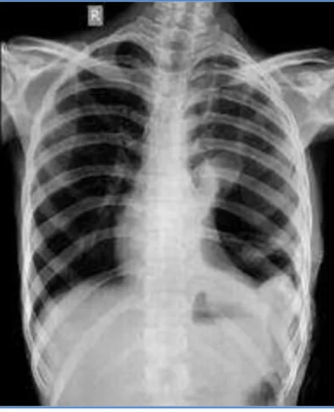
Chest radiograph (Fig. 1) confirmed the position of the tube.
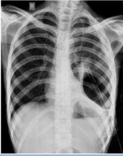
Chest radiograph taken three days after the chest tube insertion showed persistent hydropneumothorax (Fig. 2), for which the patient underwent a contrast enhanced computed tomography of thorax (CECT).
Contrast enhanced computed tomography of thorax revealed herniation of fundus of stomach through a defect measuring ~ 3cm in the left dome of diaphragm into the left thoracic cavity with leakage of oral contrast into the left pleural cavity suggestive of gastric perforation with formation of gastropleural fistula. (Fig. 3)
The presence of food particles in the intercostal drainage tube confirmed our diagnosis. The patient underwent laparotomy with repair of the diaphragmatic defect and closure of the gastric perforation. The patient had an uneventful recovery and taking orals normally.
CASE REPORT
J of Evidence Based Med & Hlthcare, pISSN- 2349-2562, eISSN- 2349-2570/ Vol. 2/Issue 8/Feb 23, 2015 Page 1090 Other causes have been subsequently described as complications of pulmonary surgery, oesophageal surgery(3),(4),(5) and gastric bypass operations for morbid obesity. Later it was also recognized that these fistulas might occur in late postoperative phase of esophagogastrectomy, with or without presence of recurrent tumour or radiation therapy.(5),(6),(7),(8)
Performing triple contrast CT, after intravenous injection and oral and rectal administration of soluble contrast, ideally with MDCT has been advocated for demonstration of digestive tract perforation in penetrating torso trauma and hence determining the need for laparotomy even when clinical signs of peritonitis and radiographic findings of pneumoperitoneum are absent.(9)
Gastropleural fistula demands early diagnosis and treatment. Diagnosis of gastropleural fistula can be achieved preoperatively by examination of pleural fluid for pH and food residues, contrast radiology after oral administration of barium or water soluble iodinated contrast and meticulous upper gastrointestinal endoscopy.(10,11)
MDCT with high resolution multiplanar reformations allows demonstration of fistulous tract across the diaphragmatic discontinuity, all along from stomach up to pleural cavity. Surgical repair is the definitive treatment of gastropleural fistula and should be performed without any delay.(11)
CONCLUSION: In conclusion, although gastropleural fistulas are relatively uncommon, the prognosis of fistulas from the upper GI tract to the pleura seems to depend upon the delay from diagnosis to surgical intervention. It is important, therefore, to consider this diagnosis early in the patient's management.(12) Cross-sectional imaging, particularly CT provide information that allows comprehensive evaluation of most acquired GI fistulas.
CASE REPORT
J of Evidence Based Med & Hlthcare, pISSN- 2349-2562, eISSN- 2349-2570/ Vol. 2/Issue 8/Feb 23, 2015 Page 1091
REFERENCES:
1. Markowitz AM, Herter FP. Gastro-pleural fistula as a complication of esophageal hiatal hernia. Ann Surg1960; 152: 129-34.
2. Schwab RJ, Jarvik JG. Tension pneumothorax secondary to a gastropleural fistula in a traumatic diaphragmatic hernia. Chest 1991; 99: 247-9.
3. O'Keefe PA, Goldstraw P. Gastropleural fistula following pulmonary resection. Thorax 1993; 48: 1278-9.
4. Biswas IH, Raghavan C, Sevcik L. Gastropleural fistula: An unusual cause of intractable postoperative nausea and vomiting. Anesth Analg 1996; 83: 186-8.
Fig. 2: Chest radiograph taken three days after intercostal drainage tube insertion showing persistent left hydropneumothorax
Fig. 3: Axial(a), coronal(b) and sagittal (c) reformatted contrast enhanced CT showing herniation of fundus of stomach into the left thoracic cavity( black arrows) with leakage
CASE REPORT
J of Evidence Based Med & Hlthcare, pISSN- 2349-2562, eISSN- 2349-2570/ Vol. 2/Issue 8/Feb 23, 2015 Page 1092 5. Virlos I, Asimakopoulos G, Forrester-Wood C. Gastropleural fistula originating from the
lesser curve: A recognized complication, an uncommon pathway of communication.Thorac Cardiovasc Surg 2001; 49: 308-9.
6. Mussi A, Lucchi M, Davini F, Angeletti CA. Gastropleural fistula as complication of postpneumonectomy empyema. J Cardiovasc Surg (Torino) 2000; 41: 147-9.
7. Bini A, Grazia M, Petrella F, Stella F, Bazzocchi R. Spontaneous biliopneumothorax (thoracobilia) following gastropleural fistula due to stomach perforation by nasogastric tube. Ann Thorac Surg 2004; 78: 339-41.
8. Shinichi T, Soichiro F, Takeyoshi Y, Kiyoshi O. Gastropleural fistula due to gastric perforation after lobectomy for lung cancer. Inter Cardiovasc Thorac Surg 2005; 4: 420-2.
9. Shanmuganathan K, Mirvis SE, Chiu WC, Killeen KL, Scalea TM. Triple-contrast helical CT in penetrating torso trauma: a prospective study to determine peritoneal violation and the need for laparotomy. AJR Am J Roentgenol 2001; 177(6): 1247–56.
10.Darbari A, Tandon S, Singh GP. Gastropleural fistula: rare entity with unusual etiology. Ann Thorac Med 2007; 2(2): 64–5.
11.Tzeng JJ, Lai KH, Lo GH, Hsu JH, Mok KT. Gastropleural fistula caused by incarcerated diaphragmatic herniation of the stomach. Gastrointest Endosc 2001; 53(3): 382–4.
12.C.J. Warburton, P.M.A. Calverley. Gastropleural fistula due to gastric lymphoma presenting as tension pneumothorax and empyema. Eur Respir J 1997; 10: 1678–1679.
AUTHORS:
1. Vijay Kumar K. R.
2. Vijayaraghavachari T. V.
3. Adarsh K. M.
4. Riya Jeeson
5. Ashwini C.
6. Ramesh V.
PARTICULARS OF CONTRIBUTORS:
1. Associate Professor, Department of Radio-diagnosis, Bangalore Medical College & Research Institute, Bangalore.
2. Post Graduate Student, Department of
Radio-diagnosis, Bangalore Medical College & Research Institute, Bangalore.
3. Post Graduate Student, Department of
Radio-diagnosis, Bangalore Medical College & Research Institute, Bangalore.
4. Post Graduate Student, Department of
Radio-diagnosis, Bangalore Medical College & Research Institute, Bangalore.
5. Post Graduate Student, Department of
Radio-diagnosis, Bangalore Medical College & Research Institute, Bangalore.
6. Post Graduate Student, Department of
Radio-diagnosis, Bangalore Medical College & Research Institute, Bangalore.
NAME ADDRESS EMAIL ID OF THE CORRESPONDING AUTHOR:
Dr. Vijayarachavachari T. V, # 42/1, K. No. 4th Street, Narayana Pillai Street Cross, Shivaji Nagar, Bangalore-560001, Karnataka.
E-mail: [email protected]
Date of Submission: 10/02/2015.