Strana 576 VOJNOSANITETSKI PREGLED Vojnosanit Pregl 2013; 70(6): 576–579.
Correspondence to: Srdjan D. Poštiý, Clinic of Dental Prosthetic, Faculty of Dental Medicine, University of Belgrade, Rankeova 4, 11 000, Belgrade, Serbia. Phone: +381 11 2435 719/ext 455. E-mail: srdjan.postic@stomf.bg.ac.rs
O R I G I N A L A R T I C L E UDC: 616.314-007-08:616.314-77@:616.716.4-007.234-08 DOI: 10.2298/VSP1306576P
Effects of calcitonin and calcium medication in treatment of
edentulous osteoporotic mandible
Efekti primene kalcitonina i kalcijuma u le
þ
enju bezubih pacijenata sa
osteoporoznim mandibulama
Srdjan D. Poštiü
Clinic of Dental Prosthetic, Faculty of Dental Medicine, University of Belgrade, Belgrade, Serbia
Abstract
Background/Aim. In addition to damage of the bones that support the remaining teeth, degradation of osteopo-rotic oral bones also lead to a consequent reduction of sup-porting tissues and the loss of dentures retention. The aim of this study was to assess the clinical and radiographic out-comes following injection of a calcitonin and calcium solu-tion into the buccal aspects of edentulous mandibles.
Methods. The experimental group of 67 edentulous pa-tients diagnosed with osteoporosis, and the control group of 19 nonosteoporotic edentulous patients were treated with the calcitonin and calcium once solution per month. Man-dibular bone density was measured from panoramic radio-graphs, supplemented by T scores of skeletal density in the experimental group. Results. After the therapy, measure-ments showed moderate increases in bone density, compen-sating for up to 4% of the total loss of minerals and solidity of denture-bearing areas of osteoporotic mandibles. Osteo-porosis affected women earlier than men in this study.
Conclusion. Application of a calcitonin and calcium solu-tion is a suitable method of preprosthetic therapy for eden-tulous osteoporotic patients.
Key words:
osteoporosis; mandible; mouth, edentulous; calcitonin; calcium; treatment outcome.
Apstrakt
Uvod/Cilj. Degradacija osteoporoznih viliÿnih kostiju vodi ka ošteýenju kostiju koje podržavaju preostale zube, ali i po-slediÿnoj redukciji potpornog tkiva i gubitku retencije prote-ze. Cilj rada bio je da se procene kliniÿki i radiografski pa-rametri posle tretmana bezubih mandibula rastvorom kal-citonina i kalcijuma ubrizganog u bukalne recesuse. Meto-de. Ispitnu grupu ÿinilo je 67 bezubih pacijenata sa dijagno-stikovanom osteoporozom, a kontrolnu grupu 19 bezubih pacijenata sa normalnom gustinom kosti (bez osteoporoze). Svi ispitanici dobijali su rastvor kalcitonina i kalcijuma jed-nom meseÿno. Gustina kosti mandibule merena je na osno-vu ortopantomograma svih ispitivanih pacijenata, uz kom-plementarni T-nalaz gustine skeleta na sistemskom nivou kod pacijenata iz ispitne grupe. Rezultati. Po završenoj te-rapiji utvrĀen je umeren porast gustine kosti, ÿime je nado-knaĀeno oko 4% ukupnog gubitka mineralnih supstanci u regionima noseýeg tkiva osteoporoznih mandibula. Osteo-poroza je zahvatala kosti žena ranije od kostiju muškaraca u ovoj studiji. Zakljuÿak. Primena rastvora kalcitonina sa kal-cijumom predstavlja kvalitetan metod medikacije i prepro-tetske pripreme bezubih pacijenata obolelih od osteoporoze.
Kljuÿne reÿi:
osteoporoza; mandibula; bezubost; kalcitonin; kalcijum; leÿenje, ishod.
Introduction
Osteoporotic microstructural degradation and macro-scopic reduction of the oral bones have been described in the literature 1. Degradation of osteoporotic oral bones has been reported not only to damage the bone supporting the re-maining teeth, but also to lead to a consequent reduction of supporting tissues and the loss of dentures retention 2. Analy-sis of the bone layer appearance on panoramic radiographs was used to assess changes in any parameters of turnover of jaw bones 3–5.
Calcitonin accelerates influx of calcium, improving bone mineral content and bone density 6. For this research it was speculated that these positive effects on osteopenic and osteoporotic bones could finally be achieved in the mandible, as well as in other human bones.
Considering various data available on multiple drug therapy and possible effects of systemic medication, it could be of particular importance to focus on dependable ap-proaches to oral treatment of osteoporosis.
Volumen 70, Broj 6 VOJNOSANITETSKI PREGLED Strana 577
Poštiý DS. Vojnosanit Pregl 2013; 70(6): 576–579.
preprosthetic and prosthetic therapy of selected edentulous osteoporotic patients.
Methods
The study included 67 edentulous patients – 11 men, aged 64–80 (mean age 71 years) and 56 women, aged 55–79 (mean age 56 years) in the experimental group, and 19 edentulous patients – 9 men, aged 55–65 (mean age 59 year), and 10 women, aged 40–55 (mean age 53.5 year), with well-pronounced edentulous ridges and with normal bone density, as the controls. Edentulous patients were selected based on a questionnaire concerning previous medical treatments for osteoporosis, skeletal density, history of fractures of any bones, postmenopausal periods in women, calcium and mi-croelements of blood-plasma, as well as oral status of bone consistency or reduction. No patients had any malignancy or manifest destructions in the mouth.
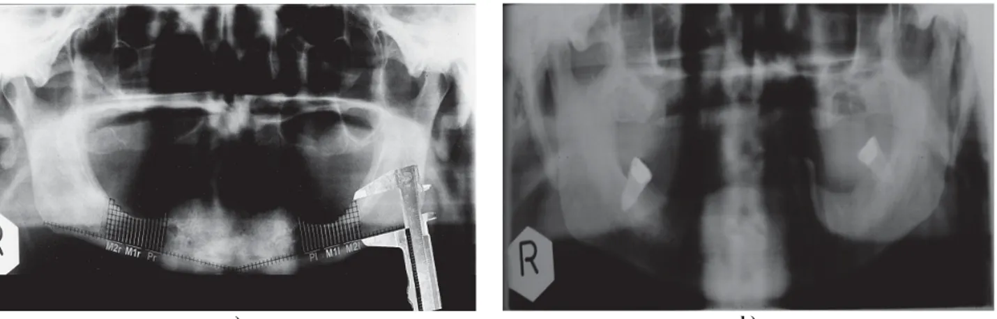
Skeletal density analysis was performed on a scanner (Lunar DPX-L, GE Healthcare, Piscataway, NJ) and T scores of the patients were obtained, indicating low bone density and systemic osteoporosis in the experimental group. Scan-ning was done for at least three of lumbar vertebral bodies in the regions of L1-L4 lumbar vertebrae of the experimental group patients. Panoramic radiographs (Orthopantomograph 10-serial no. 01492, Siemens, Germany) of every patient in the experimental group were focused on degradations in the osteoporotic jaw (Figure 1a) at baseline. Panoramic radio-graphic finding of a patient from the control group before application of calcitonin and calcium solution is shown in
Figure 1b). Calcitonin (Miacalcic, Novartis, Switzerland, and Huber, Galenika A.D., Serbia), and calcium (Calcium-gluconate Sterop, Belgium; Calcium-Sandoz, 10% calcium glubionas amp., Sandoz Switzerland) were used locally to improve solidity of edentulous mandibles. After local an-aesthetic application, up to 1.5 mL of a solution containing calcitonin and calcium (2 : 1 ration in one dose) was injected
submucosally adjacent to the mandibular bone surfaces (Fig-ure 2). A total of 5 doses were applied to both the right and
left sides of the osteoporotic mandible at monthly intervals, to each patient of both groups. This treatment was adminis-tered over a 4-month period. The final separate dose of cal-citonin and calcium solution was applied only to each patient of the experimental group at recalls, after 4 months, but not later than 6 months of follow-up. For the patients of the con-trol group calcitonin and calcium was applied after tooth ex-tractions to consolidate the buccal bone and accelerate re-covery of post-extraction wounds, but also for preventing bone loss and to compare increments of bone densities be-tween the control group and the experimental group.
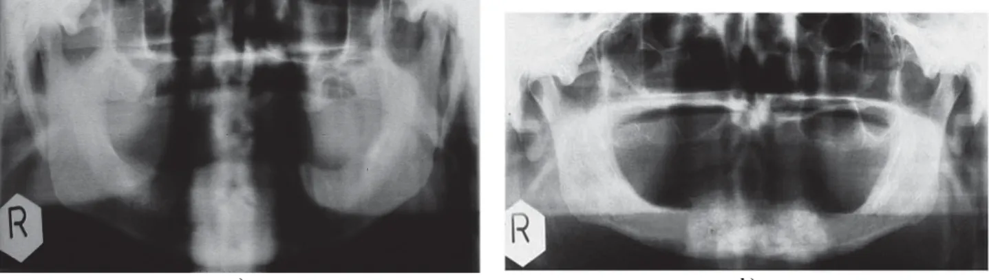
Pano-ramic radiographs of the patients were taken after each ap-plication of the solution (Figure 3). Complete dentures (CD) were fabricated for each patient of both groups. All of the patients had complete dentures during the experimental pe-riod. During the period of application of the solution con-taining calcitonin and calcium, a digital densitometer (DT II 05, U.K.) was used to analyse the bone density of
mandibu-a) b)
Fig. 1 – a) A grid positioned in the regions of particular interest for panoramic radiograph of osteoporotic edentulous ble and the determined regions – Pr, M1r, M2r, Pl, M1r, M2r (Pr – region of missing premolar to the right side of the mandi-ble; M1r – region of missing the 1st molar to the right side of the mandimandi-ble; M2r – region of missing the 2nd molar to the
right side of the mandible; Pl – region of missing premolar to the left side of the mandible; M1l – region of missing the1st molar to the left side of the mandible; M2l – region of missing the 2nd molar to the left side of the mandible); b) – Panoramic radiograph of the control group patient before application of calcitonin and calcium solution.
Fig. 2 – Submucosal injection of a calcitonin and calcium solution in the buccal aspect of osteoporotic mandible of the
Strana 578 VOJNOSANITETSKI PREGLED Volumen 70, Broj 6
Poštiý DS. Vojnosanit Pregl 2013; 70(6): 576–579. lar segments on panoramic radiographs. Measurements of the
density of the mandibles were taken over the 4 month period of application of solution. Numerical values of bone density on radiograph were shown on digital densitometer repre-senting intensity of entry of focused beam of light into the determined region of dental panoramic radiographs, and these values were expressed in arbitrary units of U/mm2.
The Student’s t-test, ANOVA, Pearson’s correlation coefficient and calculation of the incremental trend were used for mandibular density analysis.
Results
Before administration of the calcitonin and calcium so-lution, T-scores from DPX scanning were up to -2.6.for the men and women of the experimental group.
The results of medication for osteoporotic oral bones were favourable in this study. The incremental changes in density of mandibular segments were compared between the experimental and the control group at each time point, as well as between the experimental group and the control group during the therapy.
When mandibular density was compared in the experi-mental group at baseline and after the 1st month of the ther-apy, differences were significant for the segments Pr (p < 0.05) with tempo and range of increment of density (R) 1.0080 (0.80%), for Pl (p < 0.05) with R 1.0078 (0.78%), for M1r (p < 0.05) with R 1.0067 (0.67%), for M1l (p < 0.05)
with R 1.0065 (0.65%), M2r (p < 0.05) with R 1.0062 (0.62%) and M2l (p < 0.05) with R 1.0060 (0.60%). Density increased further after the 2nd month of application and was significantly increased for Pr (p < 0.05) with R 1.0087 (0.87%), for Pl (p < 0.05) with R 1.0084 (0.84%), for M1r (p
< 0.05) with R 1.0074 (0.74%), for M1l (p < 0.05) with R 1.0073 (0.73%), M2r (p < 0.05) with R 1.0066 (0.66%) and M2l (p < 0.05) with R 1.0065 (0.65%). Mandibular density comparison at baseline and after the 3rd month of application showed that it was a significant change in density for Pr (p < 0.01) with R 1.0117 (1.17%), for Pl (p < 0.01) with R 1.0114 (1.14%), for M1r (p < 0.01) with R 1.007 (1.07%), for M1l (p < 0.01) with R 1.0094 (0.94%), M2r (p < 0.05) with R 1.0083 (0.83%) and M2l (p < 0.05) with R 1.0079 (0.79%).
Following application of the calcium and calcitonin solution, the increase in density was significant for Pr, Pl (p
< 0.01), M1r, M1l, M2r and M2l segments (p < 0.05), with respect to the baseline (Table 1), compensating for up to 4% of the total loss of jaw bone mineral in the experimental group. In the control group, a statistically small increase in bone density (p < 0.05) was observed in a premolar and the 1st missing molar regions (Table 1).
Discussion
In the dental literature there are few reports on treat-ment and local medication for osteoporotic oral bones.
Based on convincing medical studies on patients, with systemic osteoporosis, the two foremost treatment approaches
a) b)
Fig. 3 – a) Panoramic radiograph of the control group patient after application of calcitonin and calcium solution; b) – Panoramic radiograph of a patient, exposing a local increment in density edentulous mandible
(for comparisson see Fig. 1).
Table 1 Mandibular bone density in the control and experimental groups at baseline and after administration of calcitonin with
ionized calcium solution
Control group Experimental group Digital value of density
ʉr SD
Digital value of density ʉr SD Segment
before therapy after therapy p
Correlation cofficient
before therapy after therapy p
Correlation cofficient Pr 1.01 r 0.6 1.83 r 0.7 < 0.05 0.578 -1.42 r 0.8 0.21 r 0.7 < 0.01 0.814 Pl 1.03 r 0.8 1.92 r 0.4 < 0.05 0.542 -1.40 r 0.9 0.19 r 0.6 < 0.01 0.793 M1r 0.83 r 0.8 1.79 r 0.8 < 0.05 0.528 -1.34 r 0.8 -0.24 r 0.9 < 0.05 0.637 M1l 0.79 r 0.9 1.76 r 0.9 < 0.05 0.519 -1.33 r 0.7 -0.21 r 0.9 < 0.05 0.619 M2r 1.61 r 0.5 1.83 r 0.3 > 0.05 0.394 -1.11 r 0.9 0.61 r 0.5 < 0.05 0.593 M2l 1.52 r 0.8 1.85 r 0.3 > 0.05 0.367 -1.11 r 0.4 0.62 r 0.6 < 0.05 0.607 Numerical values of density are in arbitrary units (U/mm2
Volumen 70, Broj 6 VOJNOSANITETSKI PREGLED Strana 579
Poštiý DS. Vojnosanit Pregl 2013; 70(6): 576–579.
could be described: the first related to application of bisphos-phonates 7, 8, while the second considered calcitonin as the primary accelerator of calcium influx to the bone 6, 7.
Making an allowance for various facts derived from studies conducted on patients undergoing systemic therapy with bisphosphonates, and a few studies on local application of bisphosphonates, as well as for the fact that certain inter-ruptions of immune-response were described as a possible complication of extended bisphosphonate usage 9, therapy with a solution containing only calcitonin and calcium, ad-ministered locally to osteoporotic jaws, seemed a rational option.
Calcitonin has been recognized as the primary sub-stance that strongly regulates the influx of calcium 6. Cal-cium influx should have been provided by mobilization of calcium ions in plasma, as well as by exchanging calcium ions from pharmacological calcium gluconate and calcium glubionas. Respecting the fact that human osteoporotic man-dible may not be very well provided with plentiful blood supply, it was assumed that calcium ions in blood vessels of the mandible would not have provided sufficient calcium in-flux to prevent osteoporotic degradation. Furthermore, addi-tional sources of calcium ions, directed towards credible lo-cal storage in close proximity to the mandible, should have a beneficial effect.
In the dental literature, there were few reports on the lo-cal application of lo-calcitonin and lo-calcium to hard oral tissues
in vivo.
On the strength of the results of this study, it appears that the solution of calcitonin and calcium should be applied to the edentulous ridge prior to positioning of a denture onto supporting tissues.
Careful selection of patients concerning the absence of infectious diseases, malignancy, serious kidney problems or blood diseases, is of ultimate priority when considering local application of calcitonin and calcium solution to oral bones.
Repeated application of calcitonin and calcium solution to jaw bone layers may significantly improve the mineral
content of bone, bone density, the condition of the bone and the potential of the area to support dentures. Osteoporotic edentulous mandibles should be the first of all oral bones to benefit from a solution containing calcitonin and calcium. Additionally, respecting the levels of concentration of cal-cium ions applied, osteoporotic maxillary bone could be treated with a solution of the same kind, over a prolonged pe-riod of remedial use.
A probable contraindication for local oral application of calcitonin and ionized calcium in solution should be a very intensive reduction of edentulous ridges with the alveolar crest positioned under the superficial surface of the mylohy-oid muscle of the edentulous mouth floor.
Calcitonin accelerated influx of calcium into the man-dibles of all the patients in both groups.
Since osteoporosis frequently affects women, mandible osteoporosis is likely to affect women rather than men. The use of calcitonin and calcium for osteoporotic toothless pa-tients should be considered the priority for local oral care. In spite of the limited value of local application, calcitonin and calcium should be the prerequisite foundation for positive bone remodelling and turnover of segments of the jaw. Re-gardless the absence of macroscopically distinct bone regen-eration, local application of calcitonin with calcium could be crucial for success at the first point of positive bone remod-elling and turnover, and for laying the foundation for later restorative procedures in oral rehabilitation of osteoporotic patients.
Conclusion
The use of calcitonin and calcium solution should be considered the priority in preprosthetic and prosthetic ther-apy of edentulous osteoporotic patients. This medication compensated for the loss of mineral from the jaw. Applica-tion of a calcitonin and calcium soluApplica-tion seems to be a suit-able method of preprosthetic therapy for edentulous osteopo-rotic patients.
R E F E R E N C E S
1. Kleerekoper M. Osteoporosis prevention and therapy: preserving
and building strength through bone quality. Osteoporos Int 2006; 17(12): 1707–15.
2. von Wowern N. General and oral aspects of osteoporosis: a
re-view. Clin Oral Investig 2001; 5(2): 71–82.
3. Horner K, Devlin H. Clinical bone densitometric study of
man-dibular atrophy using dental panoramic tomography. J Dent 1992; 20(1): 33–7.
4. Yaûar F, Akgünlü F. The differences in panoramic mandibular
indices and fractal dimension between patients with and with-out spinal osteoporosis. Dentomaxillofac Radiol 2006; 35(1): 1–9.
5. Mohammad AR, Alder M, McNally MA. A pilot study of
pano-ramic film density at selected sites in the mandible to predict osteoporosis. Int J Prosthodont 1996; 9(3): 290î4.
6. Chesnut CH 3rd, Azria M, Silverman S, Engelhardt M, Olson M,
Mindeholm L. Salmon calcitonin: a review of current and future
therapeutic indications. Osteoporos Int 2008; 19(4): 479î91.
7. Postic S. Therapy and prosthetic management of edentulous
osteoportic mandibles. Belgrade: Zadužbina Andrejevic; 2010. p. 46î58. (Serbian)
8. Cardona JM, Pastor E. Calcitonin versus etidronate for the
treatment of postmentopausal osteoporosis: A meta analysis of published clinical trials. Osteoporos Int 1997; 7(3): 165–74.
9. Ruggiero S, Mehrotra B. Bisphosphonate related osteonecrosis of
the jaw: diagnosis, prevention and treatment. Annu Rev Med 2008; 60: 85–96.