Letters to the Editor
Radiol Bras. 2015 Nov/Dez;48(6):399–403
400
http://dx.doi.org/10.1590/0100-3984.2015.0075
Maurício Fabro1, Sara Raquel Madalosso Fabro1, Rafael Santiago Oliveira Sales1, Cesar Augusto Machado1, Gustavo Lopes de Araújo1
1. Hospital Santa Catarina de Blumenau, Blumenau, SC, Brazil. Mailing Address: Dr. Maurício Fabro. Rua Tobias Barreto, 266, ap. 304, Vila Nova. Blumenau, SC, Brazil, 89035-070. E-mail: [email protected].
There is no defined treatment for PVT(1,3), and possible
treat-ment strategies include use of anticoagulant drugs(1–4,6),
NSAIDs(1,3,6), elastic socks(3,6) and rest(6). However, the different
therapies have shown similar results.
The most important complications of PVT include throm-bosis extension into deep veins in the leg(7) and occurrence of
pul-monary embolism(1).
Amongst the differential diagnosis of PVT, plantar fasciitis(2,4,5),
tendinous involvement(3,5), bursitis(5), Morton’s neuroma(4,5), stress
fractures(2,4,5), sesamoiditis(5) and ganglion cysts(5). No
descrip-tion of death associated with PVT is found in the literature.
REFERENCES
1. Barros M, Nascimento I, Barros T, et al. Plantar vein thrombosis and pulmonary embolism. Phlebology. 2015;30:66–9.
2. Bruetman JE, Andrews JA, Finn BC, et al. Plantar vein thrombosis as a cause of local pain. Medicina (B Aires). 2014;74:87–8.
3. Czihal M, Röling J, Rademacher A, et al. Clinical characteristics and course of plantar vein thrombosis: a series of 22 cases. Phlebology. 2014. [Epub ahead of print].
4. Karam L, Tabet G, Nakad J,et al. Spontaneous plantar vein thrombosis: state of the art. Phlebology. 2013;28:432–7.
5. Miranda FC, Carneiro RD, Longo CH, et al. Tromboflebite plantar: achados em ressonância magnética. Rev Bras Ortop. 2012;47:765–9. 6. Geiger C, Rademacher A, Chappell D, et al. Plantar vein thrombosis due to busy night duty on intensive care unit. Clin Appl Thromb Hemost. 2011;17:232–4.
7. Barros MV, Labropoulos N. Plantar vein thrombosis – evaluation by ul-trasound and clinical outcome. Angiology. 2010;61:82–5.
8. Bernathova M, Bein E, Bendix N, et al. Sonographic diagnosis of plantar vein thrombosis: report of 3 cases. J Ultrasound Med. 2005;24:101–3. 9. Siegal DS, Wu JS, Brennan DD, et al. Plantar vein thrombosis: a rare
cause of plantar foot pain. Skeletal Radiol. 2008;37:267–9.
Pulmonary neoplasia mimicking fungus ball
Neoplasia pulmonar simulando bola fúngica
Dear Editor,
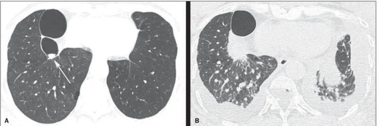
We report the case of a 74-year-old man smoking 80 ciga-rette packages per year, with history of pulmonary tuberculosis for 50 years. Two years ago, the patient underwent chest computed tomography that demonstrated centrilobular and paraseptal em-physema, besides sparse bullae, the largest one located in the right lower lobe, with a small nodular mass inside, measuring about 0.8 cm in diameter (Figure 1A).
The patient didn’t return for follow-up and after two years pre-sented with progressive dyspnea whose onset had occurred two months ago, in association with cough, weight loss and pain in the lower third of the right hemithorax. A new chest computed tomography demonstrated a mass with spiculated margins, adja-cent to the posterior portion of the largest bulla, occupying the whole bulla where the nodular mass had been seen at the previ-ous computed tomography images (Figure 1B). Also, interstitial thickening suggestive of carcinomatous lymphangitis was ob-served, besides bilateral pleural effusion.
Pericardium biopsy and cytological analysis of pleural effu-sion revealed adenocarcinoma, raising the hypothesis of lung ad-enocarcinoma with metastasis to the pleura and pericardium. A chemotherapy protocol with gemcitabine and carboplatin was ini-tiated. The patient presented worsening of the respiratory condi-tion, progressing to death after two months.
Lung cancer frequently presents like a nodule or solitary lung mass(1,2). However, the disease presentation forms are quite
vari-able and some typical findings may be observed. One of such find-ings is growth from a preexisting cystic mass, mimicking a fun-gus ball. Thus, a cystic image showing either focal or diffuse wall thickening progressing to a nodular mass should include lung tumor in the differential diagnosis(3), particularly in cases where
the nodule is attached to the wall and does not move with change in decubitus.
Other conditions which may present the finding of fungus ball include Rasmussen aneurysms, hydatid cysts, abscesses and intra-cavitary hematomas, besides fungal diseases themselves (aspergillo-sis, nocardia(aspergillo-sis, actinomyco(aspergillo-sis, candidia(aspergillo-sis, coccidioidomycosis)(2,4).
As the neoplasm develops in previous pulmonary lesions, it is found especially in fibroatelectatic or granulomatous areas
result-Figure 1. HRCT scan at the level of the lung bases (A) showing two bullae at right, with a small nodular mass measuring about 0.8 cm in diameter inside the small bulla (arrow). On B, scan acquired two years later, with a section of the same region, showing the presence of a mass with spiculated borders, adjacent to the posterior portion of the largest bulla, occupying the small bulla where the nodular mass had been seen at the previous CT images. Also, observe the presence of interstitial thickening suggestive of carcinomatous lymphangitis, besides bilateral pleural effusion.
Letters to the Editor
Radiol Bras. 2015 Nov/Dez;48(6):399–403
401
http://dx.doi.org/10.1590/0100-3984.2015.0119 ing from sequelae, generally associated with tuberculosis. The
occurrence of lung cancer in cavities mimicking fungus ball or air crescent sign is quite rare(1,2,5). The tumor tends to infiltrate
in the adjacent pulmonary parenchyma causing a paracicatricial effect, and may lead to emphysematous or cystic changes adja-cent to the neoplastic process(1).
In conclusion, lung cancer must be considered in the differ-ential diagnosis for patients who present with a fungus ball-like le-sion, particularly in cases where the nodule is fixed to the cavity wall.
REFERENCES
1. Wang LF, Chu H, Chen YM, et al. Adenocarcinoma of the lung present-ing as a mycetoma with an air crescent sign. Chest. 2007;131:1239–42. 2. Gazzoni FF, Severo LC, Marchiori E, et al. Pulmonary diseases with imaging findings mimicking aspergilloma. Lung. 2014;192:347–57.
Bruno Fernandes Cavalcante1, Gláucia Zanetti1, Edson Marchiori1
1. Department of Radiologiy – Universidade Federal do Rio de Janeiro (UFRJ), Rio de Janeiro, RJ, Brazil. Endereço para correspondência: Dr. Edson Marchiori. Rua Thomaz Cameron, 438, Valparaíso. Petrópolis, RJ, Brazil, 25685-120. E-mail: [email protected].
3. Truong MT, Ko JP, Rossi SE, et al. Update in the evaluation of the soli-tary pulmonary nodule. Radiographics. 2014;34:1658–79.
4. Watanabe H, Uruma T, Tsunoda T, et al. Lung metastasis of transi-tional cell cancer of the urothelium, with fungus ball-like shadows closely resembling aspergilloma: a case report and review of the literature. Oncol Lett. 2014;8:95–8.
5. Bandoh S, Fujita J, Fukunaga Y, et al. Cavitary lung cancer with an aspergilloma-like shadow. Lung Cancer. 1999;26:195–8.
Extramedullary plasmacytoma in the right pulmonary hilum
Plasmocitoma extramedular no hilo pulmonar direito
Dear Editor,
A 53-year-old black, asymptomatic man, driver, being as-sessed to be released for physical activity. The patient denied smoking as well as having comorbidities.
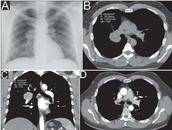
Chest radiography performed on February 1st, 2011 showed ovoid opacity in the right hilar region, with no other abnormality (Figure 1A). Chest computed tomography (CT) performed on March 13, 2011 identified circumscribed round opacity with soft parts attenuation in the right hilar region, presenting enhance-ment after intravenous contrast agent injection, adjacent to the ipsilateral main pulmonary artery and its branches. Absence of other findings (Figures 1B and 1C).
Lesion biopsy result: macro/microscopy – hypercellular light-brownish fragments showing well-differentiated plasmacytoid cells
with small, eccentric and hyperchromatic nuclei;
immunohis-tochemical analysis – positive for CD138 and lambda antibodies; and negative for CD3, CD20, AE1/AE3 and kappa antibodies.
The investigation proceeded with abdominal CT (on May 16, 2011) that showed the presence of a liver cyst and signs of fat infiltration into the liver; normal blood count; negative Bence-Jones proteinuria; protein electrophoresis with no abnormalities; absence of noteworthy findings at bone scintigraphy and bone marrow aspiration.
Radiotherapy was the treatment of choice, with satisfactory response.
Chest CT performed on November 9, 2012 (Figure 1D) and other radiological studies with no suspect finding of disease re-currence/progression until May 20, 2015.
Diagnosis: extramedullary plasmacytoma (EMP) in the pul-monary hilum.
Plasmacytoma are primarily classified into solitary bone marrow/bone plasmacytoma (solitary myeloma), extramedullary plasmacytoma or one of multiple myeloma components(1,2). Such