Inhibition of Bone Morphogenetic Protein
Signal Transduction Prevents the Medial
Vascular Calcification Associated with Matrix
Gla Protein Deficiency
Rajeev Malhotra1*, Megan F. Burke1, Trejeeve Martyn2, Hannah R. Shakartzi2, Timothy E. Thayer1, Caitlin O’Rourke2, Pingcheng Li2, Matthias Derwall2,3, Ester Spagnolli2, Starsha A. Kolodziej2, Konrad Hoeft2, Claire Mayeur2, Pawina Jiramongkolchai2, Ravindra Kumar4, Emmanuel S. Buys2, Paul B. Yu5, Kenneth D. Bloch1,2, Donald B. Bloch2,6
1Cardiovascular Research Center and Cardiology Division of the Department of Medicine, Massachusetts General Hospital, Harvard Medical School, Boston, MA, United States of America,2Anesthesia Center for Critical Care Research of the Department of Anesthesia, Critical Care, and Pain Medicine, Massachusetts General Hospital, Harvard Medical School, Boston, MA, United States of America,3Department of Anesthesiology, Uniklinik Aachen, RWTH Aachen University, Aachen, Germany,4Acceleron Pharma, Inc. Cambridge, MA, United States of America,5Cardiovascular Division, Brigham and Women’s Hospital, Harvard Medical School, Boston, MA, United States of America,6Center for Immunology and Inflammatory Diseases and the Division of Rheumatology, Allergy, and Immunology of the Department of Medicine, Massachusetts General Hospital, Harvard Medical School, Boston, MA, United States of America
Abstract
Objective
Matrix Gla protein (MGP) is reported to inhibit bone morphogenetic protein (BMP) signal transduction. MGP deficiency is associated with medial calcification of the arterial wall, in a process that involves both osteogenic transdifferentiation of vascular smooth muscle cells (VSMCs) and mesenchymal transition of endothelial cells (EndMT). In this study, we investi-gated the contribution of BMP signal transduction to the medial calcification that develops in MGP-deficient mice.
Approach and Results
MGP-deficient mice (MGP-/-) were treated with one of two BMP signaling inhibitors,
LDN-193189 or ALK3-Fc, beginning one day after birth. Aortic calcification was assessed in 28-day-old mice by measuring the uptake of a fluorescent bisphosphonate probe and by staining tissue sections with Alizarin red. Aortic calcification was 80% less in MGP-/-mice
treated with LDN-193189 or ALK3-Fc compared with vehicle-treated control animals (P<0.001 for both). LDN-193189-treated MGP-/-mice survived longer than vehicle-treated
MGP-/-mice. Levels of phosphorylated Smad1/5 and Id1 mRNA (markers of BMP signaling)
did not differ in the aortas from MGP-/-and wild-type mice. Markers of EndMT and
OPEN ACCESS
Citation:Malhotra R, Burke MF, Martyn T, Shakartzi
HR, Thayer TE, O’Rourke C, et al. (2015) Inhibition of
Bone Morphogenetic Protein Signal Transduction Prevents the Medial Vascular Calcification Associated with Matrix Gla Protein Deficiency. PLoS ONE 10(1): e0117098. doi:10.1371/journal.pone.0117098
Academic Editor:Elena Aikawa, Brigham and
Wom-en’s Hospital, Harvard Medical School, UNITED
STATES
Received:October 6, 2014
Accepted:December 18, 2014
Published:January 20, 2015
Copyright:© 2015 Malhotra et al. This is an open access article distributed under the terms of the
Creative Commons Attribution License, which permits unrestricted use, distribution, and reproduction in any medium, provided the original author and source are credited.
Data Availability Statement:All relevant data are within the paper and its Supporting Information files.
osteogenesis were increased in MGP-/-aortas, an effect that was prevented by
LDN-193189. Calcification of isolated VSMCs was also inhibited by LDN-LDN-193189.
Conclusions
Inhibition of BMP signaling leads to reduced vascular calcification and improved survival in MGP-/-mice. The EndMT and osteogenic transdifferentiation associated with MGP
deficien-cy is dependent upon BMP signaling. These results suggest that BMP signal transduction has critical roles in the development of vascular calcification in MGP-deficient mice.
Introduction
Calcification of the intimal and medial layers of the arterial wall is an important risk factor for
cardiovascular events [1,2,3,4]. Intimal and medial calcification are the results of different
un-derlying pathogenic mechanisms [5,6]. Intimal calcification is preceded by subintimal lipid
de-position and macrophage accumulation whereas medial calcification is not associated with lipid deposition or inflammation and results from metabolite-induced upregulation of
osteo-genic gene programs in the vasculature [5,6]. The processes of intimal and medial vascular
cal-cification have been likened to bone formation, of which there are two types: Intimal
atherosclerotic calcification displays similarities to endochondral ossification, involving chon-drogenesis prior to bone formation; medial vascular calcification is similar to intramembra-nous bone formation in which bone derives from mesenchymal stem cells that have differentiated directly into osteoblasts [7,8].
Matrix Gla protein (MGP) is an extracellular polypeptide that inhibits arterial calcification
[9]. Mutations in theMGPgene are associated with Keutel syndrome [10], a rare autosomal
re-cessive disease characterized by calcification of the coronary, cerebral, hepatic, and renal
arteri-al beds [10,11,12,13]. Common sequence variants in theMGPgene are associated with
increased risk and progression of coronary calcification in humans [14,15]. MGP requires
γ-carboxylation of glutamic acid residues for activity, a process that depends on vitamin K as a
cofactor and is inhibited by warfarin [9,16]. Mice lacking both copies of themgpgene
sponta-neously develop medial arterial calcification beginning at 2 weeks of age. Vascular calcification
progresses over time and results in aortic rupture by 6–8 weeks of age [9].
At least two mechanisms have been proposed to explain the ability of MGP to inhibit vascu-lar calcification: MGP binds to calcium ions, as well as to hydroxyapatite crystals, and may
thereby directly inhibit crystal growth [17,18,19,20,21,22]; MGP may also sequester bone
morphogenetic protein (BMP)-2, BMP-4, and BMP-7 and reduce BMP signaling [23,24,25].
More than twenty ligands of the BMP family bind to heteromeric complexes of BMP type I
and type II serine-threonine kinase receptors [26,27]. BMP type II receptors phosphorylate
BMP type I receptors, which in turn phosphorylate the cytosolic BMP effector proteins, Smads 1, 5, and 8 (Smad 1/5/8). Phosphorylated Smads 1/5/8 translocate to the nucleus together with
Smad 4, where they activate specific targets, including the inhibitor of DNA binding (Id) genes
[26]. BMPs are potent osteogenic factors that are required for osteoblast differentiation and
bone formation [28].
BMP signaling has been implicated in intimal arterial wall calcification [7,29]. Increased
BMP-2 expression was observed in human atherosclerotic plaques [29]. Transgenic
over-expression of BMP-2 in ApoE-deficient mice accelerated the development of intimal
calcifica-tion [30]. Furthermore, BMP signaling promotes inflammation in atherosclerotic lesions,
Morphogenetic Protein Signaling in the Pathogenesis of Pulmonary and Systemic Vascular Diseases, PBY and KDB); the National Institute of Arthritis and Mus-culoskeletal and Skin Diseases (R01AR057374, PBY); and the National Institute of Diabetes and Di-gestive and Kidney Diseases (R01DK082971, KDB). The funders had no role in study design, data collec-tion and analysis, decision to publish, or preparacollec-tion of the manuscript.
Competing Interests:Dr. Ravindra Kumar is an em-ployee of Acceleron Pharma Inc. and has stock own-ership in the company. This does not alter the
authors’adherence to PLOS ONE policies on sharing
data and materials. The Massachusetts General Hos-pital has applied for patents related to pharmacologic BMP signaling inhibitors. These patents include: 1. In-hibitors of the bmp signaling pathway (Application # CA 2718403, PCT/US2009/001606). Drs. Yu and K. Bloch may be entitled to royalties. 2. Compositions and methods for cardiovascular disease (Application # PCT/US2012/022119). Drs. Malhotra, Derwall, Yu, and K. Bloch may be entitled to royalties. 3. Bmp in-hibitors and methods of use thereof (Application # PCT/US2014/020360). Drs. Yu and K. Bloch may be entitled to royalties. These patents do not alter the
au-thors’adherence to PLOS ONE policies on sharing
which indirectly promotes the development of intimal calcification [31]. Within calcified inti-mal vascular lesions, vascular smooth muscle cells (VSMCs) undergo accelerated proliferation, lose their smooth muscle contractile phenotype, and undergo transdifferentiation to an
osteo-genic phenotype [7,32,33].In vitrostudies demonstrated a relationship between BMP
signal-ing and the expression of factors important for VSMC osteogenic transdifferentiation
including runt-related transcription factor 2 (Runx2) [34,35,36].
The medial vascular calcification that develops in MGP-deficient mice is also characterized by a transdifferentiation of aortic VSMCs to osteogenic cells. This transdifferentiation is
associ-ated with both a loss of smooth muscle cell markers (including myocardin,α-smooth muscle
actin (SMA), transgelin (tagln), and calponin), and an increase in osteogenic markers such as
Runx2 and osteopontin (OPN) [37,38,39]. Runx2 is required for VSMC transdifferentiation
and osteogenic activity [38,40,41]. The potential role of BMP signaling in the loss of VSMC
phenotype, the increase in expression of osteogenic markers (Runx2 and OPN), and the medial vascular calcification associated with MGP deficiency is unknown.
The vascular endothelium provides a source of multipotent cells that contribute to vascular calcification in MGP-deficient mice, in a process termed endothelial-mesenchymal transition
(EndMT) [42,43]. Endothelial markers (VE-Cadherin and CD31) are increased and
co-expressed with markers of multipotency (nanog, Oct 3/4, and sox2) prior to transitioning to
mesenchymal cells that then express an osteogenic phenotype [42]. Depletion of MGP in
cul-tured human aortic endothelial cells is associated with increased EndMT and calcification, ef-fects that are enhanced by treatment with BMP-2, suggesting that BMP signaling is important
for EndMT associated with vascular calcification [42].
To further investigate the role of BMP signaling in the loss of VSMC phenotype, EndMT, and medial vascular calcification associated with MGP deficiency, we studied the effects of
in-hibition of BMP signal transduction in MGP-/-mice. We report that inhibition of BMP
signal-ing ussignal-ing LDN-193189, a small molecule inhibitor of BMP type I receptor kinases [44], and
ALK3-Fc, a recombinant protein that sequesters BMP ligands including BMP-2, BMP-4, and BMP-7, prevents vascular calcification associated with MGP deficiency. Long-term treatment
of MGP-/-mice with LDN-193189 improved survival. BMP signaling was essential for the
de-velopment of osteogenic cells, EndMT, and VSMC calcification associated with MGP deficiency.
Materials and Methods
Chemicals and reagents
LDN-193189 (4-[6-(4-piperazin-1-ylphenyl)pyrazolo[1,5-a]pyrimidin-3-yl]quinoline) was
synthesized as previously described [44], dissolved in water at a concentration of 0.5 mg/mL,
and titrated to a pH of 5.5 with NaOH. Recombinant ALK3-Fc was provided by Acceleron Pharma Inc. (Cambridge, MA). OsteoSense-680 and ProSense-750 were obtained from Perki-nElmer (Waltham, MA). Recombinant human BMP-2 was purchased from R&D Systems (Minneapolis, MN).
Animals
Any mice discovered to be with evidence of discomfort (manifested as immobility and/or an inability to feed) or respiratory distress (manifested by tachypnea) were promptly euthanized
with CO2gas. MGP+/-mice were generated by Dr. Karsenty and colleagues [9]. Heterozygous
mice were bred to obtain homozygous MGP-/-mice, as well as MGP+/+littermate control mice.
Animals were maintained on a standard diet. MGP-/-mice were treated intraperitoneally (i.p.)
with LDN-193189 (2.5 mg/kg, once daily), ALK3-Fc (2 mg/kg every other day), or vehicle (water) starting on the first day of life and continuing for 28 days. Wild-type mice were treated with vehicle over the same time period. Aortas were harvested two hours after the last dose of LDN-193189, ALK3-Fc, or vehicle. Livers and lungs were harvested from vehicle-treated
wild-type and MGP-/-mice.
Near-infrared imaging and quantification of aortic calcification and
inflammation
Mice were injected via the tail vein with OsteoSense-680 and ProSense-750 (150 µl each) 24
hours before euthanasia, as described previously [45,46]. Aortas were isolated and analyzedex
vivoby fluorescence reflectance imaging using an Odyssey Imaging System (LI-COR
Biotech-nology, Lincoln, NE) and software version 3.0.16 [31].
Histology and Immunofluorescence
Aortas were embedded and cryopreserved in optimal cutting-temperature medium (Sakura
Tissue-Tek, Zoeterwoude, Netherlands), and 6-µm sections were prepared [31]. To detect
cal-cification, a solution of 2 g/dL Alizarin Red in distilled water (pH 4.2) was applied to aortic sec-tions for 60 seconds, after which secsec-tions were washed with acetone and acetone-xylene (1:1) before mounting.
To detect macrophages in aortas, frozen tissue sections were fixed in cold 100% methanol and incubated with a monoclonal antibody specific for MAC-2 (Cedarlane, Burlington, NC) followed by Alexa Fluor 594-conjugated donkey anti-rat IgG antiserum (Jackson ImmunoRe-search Laboratories, West Grove, PA). The location of nuclei was identified by staining with
4’,6-diamidino-2-phenylindole (DAPI).
Preparation of mouse aortic vascular smooth muscle cells
Vascular smooth muscle cells (VSMCs) were isolated from aortas of MGP-/-mice and
wild-type littermate controls, as previously described [47]. Aortas were digested with Type 2
collage-nase (175 U/mL, Worthington) and elastase (1.25 U/mL, Sigma) for 30 min, and the adventitial layer was removed. Aortas were further digested with collagenase and elastase for 60 min, and
cells were plated and maintained in Dulbecco’s Minimum Essential Medium (DMEM,
Invitro-gen) supplemented with 10% fetal bovine serum (FBS, InvitroInvitro-gen), 100 units/ml of penicillin,
and 100 µg/ml of streptomycin at 37°C with 5% CO2. VSMC lineage was confirmed by
immu-nocytochemistry using an antibody directed againstα-smooth muscle actin (SMA, Sigma).
Ex-periments with VSMCs were performed using cells that were passaged between 2–8 times. For
gene expression experiments involving BMP-2 stimulation, cells were grown to 70% confluence followed by overnight serum starvation in DMEM with 0.1% fetal bovine serum.
Measurement of gene expression by quantitative RT-PCR
Total RNA from aortas, livers, lungs, and cultured VSMCs was extracted by the
phenol/guani-dinium method [48]. Reverse transcription was performed using Moloney murine leukemia
(Eppendorf, Hamburg, Germany) was used for real-time amplification and quantification of transcripts. Relative expression of target transcripts were normalized to levels of 18S ribosomal
RNA, determined using the relative CTmethod. Taqman gene expression assays were used to
quantify mRNA levels encoding Id1 and Runx2. Quantitative PCR was performed with SYBR green for 18S, osteopontin (OPN), myocardin, SMA, transgelin (tagln), calponin, VE-cadherin,
CD31, nanog, sox2, Oct 3/4, and MGP using the primer sequences inS1 Table.
Immunoblot techniques
Aortas, livers, lungs, and VSMCs were homogenized in RIPA buffer containing protease and phosphatase inhibitors (Sigma). Tissue and cell lysates (20 µg/lane) were separated by SDS-PAGE, transferred to polyvinylidene difluoride (PVDF) membranes (GE Amersham Biosci-ences), and probed with antibodies specific for phosphorylated SMAD1/5 (P-SMAD1/5, rabbit monoclonal, Cell Signaling, catalog #9516S), total Smad 1 (mouse monoclonal, Life Span,
cata-log #LS-C75853),α-smooth muscle actin (mouse monoclonal, Sigma, catalog #A2547), or
glyc-eraldehyde 3-phosphate dehydrogenase (GAPDH, rabbit monoclonal, Cell Signaling, catalog #5174S). Blots were incubated with horseradish peroxidase-conjugated rabbit or anti-mouse IgG, and bound secondary antibodies were visualized by chemiluminescence (ECL Plus) and quantified using a VersaDoc Imaging System (BioRad, Hercules, CA).
Depletion of MGP using siRNA
siRNA targeting MGP (siMGP) and scrambled control siRNA (siSC) were obtained from Dharmacon (SMARTpool, Thermo Scientific). Aortic VSMCs isolated from wild-type mice were transfected with siRNA using Lipofectamine RNAiMAX reagent, as described by the manufacturer (Life Technologies).
Restoration of MGP using adenovirus-mediated gene transfer
Recombinant adenovirus directing expression of MGP (Ad.MGP) was constructed using a full
length murine MGP cDNA (catalog # MMM1013–7514153, Open Biosytems) and the AdEasy
system, as previously described [49]. A control adenovirus specifying GFP was purchased from
Vector Biolabs (Ad.GFP, catalog # 1060). Ad.GFP and Ad.MGP were amplified in 293 cells. The virus was purified from cellular extracts using Adeno-X Mega Purification Kit (Clontech Laboratories, Mountain View, CA). Viral infectious forming units were quantified using the Adeno-X Rapid Titer Kit (Clontech Laboratories, Mountain View, CA). Aortic VSMCs isolated
from MGP-/-mice were infected with Ad.MGP or Ad.GFP at a multiplicity of infection of 10.
Calcium Staining
To induce calcification in isolated VSMCs, cells were treated with DMEM supplemented with
10% FCS and 2 mM sodium phosphate, as previously described [38]. Cells were fixed in 10%
formalin and incubated with von Kossa stain to detect calcification.
Statistical analysis
LDN-193189-treated MGP-/-mice compared to vehicle-treated mice. In all cases, a P<0.05 was
considered to indicate statistical significance.
Results
BMP inhibition reduces vascular calcification and improves survival in
MGP
-/-mice
Calcification in the aortas of wild-type and MGP-deficient mice was quantified using Osteo-Sense-680, a near-infrared fluorescent bisphosphonate imaging probe that incorporates into
the hydroxyapatite crystals of calcific lesions [45]. Aortas from wild-type mice exhibited little if
any OsteoSense-680 labeling consistent with the absence of calcification (Fig. 1). A strong
OsteoSense-680 signal was detected in the aortas and medium-sized arteries (e.g., carotid,
sub-clavian, and iliac arteries) of 28-day-old MGP-/-mice (Fig. 1). To confirm the presence of
vas-cular calcification in the aortas of MGP-/-mice, aortic tissue sections were stained with Alizarin
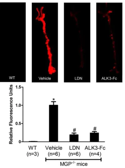
Figure 1. Pharmacologic inhibition of BMP signaling reduces osteogenic activity in the aortas of MGP-deficient mice.MGP-/-mice were treated with vehicle, LDN-193189 (LDN), or ALK3-Fc starting at day 1 of life for 28 days. Wild-type mice (WT) were treated with vehicle. On day 27, OsteoSense-680 was injected via the tail vein. Aortas were harvested 24 hours later and imaged with near-infrared fluorescence (upper panel). Fluorescence intensities of the entire aorta normalized to vehicle-treated MGP-/-mouse aortas were quantified (lower panel). Inhibition of BMP signaling reduced vascular calcification by 80% in MGP-/-mice. *P<0.001 compared to vehicle-treated WT mice. # P<0.001 compared to vehicle-treated MGP-/-mice.
Red. No aortic calcification was observed in MGP-/-mice at 7 days of age or younger, but
dif-fuse medial calcification was evident in aortas from 14-day-old MGP-/-mice (data not shown),
as previously reported [9]. In 28-day-old MGP-/-mice, there was extensive aortic calcification
with architectural distortion of the medial elastic layer. Calcification was not detected in the
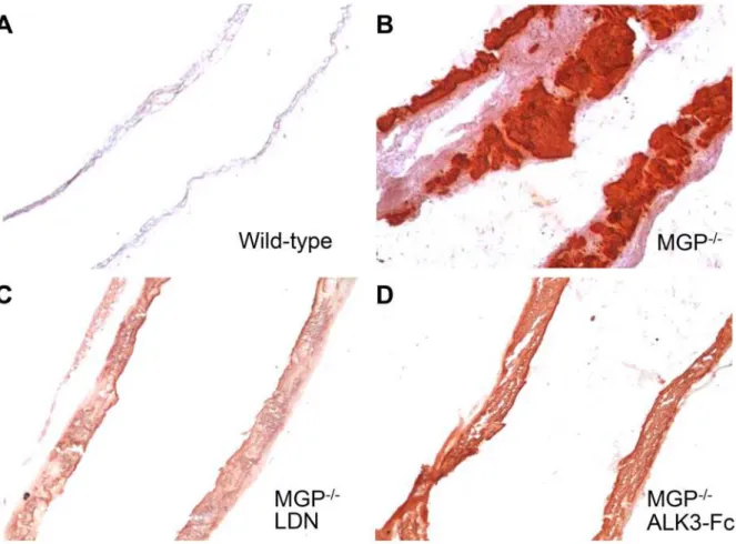
aortas of 28-day-old wild-type mice (Fig. 2A-B).
To determine whether the vascular calcification associated with MGP deficiency is
depen-dent on BMP signaling, MGP-/-mice were treated with intraperitoneal injections (i.p.) of
LDN-193189 (2.5 mg/kg, once daily), ALK3-Fc (2 mg/kg every other day), or vehicle starting at day 1 of life and continuing for 28 days. Calcification was markedly reduced in the aortas of the two treatment groups compared to the vehicle-treated animals. Mice treated with inhibitors of BMP signaling exhibited an 80% reduction in aortic calcification compared to
vehicle-treated MGP-/-mice, as measured by incorporation of OsteoSense-680 (Fig. 1, P<0.001 for
both LDN-193189 and ALK3-Fc versus vehicle). Furthermore, aortic tissue sections from
MGP-/-mice treated with either LDN-193189 or recombinant ALK3-Fc exhibited less Alizarin
Red staining than did sections from vehicle-treated MGP-/-mice (Fig. 2B–D).
To determine whether inhibition of BMP signaling in MGP deficiency improves survival,
MGP-/-mice were treated with LDN-193189 (2.5 mg/kg i.p. daily) or vehicle starting at day 1
of age. Treatment with LDN-193189 improved survival (log rank P = 0.002,Fig. 3) with a Cox
Figure 2. Vascular calcification associated with MGP deficiency is dependent on bone morphogenetic protein signaling.Aortas were harvested from mice at 28 days of age, sectioned, and stained for tissue calcium with Alizarin Red. Sections from wild-type (A) and MGP-/-(B-D) mice were
photographed at 100x magnification. Starting at day 1 of age, MGP-/-mice were treated with i.p. injections of vehicle (B), LDN-193189 (LDN, 2.5 mg/kg daily, C), or ALK3-Fc (2 mg/kg every other day,D). Inhibition of BMP signaling reduced aortic calcification in MGP-/-mice.
hazard ratio of 0.04 (95% CI 0.01–0.17). Vehicle-treated MGP-/-mice had a median survival of
40 days, whereas LDN-193189-treated MGP-/-mice exhibited a median survival of 57 days.
LDN-193189-treated MGP-/-mice also grew more rapidly than did vehicle-treated MGP
-/-mice (S1 Fig.). These results suggest that BMP signaling plays a critical role in the vascular
cal-cification and mortality associated with MGP deficiency.
MGP deficiency causes vascular calcification in the absence of
inflammation
In mouse models of atherosclerosis, we and others observed that intimal aortic calcification
was associated with the presence of BMP-dependent vascular inflammation [31,50]. To
deter-mine whether vascular calcification in MGP-deficient mice is associated with inflammation, MGP-deficient and wild-type mice were simultaneously injected with OsteoSense-680 and ProSense-750, the latter being a cathepsin-activated near-infrared fluorescent molecule that
serves as a measure of macrophage activity in blood vessels [45]. Although the MGP-/-aortas
had a strong signal for calcification compared to wild-type aortas, there was no difference in
macrophage activity between MGP-/-and wild-type mice (Fig. 4A). To further assess for the
potential presence of inflammation, aortic tissue sections were stained for MAC-2, a marker of
macrophages (Fig. 4B). MAC-2 was not detected in aortic tissue sections from 28-day-old
wild-type and MGP-/-mice, compared to a high level of MAC-2 that was observed in aortic
tis-sue sections obtained from LDLR-/-mice fed a high fat diet, a murine model of atherosclerosis
[31]. These findings indicate that MGP deficiency is a model of vascular calcification that
oc-curs in the absence of vascular inflammation. The ability of LDN-193189 and ALK3-Fc to
in-hibit the vascular calcification observed in MGP-/-mice is therefore independent of the
potential effects of BMP inhibition on vascular inflammation.
BMP signaling is not increased in MGP-deficient mice
It has been proposed that MGP inhibits vascular calcification by sequestering BMP ligands,
thereby reducing BMP signaling [23,24,51]. To consider the possibility that MGP deficiency is
associated with increased BMP signaling, levels of phosphorylated Smad 1/5 and total Smad 1
in the aortas of 7-, 14-, and 28-day-old wild-type and MGP-/-mice were measured. Levels of
phosphorylated Smad 1/5 normalized to total Smad 1 levels were not increased in the aortas of
MGP-/-mice compared to wild-type mice (Fig. 5A). To further assess the impact of MGP
Figure 3. Inhibition of BMP signaling improves survival of MGP-/-mice.MGP
-/-mice were treated once daily with LDN-193189 (LDN) or vehicle starting at day 1 of life. Kaplan-Meier survival was compared using the log rank test. MGP-/-mice treated with LDN-193189 survived longer than vehicle-treated mice.
deficiency on aortic BMP signaling, Id1 mRNA levels in the aortas of wild-type and MGP
-/-mice at 1, 7, 14, and 28 days of age were measured (Fig. 5B). Id1 mRNA levels did not change
over time in aortas from either wild-type or MGP-/-mice, and Id1 mRNA levels did not differ
between the two genotypes. To determine whether MGP deficiency alters BMP signaling in or-gans other than the aorta, Smad 1/5 phosphorylation and Id1 gene expression were measured
in the livers and lungs of MGP-/-and wild-type mice at 14 days of age. MGP gene expression
was more than 100-fold greater in wild-type mouse lung than in liver (S2A Fig.). Across this
broad range of MGP gene expression, Id1 mRNA levels and Smad 1/5 phosphorylation did not
differ between MGP-/-and wild-type mice (S2A-B Fig.). Taken together, these observations
suggest that, although vascular calcification in MGP-deficient mice is dependent on BMP
Figure 4. Vascular calcification associated with MGP deficiency occurs in the absence of vascular inflammation.(A) At 27 days of age, OsteoSense-680 and Prosense-750 were injected via the tail vein of wild-type (WT) and MGP-/-mice. Aortas were harvested 24 hours later and imaged. Although aortas from MGP-/-mice exhibited extensive vascular calcification, this calcification was not associated with increased macrophage activity. (B) Aortas were harvested from WT and MGP-/-mice at 28 days of age, sectioned, and stained for macrophages with an antibody directed towards MAC-2. Aortas from LDLR-/-mice on a high fat diet were used as a positive control. Nuclei were stained with DAPI. Similar to WT mice, macrophages were not detected by immunohistochemistry in the aortas of MGP-/-mice.
signaling, basal BMP signaling is not increased in the aortas, livers, and lungs of MGP-/-mice compared to wild-type mice.
MGP-deficiency does not alter BMP signaling in isolated vascular
smooth muscle cells
Although increased BMP signaling was not observed in the whole aortas of MGP-/-mice, it was
possible that individual cells types within the vasculature such as VSMCs might exhibit
in-creased BMP signaling in the absence of MGP [23,24,51]. To determine whether MGP
defi-ciency enhances the response of VSMCs to BMP ligands, VSMCs were isolated from the aortas
of wild-type and MGP-/-mice and were incubated in the presence or absence of BMP-2. At
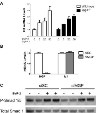
baseline, Id1 mRNA levels did not differ between wild-type and MGP-/-VSMCs (Fig. 6A).
In-cubation with varying concentrations of BMP-2 increased Id1 mRNA to similar levels in
wild-type and MGP-/-VSMCs. In complementary experiments, siRNA specific for MGP affected
neither basal Id1 gene expression nor basal or BMP-2-stimulated Smad 1/5 phosphorylation in
isolated aortic VSMCs derived from wild-type mice (Fig. 6B–C). These results demonstrate
that MGP deficiency does not augment the sensitivity of isolated VSMCs to BMP signaling.
Figure 5. BMP signaling is not increased in aortas of MGP-/-mice.(A) Protein lysates were isolated from the aortas of 7-, 14-, and 28-day-old WT and MGP-/-mice. Each lane represents protein isolated from four pooled aortas. PVDF membranes were incubated with antibodies directed against phosphorylated Smad 1/5 (P-Smad 1/5) and total Smad 1. The ratio of P-Smad 1/5 to total Smad 1 was the same in aortas derived from WT and MGP-/-mice. (B) RNA was isolated from aortas of WT and MGP-/-mice at 1, 7, 14, and 28 days of (n = 6–8 in each group, as indicated). No difference in aortic Id1 mRNA levels was observed between MGP -/-and WT mice.
BMP signaling does not cause the loss of VSMC phenotype associated
with MGP deficiency
Vascular calcification in MGP-deficient mice is associated with a loss of VSMC phenotype
[37,41]. To determine whether this loss of phenotype associated with MGP deficiency is
de-pendent on BMP signaling, we measured levels of mRNAs encoding VSMC markers in aortas
harvested from 7- and 14-day-old wild-type and MGP-/-mice, in the absence and presence of
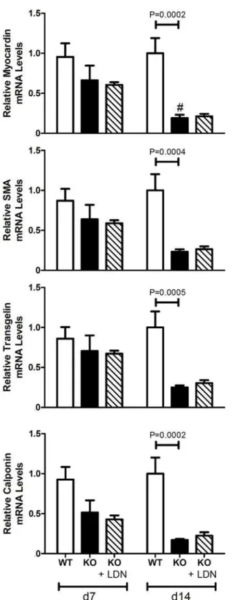
LDN-193189. In wild-type mice, levels of four VSMC markers (myocardin, SMA, tagln, and
calponin) did not differ in aortas of 7- and 14-day-old animals (Fig. 7). In contrast, expression
of myocardin was less in the aortas of 14-day-old MGP-/-mice than in those of 7-day-old
MGP-/-mice, and the expression of SMA, tagln, and calponin tended to decrease with age
(P = 0.06, 0.07, and 0.06, respectively). Levels of all four VSMC markers were less in aortas of
MGP-/-mice than in those of wild-type mice at 14 days of age. Consistent with decreased SMA
mRNA levels, we observed that SMA protein levels were less in aortas of MGP-/-mice than in
those of wild-type mice (S3 Fig.). These results show that VSMC markers decrease with age in
MGP-/-mice but not in wild-type mice, consistent with a loss of VSMC phenotype.
Figure 6. MGP deficiency does not alter basal BMP signaling or responsiveness to BMP-2 in VSMCs. (A) VSMCs were isolated from the aortas of wild-type and MGP-/-mice. VSMCs were treated without or with recombinant human BMP-2 (for 2 hours at the indicated doses). Groups were compared using a 2-way ANOVA. Both WT and MGP-/-VSMCs exhibited similar Id1 mRNA levels, both at baseline and in response to exogenous BMP-2. (B) Cultured aortic VSMCs from wild-type mice were transfected with either scrambled siRNA (siSC) or siRNA targeting MGP (siMGP) at 20 nM. RNA was isolated from cells after 4 days. siMGP decreased MGP mRNA levels in WT VSMCs by>95% compared with siSC-treated cells. However, depletion
of MGP in WT VSMCs did not alter Id1 mRNA levels.**P<0.0001 compared to siSC-treated VSMCs. (C) VSMCs isolated from wild-type mice were treated with 20 nM of either scrambled siRNA (siSC) or siRNA specific for MGP (siMGP). Cells were incubated with or without BMP-2 (20 ng/mL) for 1 h prior to protein harvest. Western blots were probed with antibodies specific for phosphorylated Smad 1/5 (P-Smad 1/5) and total Smad 1. Depletion of MGP in WT VSMCs did not alter the ratio of P-Smad 1/5 levels to total Smad 1 levels, both at baseline and in response to exogenous BMP-2.
Figure 7. Aortic expression of VSMC markers in wild-type and MGP-/-mice.RNA was isolated from aortas of WT and MGP-/-mice and from LDN-193189-treated MGP-/-mice at 7 and 14 days of age (n = 4
–8 in each group). Levels of mRNAs encoding myocardin,αsmooth muscle actin (SMA), transgelin, and calponin
are depicted. The aortas of 14-day-old MGP-/-mice have decreased expression of VSMC markers compared to WT mice. Treatment with LDN-193189 did not restore the expression of VSMC markers to WT levels. # P<0.05 compared to 7-day-old MGP-/-mice.
To determine whether BMP signaling is necessary for the loss of VSMC phenotype in the
aortas of MGP-/-mice, we measured the levels of myocardin, SMA, tagln, and calponin mRNA
in the aortas of MGP-/-mice treated with LDN-193189 beginning at 1 day of age. Inhibition of
BMP signaling with LDN-193189 did not normalize expression of the markers of VSMC
phe-notype in aortas of 14-day-old MGP-/-mice compared to wild-type mice (Fig. 7). These results
suggest that BMP signaling does not cause the loss of VSMC phenotype observed in the aortas
of MGP-/-mice.
BMP signaling is required for the induction of osteogenic markers in the
aortas of MGP
-/-mice
The medial vascular calcification that develops in MGP-deficient mice is characterized by a phenotypic switch of aortic VSMCs to osteogenic cells. To investigate the effects of BMP inhi-bition on aortic osteogenic transdifferentiation, we measured aortic levels of mRNAs encoding
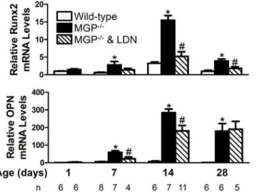
osteogenic markers, Runx2 and OPN, in 1-, 7-, 14-, and 28-day-old MGP-/-and wild-type
mice, both in the absence and presence of LDN-193189 (Fig. 8). Aortic Runx2 mRNA levels
did not change over time in wild-type mice. In contrast, aortic expression of Runx2 increased
with age in the MGP-/-mice, peaking at 14 days. Runx2 gene expression was greater in MGP
-/-aortas at 7, 14, and 28 days of age than in -/-aortas from age-matched wild-type mice. Similar to Runx2, aortic OPN gene expression did not change over time in wild-type mice but increased
with time in MGP-/-mice, peaking at 14 days. OPN mRNA levels were greater in MGP-/-aortas
at 7, 14, and 28 days of age than in aortas from wild-type mice.
To determine whether increased aortic Runx2 and OPN in MGP-deficient mice is depen-dent on BMP signaling, we examined the expression of mRNAs encoding Runx2 and OPN in
aortas of MGP-/-mice treated with LDN-193189. To confirm that LDN-193189 was able to
in-hibit aortic BMP signaling, levels of Id1 mRNA were measured. Treatment with LDN-193189
reduced Id1 gene expression by 70–80% in the aortas of 7-, 14-, and 28-day old MGP-/-mice
(S4 Fig.). Inhibition of BMP signaling was associated with a 70% and 50% reduction in aortic
Figure 8. BMP signaling is required for the increased aortic expression of osteogenic markers associated with MGP deficiency.RNA was isolated from aortas of WT and MGP-/-mice at 1, 7, 14, and 28 days of age and from LDN-193189-treated MGP-/-mice at 7, 14, and 28 days of age (n = 4
–11 in each group, as indicated). Expression of genes encoding Runx2 and osteopontin (OPN) was measured. MGP-/-mice had increased levels of aortic Runx2 and OPN mRNA compared to WT mice. Treatment of MGP-/-mice with LDN-193189 reduced aortic Runx2 and OPN mRNA levels.*P<0.001 compared to WT mice of same age.
# P<0.05 compared to age-matched MGP-/-mice treated with vehicle.
Runx2 mRNA levels at 14 and 28 days of age, respectively (Fig. 8). Treatment with
LDN-193189 reduced aortic OPN mRNA levels in 7- and 14-day-old MGP-/-mice by 60% and 40%,
respectively. These results suggest that the observed increase in Runx2 and OPN in the aortas of MGP-deficient mice is dependent, at least in part, on BMP signaling.
BMP signal transduction is required for VSMC calcification observed in
MGP deficiency
The process of vascular calcification involves the initial loss of VSMC phenotype followed by
the development of osteogenic cells and subsequent calcification [38]. Although we observed
that BMP inhibition did not prevent loss of VSMC phenotype in the aortas of MGP-/-mice,
BMP signaling was required for the induction of osteogenic cells. We next investigated whether
calcification of MGP-deficient VSMCs is dependent on BMP signalingin vitro. Calcification
was induced in isolated VSMCs by growing the cells in DMEM supplemented with 10% FCS
and 2 mM sodium phosphate [38]. Expression of MGP in MGP-/-VSMCs using an adenovirus
vector reduced calcification by 70%, as detected by von Kossa stain, compared with adenovirus
control-treated cells (Fig. 9A–B, F). Treatment of wild-type VSMCs with siRNA directed
against MGP (siMGP) increased calcification compared to treatment with control scrambled
siRNA (siSC) (Fig. 9C–D). These results indicate that, similar toin vivofindings, MGP
expres-sion modulates the calcification occurring in cultured VSMCs treated with high phosphate-containing media. Treatment with LDN-193189 inhibited the calcification induced by siMGP
in wild-type VSMCs by approximately 50% (P = 0.0003;Fig. 9D–E, 9G). Taken together, these
observations suggest that MGP inhibits calcification of isolated VSMCs and that calcification of MGP-deficient VSMCs is, at least in part, dependent on BMP signal transduction.
BMP signal transduction is required for the increased
endothelial-mesenchymal transition observed in MGP-deficient mice
A recent study identified endothelial cells in the aortas of MGP-deficient mice as a source of multipotent progenitor cells which subsequently give rise to osteogenic mesenchymal cells
[42]. To investigate the potential role of BMP signaling in the induction of EndMT, levels of
mRNAs encoding endothelial markers (VE-cadherin and CD31) and multipotency markers (nanog, Oct 3/4, and sox2) were measured in aortas harvested from 7- and 14-day-old
wild-type and MGP-/-mice treated with LDN-193189 or vehicle. In the absence of BMP inhibition,
levels of mRNAs ecoding VE-cadherin and multipotency markers were greater in aortas of
14-day-old compared with 7-day-old wild-type mice (Fig. 10). Endothelial and multipotency
markers were greater in the aortas of 14-day-old MGP-/-mice than in those of 7-day-old
MGP-/-mice. The expression of endothelial and multipotency markers was greater in the aortas
of 14-day-old MGP-/-mice compared with age-matched wild-type mice, suggesting that the
en-hanced EndMT observed in MGP-/-mice begins after the first week of life. Treatment with
LDN-193189 reduced the expression of endothelium and multipotency markers in the aortas
of 14-day-old MGP-/-mice to the levels seen in wild-type mice. These observations indicate
that BMP signaling is required for the increased EndMT associated with MGP deficiency.
Discussion
In this study, we identified an essential role for basal BMP signaling in the development of me-dial vascular calcification associated with MGP deficiency. Inhibition of basal BMP signaling
prevented vascular calcification and improved survival in MGP-/-mice. Although the loss of
the subsequent expression of osteogenic factors in the aortas of MGP-/-mice was dependent on
BMP signaling and ourin vitrodata implicate BMP signaling as a cause of calcification in
iso-lated VSMCs. BMP signaling was also required for the increased EndMT associated with MGP deficiency. Thus, this study has identified BMP-dependent and BMP-independent aspects of vascular cell phenotype and calcification associated with MGP deficiency.
Prior studies demonstrated a role for BMP signaling in the development of atherosclerosis
and intimal calcification [31,50]. Transgenic over-expression of BMP-2 under the direction of
the SMA promoter accelerated intimal calcification in ApoE-deficient mice with
atherosclero-sis [30]. Furthermore, inhibition of BMP signaling in low-density lipoprotein receptor
(LDLR)-deficient mice fed a high-fat diet reduced intimal calcification [31]. This previous
work demonstrated a role for BMP signaling in regulating LDL levels and macrophage
recruit-ment to the vessel wall [31]. However, the MGP-/-mouse model differs from atherosclerotic
Figure 9. Restoration of MGP levels decreases calcification of MGP-/-vascular smooth muscle cells while siRNA-mediated depletion of MGP increases calcification of wild-type vascular smooth muscle cells in a BMP-dependent manner.Cultured aortic VSMCs isolated from MGP-/-mice were infected with either (A) a control adenovirus (Ad.GFP) or (B) an adenovirus expressing MGP (Ad.MGP) at a multiplicity of infection of 10 and placed in DMEM supplemented with 10% FBS and 2 mM sodium phosphate. Cultured aortic VSMCs isolated from wild-type mice were transfected with either (C) scrambled siRNA (siSC) or (D & E) siRNA targeting MGP (siMGP) at 20 nM and placed in DMEM supplemented with 10% FBS and 2 mM sodium phosphate. Cells were also treated without (C & D) or with (E) 100 nM LDN-193189 (LDN). Cells were stained after 7 days using the von Kossa method. Serial fields of view were photographed for each condition and von Kossa stain was quantified using image J software after background subtraction (F & G). In (F), *P = 0.03 compared to Ad.GFP. In (G),**P<0.0001 compared to siSC-treated cells. #P = 0.0003 compared to siMGP + control. Restoration of MGP
expression reduced phosphate-induced calcification of MGP-/-VSMCs, while depletion of MGP increased calcification of WT VSMCs and this calcification was partially inhibited by treatment with LDN-193189.
Figure 10. Aortic expression of endothelial and multipotency markers in wild-type and MGP-/-mice. RNA was isolated from aortas of WT and MGP-/-mice and from LDN-193189-treated MGP-/-mice at 7 and 14 days of age (n = 4–8 in each group). Levels of mRNAs encoding endothelial markers (VE-Cadherin and CD31) and multipotency markers (nanog, Oct 3/4, and Sox2) are depicted. The aortas of 14-day-old MGP -/-mice have increased endothelial and multipotency markers compared to WT -/-mice. Treatment with LDN-193189 normalized the endothelial and multipotency markers to WT levels in MGP-/-mice.
*P0.01 compared to 7-day-old WT mice. # P<0.05 compared to 7-day-old MGP-/-mice.
mouse models in that calcification develops in the medial layer of arteries and occurs in the ab-sence of vascular inflammation. Two inhibitors of BMP signaling with different modes of ac-tion, LDN-193189 (which targets the BMP type I receptor) and ALK3-Fc (which targets BMP ligands), both inhibited vascular calcification. Inhibition of BMP signaling also improved sur-vival and growth in MGP-deficient mice. These results identify an important role for BMP sig-naling in medial vascular calcification and demonstrate that inhibition of BMP sigsig-naling can ameliorate calcification of the medial arterial layer that occurs in the absence of inflammation.
A previous report suggested that levels of phosphorylated BMP-responsive Smads are
great-er in the aortas of MGP-/-mice than in those of wild-type mice [50]. In this study, we did not
observe increased aortic levels of phosphorylated Smad 1/5 or Id1 mRNA in MGP-/-compared
to wild-type mice. We also did not detect a difference in basal BMP signaling in the livers (ex-pressing low levels of MGP in wild-type mice) and lungs (ex(ex-pressing higher levels of MGP in
wild-type mice) of MGP-/-and type mice. In studies of aortic VSMCs isolated from
wild-type and MGP-/-mice, there was no difference in Smad 1/5 phosphorylation or Id1 gene
ex-pression at baseline or after stimulation with BMP-2. Furthermore, depletion of MGP from wild-type aortic VSMCs using siRNA did not alter levels of Smad 1/5 phosphorylation or Id1
mRNA. Similar to ourin vivofindings, inhibition of basal BMP signaling in aortic VSMCs
de-pleted of MGP with siRNA prevented the calcification induced by high phosphate conditions. Taken together, the findings suggest that MGP deficiency results in vascular calcification via a mechanism that requires basal, but not increased BMP signaling.
MGP appears to have an important role in the regulation of VSMC phenotypein vivo
[21,38,41,42]. Speer and colleagues reported that VSMCs in MGP-deficient mice lose their
smooth muscle phenotype and transdifferentiate into osteogenic cells [41]. This study
con-firmed that MGP deficiency was associated with reduced aortic levels of VSMC markers. How-ever, inhibition of BMP signaling starting at day 1 of life did not prevent the loss of VSMC
phenotype in MGP-/-mice. These results suggest that, in the absence of MGP, the loss of
VSMC phenotype occurs via a mechanism that is independent of BMP signaling.
To investigate the role of BMP signaling in the induction of the osteogenic phenotype in the
vasculature of MGP-/-mice, we examined the impact of LDN-193189 on aortic expression of
Runx2 and OPN. Runx2 is an essential regulator of the osteogenic differentiation of vascular
cells [52], and VSMC-specific deficiency of Runx2 protects ApoE-deficient mice from high-fat
diet-induced vascular calcification [53]. OPN is a marker of osteogenesis and vascular
calcifica-tion in MGP-deficient mice [54]. Compared to wild-type mice, MGP-/-mice had increased
aor-tic Runx2 and OPN mRNA levels beginning at 7 days of age. Treatment of MGP-/-mice with
LDN-193189 decreased aortic Runx2 mRNA levels in 14- and 28-day-old mice, and treatment with the BMP inhibitor reduced aortic OPN mRNA levels in 7- and 14-day-old mice. These
re-sults suggest that inhibition of BMP signaling reduces vascular calcification in MGP-/-mice by
preventing development of an osteogenic phenotype.
In MGP-/-mice, endothelial cells undergo EndMT prior to differentiation into osteogenic
cells [42]. MGP deficiency is associated with increased aortic expression of markers of
endothe-lial cells (VE-cadherin and CD31) and multipotent cells (Nanog, Oct 3/4, Sox2). Treatment of
MGP-/-mice with LDN-193189 reduced aortic expression of endothelial and multipotency
markers to levels seen in wild-type mice. These observations suggest that the EndMT associat-ed with MGP deficiency is dependent on BMP signaling. The results are complementary to the findings of Yao et al. who demonstrated that BMP signaling augments EndMT in cultured
MGP-deficient human aortic endothelial cells [42].
once daily dosing of LDN-193189 may provide only intermittent inhibition of vascular BMP signaling, delaying but not fully preventing vascular calcification. Alternatively, because treat-ment with LDN-193189 did not prevent loss of the VSMC phenotype, it is possible that less ef-ficient, BMP-independent pathways may contribute to subsequent osteogenesis.
In summary, our work has identified the BMP signaling pathway as an important mediator of calcification of the vascular media and inhibition of BMP signaling can reduce vascular
calci-fication and improve survival in MGP-/-mice. MGP deficiency does not increase aortic BMP
signaling; basal BMP signaling is sufficient to permit vascular calcification. While BMP signal-ing is not required for the loss of VSMC phenotype associated with MGP deficiency, the devel-opment of an osteogenic phenotype and subsequent calcification of VSMCs is BMP signaling-dependent. The increased endothelial-to-mesenchymal transition observed in MGP deficiency also depends on BMP signaling. Because BMP signaling appears to have a critical role in several steps in the pathogenesis of medial artery calcification, inhibition of the BMP pathway may prove to be an effective approach for the prevention or treatment of this vascular disease.
Supporting Information
S1 Table. Primer sequences.The forward and reverse primer sequences used for gene expres-sion analysis in this study.
(DOC)
S1 Fig. Treatment with LDN-193189 improves growth of MGP-/-mice.MGP-/-mice were treated with daily i.p. injections of LDN-193189 (LDN) or vehicle starting at day 1 of life and weighed weekly. Increased body weight was detected in LDN-193189-treated compared with
vehicle-treated MGP-/-mice beginning at 21 days. Data are presented as mean ± standard
devi-ation (n = 10 in each group).P<0.001 compared to vehicle-treated group of same age.
(TIF)
S2 Fig. BMP signaling does not differ in the livers and lungs of MGP-/-mice compared to wild-type mice.(A) MGP mRNA levels were more than 100-fold greater in the lungs
com-pared with the livers of wild-type mice (left panel).P<0.001 compared to liver MGP mRNA
levels. No difference in Id1 mRNA levels was detected between WT and MGP-/-mice, both in
the livers and lungs (n = 6 in each group, right panel). (B) Smad 1/5 phosphorylation (P-Smad
1/5) and total Smad 1 levels were measured in the livers and lungs of wild-type (n = 6) and
MGP-/-mice (n = 6) at 14 days of age. There was no difference in the ratio of P-Smad 1/5 to
total Smad 1 protein levels in WT and MGP-/-mice, in either liver or lung.
(TIF)
S3 Fig. MGP deficiency is associated with decreased levels of smooth muscle actin.Protein
lysates were harvested from aortas of WT and MGP-/-mice. PVDF membranes were treated
with antibodies directed against SMA and GAPDH. Aortas from MGP-/-mice have reduced
SMA protein levels compared to those of WT mice. (TIF)
S4 Fig. LDN-193189 inhibits aortic BMP signaling in MGP-/-mice.RNA was isolated from
aortas of MGP-/-mice treated with either LDN-193189 or vehicle at 7, 14, and 28 days of age
(n = 4–11 in each group, as indicated). Treatment of MGP-/-mice with LDN-193189 reduced
aortic Id1 mRNA levels by 70–80%. # P<0.05 compared to age-matched MGP-/-mice treated
Acknowledgments
We are grateful to Dr. Gerard Karsenty (Columbia University, NY) and Dr. Terete Borras
(Univ. of North Carolina, NC) for providing us with MGP+/-heterozygous mice and to
Accel-eron Pharma Inc. (Cambridge, MA) for providing ALK3-Fc.
Author Contributions
Conceived and designed the experiments: RM MFB TM HRS MD ESB PBY KDB DBB. Per-formed the experiments: RM MFB TM HRS TET CO PL MD ES SAK KH CM PJ. Analyzed the data: RM MFB TM HRS TET CO. Contributed reagents/materials/analysis tools: RM RK PBY KDB DBB. Wrote the paper: RM MFB TM HRS TET CO PL MD ES SAK KH CM PJ RK ESB PBY KDB DBB.
References
1. Abedin M, Tintut Y, Demer LL (2004) Vascular calcification: mechanisms and clinical ramifications. Arterioscler Thromb Vasc Biol 24: 1161–1170. doi:10.1161/01.ATV.0000133194.94939.42PMID: 15155384
2. Huang H, Virmani R, Younis H, Burke AP, Kamm RD, et al. (2001) The impact of calcification on the bio-mechanical stability of atherosclerotic plaques. Circulation 103: 1051–1056. doi:10.1161/01.CIR.103. 8.1051PMID:11222465
3. Vengrenyuk Y, Carlier S, Xanthos S, Cardoso L, Ganatos P, et al. (2006) A hypothesis for vulnerable plaque rupture due to stress-induced debonding around cellular microcalcifications in thin fibrous caps. Proc Natl Acad Sci U S A 103: 14678–14683. doi:10.1073/pnas.0606310103PMID:17003118
4. Virmani R, Burke AP, Farb A, Kolodgie FD (2006) Pathology of the vulnerable plaque. J Am Coll Cardiol 47: C13–18. doi:10.1016/j.jacc.2005.10.065PMID:16631505
5. Amann K (2008) Media calcification and intima calcification are distinct entities in chronic kidney dis-ease. Clin J Am Soc Nephrol 3: 1599–1605. doi:10.2215/CJN.02120508PMID:18815240
6. Otsuka F, Sakakura K, Yahagi K, Joner M, Virmani R (2014) Has our understanding of calcification in human coronary atherosclerosis progressed? Arterioscler Thromb Vasc Biol 34: 724–736. doi: 10.1161/ATVBAHA.113.302642PMID:24558104
7. Hruska KA, Mathew S, Saab G (2005) Bone morphogenetic proteins in vascular calcification. Circ Res 97: 105–114. doi:10.1161/01.RES.00000175571.53833.6cPMID:16037577
8. Vattikuti R, Towler DA (2004) Osteogenic regulation of vascular calcification: an early perspective. Am J Physiol Endocrinol Metab 286: E686–696. doi:10.1152/ajpendo.00552.2003PMID:15102615
9. Luo G, Ducy P, McKee MD, Pinero GJ, Loyer E, et al. (1997) Spontaneous calcification of arteries and cartilage in mice lacking matrix GLA protein. Nature 386: 78–81. doi:10.1038/386078a0PMID: 9052783
10. Keutel J, Jorgensen G, Gabriel P (1971) [A new autosomal-recessive hereditary syndrome. Multiple pe-ripheral pulmonary stenosis, brachytelephalangia, inner-ear deafness, ossification or calcification of cartilages]. Dtsch Med Wochenschr 96: 1676–1681 passim. doi:10.1055/s-0028-1110200PMID: 5099199
11. Munroe PB, Olgunturk RO, Fryns JP, Van Maldergem L, Ziereisen F, et al. (1999) Mutations in the gene encoding the human matrix Gla protein cause Keutel syndrome. Nat Genet 21: 142–144. doi: 10.1038/5102PMID:9916809
12. Meier M, Weng LP, Alexandrakis E, Ruschoff J, Goeckenjan G (2001) Tracheobronchial stenosis in Keutel syndrome. Eur Respir J 17: 566–569. doi:10.1183/09031936.01.17305660PMID:11405537
13. Proudfoot D, Shanahan CM (2006) Molecular mechanisms mediating vascular calcification: role of ma-trix Gla protein. Nephrology (Carlton) 11: 455–461. doi:10.1111/j.1440-1797.2006.00660.x
14. Crosier MD, Booth SL, Peter I, Dawson-Hughes B, Price PA, et al. (2009) Matrix Gla protein polymor-phisms are associated with coronary artery calcification in men. J Nutr Sci Vitaminol (Tokyo) 55: 59–65. doi:10.3177/jnsv.55.59
16. Johnson RC, Leopold JA, Loscalzo J (2006) Vascular calcification: pathobiological mechanisms and clinical implications. Circ Res 99: 1044–1059. doi:10.1161/01.RES.0000249379.55535.21PMID: 17095733
17. O'Young J, Liao Y, Xiao Y, Jalkanen J, Lajoie G, et al. (2011) Matrix Gla protein inhibits ectopic calcifi-cation by a direct interaction with hydroxyapatite crystals. J Am Chem Soc 133: 18406–18412. doi: 10.1021/ja207628kPMID:21961692
18. Yagami K, Suh JY, Enomoto-Iwamoto M, Koyama E, Abrams WR, et al. (1999) Matrix GLA protein is a developmental regulator of chondrocyte mineralization and, when constitutively expressed, blocks en-dochondral and intramembranous ossification in the limb. J Cell Biol 147: 1097–1108. doi:10.1083/jcb. 147.5.1097PMID:10579728
19. Lomashvili KA, Wang X, Wallin R, O'Neill WC (2011) Matrix Gla protein metabolism in vascular smooth muscle and role in uremic vascular calcification. J Biol Chem 286: 28715–28722. doi:10.1074/jbc. M111.251462PMID:21705322
20. Roy ME, Nishimoto SK (2002) Matrix Gla protein binding to hydroxyapatite is dependent on the ionic environment: calcium enhances binding affinity but phosphate and magnesium decrease affinity. Bone 31: 296–302. doi:10.1016/S8756-3282(02)00821-9PMID:12151082
21. Schurgers LJ, Uitto J, Reutelingsperger CP (2013) Vitamin K-dependent carboxylation of matrix Gla-protein: a crucial switch to control ectopic mineralization. Trends Mol Med 19: 217–226. doi:10.1016/j. molmed.2012.12.008PMID:23375872
22. Goiko M, Dierolf J, Gleberzon JS, Liao Y, Grohe B, et al. (2013) Peptides of matrix gla protein inhibit nu-cleation and growth of hydroxyapatite and calcium oxalate monohydrate crystals. PLoS One 8: e80344. doi:10.1371/journal.pone.0080344PMID:24265810
23. Yao Y, Zebboudj AF, Shao E, Perez M, Bostrom K (2006) Regulation of bone morphogenetic protein-4 by matrix GLA protein in vascular endothelial cells involves activin-like kinase receptor 1. J Biol Chem 281: 33921–33930. doi:10.1074/jbc.M604239200PMID:16950789
24. Zebboudj AF, Imura M, Bostrom K (2002) Matrix GLA protein, a regulatory protein for bone morphoge-netic protein-2. J Biol Chem 277: 4388–4394. doi:10.1074/jbc.M109683200PMID:11741887
25. Wallin R, Cain D, Hutson SM, Sane DC, Loeser R (2000) Modulation of the binding of matrix Gla protein (MGP) to bone morphogenetic protein-2 (BMP-2). Thromb Haemost 84: 1039–1044. PMID:11154111
26. Cai J, Pardali E, Sanchez-Duffhues G, ten Dijke P (2012) BMP signaling in vascular diseases. FEBS Lett 586: 1993–2002. doi:10.1016/j.febslet.2012.04.030PMID:22710160
27. Bragdon B, Moseychuk O, Saldanha S, King D, Julian J, et al. (2011) Bone morphogenetic proteins: a critical review. Cell Signal 23: 609–620. doi:10.1016/j.cellsig.2010.10.003PMID:20959140
28. Abe E, Yamamoto M, Taguchi Y, Lecka-Czernik B, O'Brien CA, et al. (2000) Essential requirement of BMPs-2/4 for both osteoblast and osteoclast formation in murine bone marrow cultures from adult mice: antagonism by noggin. Journal of bone and mineral research: the official journal of the American Society for Bone and Mineral Research 15: 663–673. doi:10.1359/jbmr.2000.15.4.663
29. Bostrom K, Watson KE, Horn S, Wortham C, Herman IM, et al. (1993) Bone morphogenetic protein ex-pression in human atherosclerotic lesions. J Clin Invest 91: 1800–1809. doi:10.1172/JCI116391 PMID:8473518
30. Nakagawa Y, Ikeda K, Akakabe Y, Koide M, Uraoka M, et al. (2010) Paracrine osteogenic signals via bone morphogenetic protein-2 accelerate the atherosclerotic intimal calcification in vivo. Arterioscler Thromb Vasc Biol 30: 1908–1915. doi:10.1161/ATVBAHA.110.206185PMID:20651281
31. Derwall M, Malhotra R, Lai CS, Beppu Y, Aikawa E, et al. (2012) Inhibition of bone morphogenetic pro-tein signaling reduces vascular calcification and atherosclerosis. Arterioscler Thromb Vasc Biol 32: 613–622. doi:10.1161/ATVBAHA.111.242594PMID:22223731
32. Hedin U, Roy J, Tran PK, Lundmark K, Rahman A (1999) Control of smooth muscle cell proliferation– the role of the basement membrane. Thromb Haemost 82 Suppl 1: 23–26.
33. Sage AP, Tintut Y, Demer LL (2010) Regulatory mechanisms in vascular calcification. Nat Rev Cardiol 7: 528–536. doi:10.1038/nrcardio.2010.115PMID:20664518
34. Lee KS, Kim HJ, Li QL, Chi XZ, Ueta C, et al. (2000) Runx2 is a common target of transforming growth factor beta1 and bone morphogenetic protein 2, and cooperation between Runx2 and Smad5 induces osteoblast-specific gene expression in the pluripotent mesenchymal precursor cell line C2C12. Mol Cell Biol 20: 8783–8792. doi:10.1128/MCB.20.23.8783-8792.2000PMID:11073979
35. Rusanescu G, Weissleder R, Aikawa E (2008) Notch signaling in cardiovascular disease and calcifica-tion. Curr Cardiol Rev 4: 148–156. doi:10.2174/157340308785160552PMID:19936191
37. Steitz SA, Speer MY, Curinga G, Yang HY, Haynes P, et al. (2001) Smooth muscle cell phenotypic tran-sition associated with calcification: upregulation of Cbfa1 and downregulation of smooth muscle lineage markers. Circ Res 89: 1147–1154. doi:10.1161/hh2401.101070PMID:11739279
38. Speer MY, Li X, Hiremath PG, Giachelli CM (2010) Runx2/Cbfa1, but not loss of myocardin, is required for smooth muscle cell lineage reprogramming toward osteochondrogenesis. J Cell Biochem 110: 935–947. doi:10.1002/jcb.22607PMID:20564193
39. Speer MY, McKee MD, Guldberg RE, Liaw L, Yang HY, et al. (2002) Inactivation of the osteopontin gene enhances vascular calcification of matrix Gla protein-deficient mice: evidence for osteopontin as an inducible inhibitor of vascular calcification in vivo. J Exp Med 196: 1047–1055. doi:10.1084/jem. 20020911PMID:12391016
40. Byon CH, Javed A, Dai Q, Kappes JC, Clemens TL, et al. (2008) Oxidative stress induces vascular cal-cification through modulation of the osteogenic transcription factor Runx2 by AKT signaling. J Biol Chem 283: 15319–15327. doi:10.1074/jbc.M800021200PMID:18378684
41. Speer MY, Yang HY, Brabb T, Leaf E, Look A, et al. (2009) Smooth muscle cells give rise to osteochon-drogenic precursors and chondrocytes in calcifying arteries. Circ Res 104: 733–741. doi:10.1161/ CIRCRESAHA.108.183053PMID:19197075
42. Yao Y, Jumabay M, Ly A, Radparvar M, Cubberly MR, et al. (2013) A role for the endothelium in vascu-lar calcification. Circ Res 113: 495–504. doi:10.1161/CIRCRESAHA.113.301792PMID:23852538
43. Medici D, Shore EM, Lounev VY, Kaplan FS, Kalluri R, et al. (2010) Conversion of vascular endothelial cells into multipotent stem-like cells. Nat Med 16: 1400–1406. doi:10.1038/nm.2252PMID:21102460
44. Cuny GD, Yu PB, Laha JK, Xing X, Liu JF, et al. (2008) Structure-activity relationship study of bone morphogenetic protein (BMP) signaling inhibitors. Bioorg Med Chem Lett 18: 4388–4392. doi: 10.1016/j.bmcl.2008.06.052PMID:18621530
45. Aikawa E, Nahrendorf M, Figueiredo JL, Swirski FK, Shtatland T, et al. (2007) Osteogenesis associates with inflammation in early-stage atherosclerosis evaluated by molecular imaging in vivo. Circulation 116: 2841–2850. doi:10.1161/CIRCULATIONAHA.107.732867PMID:18040026
46. Aikawa E, Nahrendorf M, Sosnovik D, Lok VM, Jaffer FA, et al. (2007) Multimodality molecular imaging identifies proteolytic and osteogenic activities in early aortic valve disease. Circulation 115: 377–386. doi:10.1161/CIRCULATIONAHA.106.654913PMID:17224478
47. Rong JX, Shapiro M, Trogan E, Fisher EA (2003) Transdifferentiation of mouse aortic smooth muscle cells to a macrophage-like state after cholesterol loading. Proc Natl Acad Sci U S A 100: 13531–13536. doi:10.1073/pnas.1735526100PMID:14581613
48. Chomczynski P, Sacchi N (1987) Single-step method of RNA isolation by acid guanidinium thiocyanate-phenol-chloroform extraction. Anal Biochem 162: 156–159. doi:10.1016/0003-2697(87)90021-2PMID: 2440339
49. Luo J, Deng ZL, Luo X, Tang N, Song WX, et al. (2007) A protocol for rapid generation of recombinant adenoviruses using the AdEasy system. Nat Protoc 2: 1236–1247. doi:10.1038/nprot.2007.135PMID: 17546019
50. Yao Y, Bennett BJ, Wang X, Rosenfeld ME, Giachelli C, et al. (2010) Inhibition of Bone Morphogenetic Proteins Protects Against Atherosclerosis and Vascular Calcification. Circ Res 107: 485–494. doi: 10.1161/CIRCRESAHA.110.219071PMID:20576934
51. Zebboudj AF, Shin V, Bostrom K (2003) Matrix GLA protein and BMP-2 regulate osteoinduction in calci-fying vascular cells. J Cell Biochem 90: 756–765. doi:10.1002/jcb.10669PMID:14587031
52. Shao JS, Cai J, Towler DA (2006) Molecular mechanisms of vascular calcification: lessons learned from the aorta. Arterioscler Thromb Vasc Biol 26: 1423–1430. doi:10.1161/01.ATV.0000220441. 42041.20PMID:16601233
53. Sun Y, Byon CH, Yuan K, Chen J, Mao X, et al. (2012) Smooth muscle cell-specific runx2 deficiency in-hibits vascular calcification. Circ Res 111: 543–552. doi:10.1161/CIRCRESAHA.112.267237PMID: 22773442