O
RIGINALA
RTICLE Revista Brasileira de FisioterapiaElectromyographic analysis and strength of the
wrist extensor muscle group during induced
ischemia
Análise eletromiográfica e força do grupo muscular extensor do punho durante
isquemia induzida
Bandeira CCA, Berni KCS, Rodrigues-Bigaton D
Abstract
Objective:To analyze the effect of induced ischemia on the parameters of electromyographic signals and the strength of the wrist extensor muscle group (WEMG) in healthy women. Methods: Thirteen right-handed sedentary subjects aged 23.38±2.32 years old, with body mass index (BMI) of 20.68±1.87kg/m2, took part. To determine WEMG strength, three maximal voluntary isometric contractions
(MVIC) were performed using a load cell for 15 seconds, with 2 minutes intervals between contractions. The entire procedure was repeated on three nonconsecutive days. Ischemia was induced for 5 minutes using a sphygmomanometer placed on the dominant arm and inflated until blood flow was absent, as confirmed by Doppler ultrasound. The EMG1000 module (Lynx)was used with differential surface electrodes (Lynx®) to record the electromyographic signal of the WEMG. Three MVIC were recorded for 15 seconds, with 30 seconds intervals between them, under the following conditions: pre-ischemia, ischemia, immediate post-ischemia (post-1) and later post-ischemia (post-2: 10 minutes after the onset of ischemia). The MATLAB 6.5.1 software was used to analyze the parameters for the electromyographic signal, the root mean square (RMS) and the median frequency of the signal power spectrum. For statistical analysis, two-way ANOVA and the Friedman test were used. Results: Ischemia caused a significant reduction (p<0.05) in WEMG strength. However, there were no significant changes in the RMS electromyographic parameters (p=0.05) or the median frequency of the signal power spectrum (p=0.09). Conclusion: Induced ischemia caused WEMG fatigue in relation to muscle strength production. However, it did not cause electromyographic fatigue in the evaluated muscle group.
Key words: electromyography; strength; ischemia.
Resumo
Objetivo: Avaliar o efeito daisquemia induzida sobre os parâmetros do sinal eletromiográfico e a força do grupo muscular extensor do punho (GMEP) em mulheres saudáveis. Métodos: Participaram 13 voluntárias, destras, sedentárias, com idade de 23,38±2,32 anos e índice de massa corporal (IMC) de 20,68±1,87kg/m2. Para determinar a força do GMEP, foram realizadas 3 contrações isométricas
voluntárias máximas (CIVM), utilizando-se uma célula de carga por 15 segundos, com intervalos de 2 minutos entre cada contração, sendo todo procedimento repetido por 3 dias não consecutivos. A isquemia foi realizada por 5 minutos, utilizando um esfigmomanômetro posicionado no braço dominante e inflado até a ausência do fluxo sanguíneo, confirmada pelo ultrassom Doppler. Para coleta do sinal eletromiográfico do GMEP, utilizou-se o equipamento EMG1000 (Lynx)com eletrodo de superfície diferencial (Lynx). Foram coletadas 3 CIVM por 15 segundos, com intervalo de 30 segundos entre elas, nas situações de pré-isquemia; isquemia; pós-isquemia imediata (pós-1) e pós-isquemia tardia (pós-2 - após 10 minutos do início da isquemia). Para análise dos parâmetros do sinal eletromiográfico, root mean square (RMS), e frequência mediana do espectro de potência do sinal foi utilizado o software MATLAB 6.5.1. Para análise estatística, foram utilizados os testes de Friedman e ANOVA two-way. Resultados: A isquemia promoveu redução significativa (p<0,05) da força do GMEP. Entretanto, não provocou alterações significativas nos parâmetros eletromiográficos RMS (p=0,05) e frequência mediana do espectro de potência do sinal (p=0,09). Conclusão: A isquemia induzida promoveu fadiga do GMEP quando relacionada à produção da força muscular. Porém, não provocou fadiga eletromiográfica do grupo muscular avaliado.
Palavras-chave:eletromiografia; força; isquemia.
Received: 18/02/2008 – Revised: 14/05/2008 – Accepted: 26/09/2008
College of Health Sciences, Universidade Metodista de Piracicaba (UNIMEP), Piracicaba (SP), Brazil
Introduction
It is common knowledge that, under ischemic conditions, the absence of adequate blood low leads to the diminishment of muscle strength production1. In this context, the sustained
repetitive activities of the upper limbs, which are present in oc-cupational activities, can cause a local reduction in blood low and tissue oxygenation, thus provoking fatigue, pain, and func-tional deicit2. According to Murphy et al.3, the wrist extensor
muscles, particularly the extensor carpi radialis brevis, are the most commonly afected in prolonged and/or repetitive static activities.
Reduced blood low4 and muscle oxygenation5 are
associ-ated with fatigue. Murphy et al.6evaluated the efect of ischemia
on extensor carpi radialis fatigue in 8 healthy individuals and found that a 3 to 5-minute ischemia led to muscle fatigue. In the aforementioned study, the authors evaluated muscle fa-tigue through the reduction in strength.
Muscle fatigue, whether produced by ischemia or not, can also be evaluated by means of surface electromyography (EMGs), as it is a non-invasive method of muscle function evaluation that detects all electrical potentials from the active motor units by means of electrodes placed on the skin7.
When assessed by means of EMGs, muscle fatigue is also known as electromyographic fatigue and, by deinition, such a condition occurs when there is an increase in the amplitude of the electromyographic signal (observed through the root mean square (RMS) values, suggesting an additional motor recruitment), concomitant to a change in the power spectrum of the electromyopraphic signal toward the lower frequencies (observed through the median frequency values due to the re-duction in the discharge frequency of the motor units)8.
Recent studies9 have used EMGs to evaluate muscle activity
in situations of induced ischemia and reported a reduction in amplitude as well as a change in the power spectrum of the electromyographic signal toward the lower frequencies. his is caused by a pH diminishment and by a change in the electro-lytic balance (imbalance of the Na+/K+ pump) along the muscle
iber membrane, a fact that alters the conduction velocity and the discharge frequency of the motor unit and may cause fa-tigue or muscle strength reduction6.
However, the behavior of electromyographic parameters in an ischemic condition has yet to be well deined in the literature because some studies show changes in the parameters of the electromyographic signals9, whereas others do not10. Another
fact to be taken into account is that some studies4,5
demon-strate that ischemia is related to muscle fatigue, which, when evaluated through electromyography, promotes an increase in electromyographic signal amplitude. his was not observed by Chung, Callahan and Kent-Braun9.
Based on the aforementioned studies, the hypothesis of the present study is that induced ischemia promotes the fatigue of the wrist extensor muscle group in healthy women. his condi-tion was observedthrough the reduction in strength produc-tion levels as well as through the increase in the amplitude and the change in the power spectrum of the electromyographic signal toward the lower frequencies, thus characterizing elec-tromyographic fatigue.
Understanding muscle behavior under ischemic condi-tions, such as occupational activities, allows the development of better methods of evaluation and treatment of the dysfunc-tions characterized by ischemia, which is why this study was carried out. herefore, the aim of this study was to analyze the parameters of the electromyographic signal and the strength of the wrist extensor muscle group (WEMG) during induced ischemia in healthy women.
Methods
he power of the sample was evaluated based on the RMS electromyographic parameter, obtained in a pilot study with 6 participants. he minimal size of the sample (n=9) was deter-mined by the t test for paired samples (mean of the diference between the groups 17.86; standard deviation of the diference, 14.26; power of 0.95, α=0.05, and unilateral test), in which the RMS parameter obtained in the pre-ischemic and ischemic conditions was used. he calculations were processed by the BioEstat 4.0.
Fourteen participants were selected for this study (23.38±2.32 years old; 53.75±7.56kg; 1.61±0.06m, BMI 20.68±1.87kg/m2). hey were healthy, sedentary, according
to the International Physical Activity Questionnare-IPAQ11,
and without orthopedic, neurological, or upper-limb vascular dysfunctions. We opted to evaluate female individuals only, as several studies suggest that women have a higher resistance to fatigue than men in submaximal12 and maximal13 isometric
contractions. Women under 20 and over 30 were excluded from the study, as well as those who were left-handed; who had upper-limb dysfunctions; a BMI under 18.5 and over 25; and those not classiied as sedentary, according to the IPAQ. All participants signed a consent form, and the research was carried out according to Resolution 196/96 of the National Health Council and approved by the Research Ethics Commit-tee of Universidade Metodista de Piracicaba, under protocol nº 83/2006.
In order to determine the real strength production of the WEMG, a muscle strength baseline was established prior to data collection, for three nonconsecutive days, during which we obtained the mean of the maximal voluntary isometric
contraction value (MVIC), employed as a reference value for strength production at the time of electromyographic signal collection.
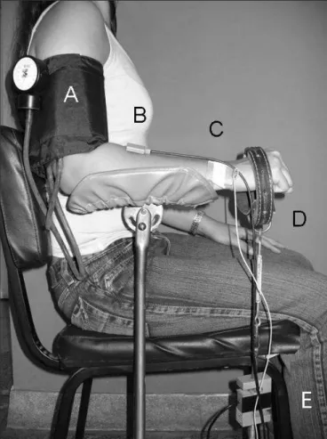
he strength measurement (kgf) of the WEMG was taken by means of a load cell (model MM-100, Kratos, São Paulo, SP, Brazil). he load cell was placed perpendicularly, with one of the ends attached to the participant’s hand by means of a metal chain and a leather strap, while the other end was ixed to the loor, making it possible to adjust the length of the chain for each participant (Figure 1D and 1E). he participant was seated with shoulders in a neutral position, elbow at 90°, in the prone position, forearm resting against a support, ingers lexed. his same position was used later to collect the electro-myographic signal (Figure 1).
he participant performed 3 MVICs for 15 seconds, with a 2-minute interval between the contractions at each base-line. hroughout this process, the participant received verbal encouragement and visual feedback, in which the participant observed a line in the computer monitor and was requested to raise this line – representing strength – as high as possible. he data was recorded after the stabilization of the muscle strength.
he ischemia was induced for 5 minutes, by using a Pres-sure N/C sphygmomanometer placed on the dominant arm (Figure 1A) and inlated until the blood low in the brachial artery was absent, conirmed by the Doppler ultrasound (Nico-let Vascular Versalab), with a 8 MHz transducer. To avoid any irreversible circulatory changes, whether functional, metabolic or muscular, the maximal time-length of ischemia was kept under 2 hours14.
To collect the electromyographic signal, the EMG1000 signal acquisition module was used (Lynx,São Paulo, SP, Brazil), as well as a differential surface electrode (Lynx, São Paulo, SP, Brazil), which follow the ISEK and SENIAM guidelines.
he EMG1000 (Lynx,São Paulo, SP, Brazil) signal acqui-sition module has a 109 Ohms impedance, a digital/analog
converter with a 16-bit resolution and an input band ranging from ±1V to ±10V, with an acquisition frequency of 2000Hz, a Butterworth-type ilter with a high-pass of 20Hz, and a low pass of 1000Hz. he EMG1000 (Lynx,São Paulo, SP, Brazil) was connected to a Pentium III desktop computer. he signal acquisition system was connected to a 12-volt battery with a capacity of 10-ampère-hour (AH), connected to a computer through optical iber in order to cancel out the interference coming from the wiring on the electromyograph, as described by Guirro, Forti and Rodrigues-Bigaton15.
he diferential surface electrode (consisting of two pure silver poles, 10mm long, 1mm wide and placed 10mm from one another, with a pre-ampliier circuit with a 20-time boost
(±1%), IRMC >100 dB, and signal/ratio <3µV RMS) was po-sitioned perpendicularly to the ibers of the WEMG, in the dorsal region of the arm, approximately 5cm from the elbow, on the muscle mass which emerged when the participant was asked to perform the counter-resistance wrist extension16
(Figure 1B). Before the electrode was attached, the skin was shaved and cleaned with alcohol at 70%. he reference elec-trode (30x40mm), made from a metallic plate, was positioned in the ulnar styloid process on the same side that was being evaluated (Figure 1C). To obtain the digital signal, and also to store the data into iles, we used the Aqdados software (Lynx, São Paulo, SP, Brazil), version 7.02 for Windows.
he collection of the electromyographic signal was car-ried out during three MVICs for 15 seconds, with 30-second intervals, in the following situations: pre-ischemia (under nor-mal blood low conditions); ischemia (in which the collection began 5 minutes after the sphygmomanometer was inlated and the absence of blood low was conirmed by the Doppler ultrasound); immediate post-ischemia (post-1; in which the contractions were started upon removing the sphygmoma-nometer and conirming the blood reperfusion by the Doppler
(A) Sphygmomanometer; (B) Differential surface electrode; (C) Reference electrode; (D) Leather belt; and (E) Load cell.
Figure 1. Position of the participant for electromyographic signal collection and determination of the wrist extensor muscle group strength.
ultrasound); later post-ischemia (post-2; 10 minutes after the onset of the ischemia).
he electromyographic signal was processed in the do-mains of time and frequency. For the analysis of the time domain, the RMS value was calculated because, according to De Luca17, this is the processing modality which best
repre-sents the amplitude of the electromyographic signal in vol-untary muscle contractions. For the analysis of the frequency domain, the Fast Fourier Transform (FFT) was applied to the electromyographic signal to generate the power spectral density. To achieve that, we used 512-point Hanning windows with 256 ms and a 50% overlap. he median frequency was used as suggested by Stulen and De Luca18, who claim that
this statistical parameter has the function of splitting the power spectrum into two isoenergetic regions, and that it is the one which best relects the physiological changes that oc-cur in the muscle during the sustained contractions17, such as
the conduction velocity of the muscle ibers, and the recruit-ment of the motor unit19.
he electromyographic signal was processed by the of-line
analysis, in the Matlab 6.5.1 software, using speciic functions to evaluate the quality of the acquired signal and to obtain the RMS values and the median frequency. he electromyographic signal was not normalized because, according to Soderberg and Knutson20, if in a given experimental procedure the individuals
are their own control, and the comparisons are performed on the same day and muscle without removing the electrode, the normalization is not necessary. he experimental procedure of the present study takes the aforementioned guidelines into account.
For the data analysis, programs such as Statistical Package for Social Science for Personal Computer (SPSS/PC version 11.0) and BioEstat 4.0 were used, and the Shapiro-Wilk test was applied to determine the normality of the sample, followed by the Friedman test and two-way ANOVA. A level of signii-cance of 5% was set for the analysis of the variables.
Results
he muscle strength values were expressed by mean and standard deviation, and the RMS values and median frequency, by median (MED) and interquartile interval (AIQ).
Figure 2 shows that the ischemia promoted a reduction in the strength of the WEMG (8.63±1.98 kgf in pre-ischemia, and 4.79±1.64 kgf in ischemia, p<0.01). In the post-1 and post-2 situations, there was a statistically signiicant increase in muscle strength when compared with the ischemia situation (7.44±1.65 kgf in the post-1, and 7.13±1.18 kgf in the post-2, p<0.05). For the RMS (µv), there was no signiicant change
(p=0.05) in the ischemia situations (MED:50.09; AIQ:68.72), post-1 (MED:30.99; AIQ:87.19) and post-2 (MED:30.35; AIQ:95.39), as shown in Figure 3.
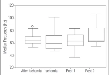
With regard to the median frequency (Figure 4), no sig-niicant changes were observed (p=0.09) in the situations of is-chemia (MED:59.89; AIQ:21.32), post-1 (MED:63.47; AIQ:17.25) and post-2 (MED:63.47; AIQ:22.13).
Discussion
Ischemia can cause a series of changes in the metabolic and enzymatic processes in the muscle, and the reversibility of this process is directly related to the duration of the isch-emic period21. hese changes occur due to the fall of the
in-tracellular pH and to the imbalance in the Na+/ K+ pump, with
an increase in the Na+ inlow, reduction in the intracellular
K+1,21 and inability to release the Ca2+ ions inside the muscle
iber, bringing about a reduction in the ability to produce strength22.
he present study found that 5 minutes of induced ischemia, in addition to 15 seconds of maximal voluntary isometric con-traction of the WEMG, which has a predominance of type-I muscle ibers on the dominant side22, were enough to cause
a decline in strength, though it was immediately recovered after the return of the blood low, reaching levels close to the strength value produced in the pre-ischemic phase. However, even after 5 minutes of blood reperfusion, the strength values did not return to their initial parameters.
Similar results were observed in the gastrocnemius muscle of dogs submitted to an ischemic period of 2 minutes, in which a 30-second reperfusion caused a recovery of 67% of the initial value of muscle strength. Nevertheless, there is no record of total strength recovery through blood reperfusion23. Muscle
strength was analyzed in the knee extensor muscles, submitted to 5 minutes of ischemia and to 5 minutes of ischemia with iso-metric contraction, and the latter condition resulted in greater strength reduction24.
here was also a change in the muscle strength in the evaluation of maximal torque and total work of the lexor and extensor muscles of the elbow in individuals with pathological ischemia of the upper limbs by means of the isokinetic dyna-mometry. he most evident strength reduction occurred in the elbow extensor muscles1.
Concerning the amplitude of the electromyographic signal, there were no signiicant changes in the mean RMS values dur-ing or after the induced ischemia. he results of the present study are in accordance with the indings of Farina, Gazzoni and Camelia10, who in a situation of ischemia, did not observe any
signiicant change in the amplitude of the electromyographic
signal of the abductor pollicis brevis muscle, which was sub-mitted to 16 minutes of blood low occlusion. However, they disagree with the indings of Leonard et al.25, who observed a
decrease in the RMS values of the soleus muscle after 8 to 12 minutes of ischemia.
In regard to the frequency of the electromyographic signal, there were no signiicant changes in the mean values of the median frequency of the power spectrum signal during and after the induced ischemia. Contrary to that inding, Merletti, Sabbahi and De Luca27 noted that induced ischemia of the
irst dorsal interosseous muscle for 10 minutes with isometric contraction brought about a signiicant reduction in the con-duction velocity of the muscle iber, i.e. the median frequency of the power spectrum of the electromyographic signal origi-nated by the accumulation of metabolites in this condition. he vasodilation and consequent increase in the temperature reached during blood reperfusion led to a quick removal of the metabolic byproducts, resulting in the immediate restoration of the median frequency of the power spectrum of the electro-myographic signal26.
he abductor pollicis brevis muscle of 9 male individuals underwent 16 minutes of ischemia in order to analyze the con-duction velocity of isolated motor units. It was found that the conduction velocity of that muscle decreased after 13 minutes of blood low occlusion10.
By using the same muscle in isometric contractions with normal blood low and with induced ischemia for 8 minutes, we observed that the muscle strength, muscle iber conduction velocity, and therefore the median frequency of the power spec-trum of the electromyographic signal diminished during the ischemia. Under normal blood low conditions, the conduction velocity of the inactive muscle ibers was reduced in 5 minutes of muscle contraction; however, in 3 minutes of ischemia, the conduction velocity of the inactive ibers of the muscle under investigation was tripled22.
We believe that the methodology employed, as well as the type of muscle iber, and the time of ischemia may have been the causes for divergence between the present study and the previously mentioned studies. here is a discrepancy in the literature as for the type of muscle iber afected by ischemia. Studies suggest that the type-I slow ibers are the most susceptible ones as they depend on an adequate blood and oxygen supply to synthesize the ATP25. Nonetheless, there
are reports attesting that ischemia led to more pronounced changes in the conduction velocity of type-II muscle ibers, given the fact that they are quick contraction ibers and con-sequently produce more metabolic byproducts during muscle contraction27.
he WEMG consists of the extensor carpi radialis brevis, the extensor carpi radialis longus, and the extensor carpi
Figure 2. Strength (kgf) of the wrist extensor muscle group during MVIC in the pre-ischemia, ischemia, post-1 and post-2 situations; * and ** differ from pre-ischemia (p<0.05 and p<0.01, respectively) and (†) differs from ischemia (p<0.01).
Post 2 Post 1
After ischemia Ischemia 14
12 10 8 6 4 2
**
*
*
Strength
(k
gf)
Figure 3. Median RMS values (µV) of the wrist extensor muscle group during MVIC in the pre-ischemia, ischemia, post-1 and post-2 situations (p=0.05).
Post 2 Post 1
After ischemia Ischemia 200
100
0
-100
RMS
(µV)
RMS= root mean square, MVIC= maximal voluntary isometric contractions.máximas.
Figure 4. Median frequency values (Hz) of the wrist extensor muscle group during MVIC in the pre-ischemia, ischemia, post-1 and post-2 situations (p=0.09).
Post 2 Post 1
After ischemia Ischemia 120
100
80
60
40
20
Median Frequency (
Hz
ulnaris. On the dominant side, they have a predominance of type-I muscle ibers, which may be altered by genetic factors, functional demands, and intra-individual variation28.
It is known that type-I ibers are more resistant to fatigue than type-II ibers, and that the motor units are recruited ac-cording to the activation threshold. hus, because they have a smaller threshold, the type-I ibers are initially activated, and the type-II ibers are then recruited29. Considering that the
WEMG has a larger amount of oxidative ibers and that the oxygen supply to these ibers was reduced due to the ischemia, it can be suggested that the type-II muscle ibers were acti-vated at the time, and that the type-I ibers were reactiacti-vated when blood low resumed.
he clinical implications of this study are relevant, given that they emphasize that the production of muscle strength is afected by ischemia and that this must be considered in the
physical therapist’s clinical evaluation, as the diminishment of strength production, among other symptoms, may indicate an ischemic condition. Regarding the electromyographic test car-ried out by means of a diferential surface electrode, we suggest that other studies be made in order to lend weight to its use in the clinical evaluation of patients with ischemic dysfunctions.
Conclusion
It can be concluded that a 5-minute induced ischemia caused fatigue of the WEMG when related to the production of muscle strength. However, when related to the electro-myographic signal, it can be stated that induced ischemia did not cause electromyographic fatigue in the assessed muscle group.
36
References
1. Nakano L. Avaliação objetiva de isquemia de membros superiores: uso do dinamômetro isocinético [tese]. São Paulo: USP; 2005.
2. Rempel DM, Harrison RJ, Barnhart S. Work-related cumulative trauma disordens of the upper extremity. JAMA. 1992;268(6):787-8.
3. Murthy G, Kahan NJ, Hargens AR, Rempel DM. Forearm muscle oxygenation decreases with low levels of voluntary contraction. J Ortho Res. 1997;15(4):507-11.
4. Bystrom EG, Kilbom A. Physiological response in the forearm during and after isometric intermittent handgrip. Eur J Appl Physiol Occup Physiol. 1990;60(6):457-66.
5. Hogan MC, Richardson RS, Kurdak SS. Initial fall in skeletal muscle force development during ischemia is related to oxygen availability. J Appl Physiol. 1994;77(5):2380-4.
6. Murthy G, Hargens AR, Lehman S, Rempel DM. Ischemia causes muscle fatigue. J Ortho Res. 2001;19(3):436-40.
7. Farina D, Merletti R, Enoka RM. The extraction of neural strategies from the surafce EMG. J Appl Physiol. 2004;96(4):1486-95.
8. Basmajian JV, De Luca CJ. Muscle alive: their function revealed by electromyography. São Paulo: Baltimore, Willians & Willians; 1985.
9. Chung LH, Callahan DM, Kent-Braun JA. Age-related resistance to skeletal muscle fatigue is preserved during ischemia. J Appl Phisiol. 2007;103(5):1628-35.
10. Farina D, Gazzoni M, Camelia F. Conduction velocity of low-threshold motor units during ischemic contractions perfomed with surface EMG feedback. J Appl Physiol. 2005;98(4):1487-94.
11. Craig CL, Marshall AL, Sjöström M, Bauman AE, Booth ML, Ainsworth BE et al. International Physical Activity Questionnaire:12-country reliability and validity. Med Sci Sport Exerc. 2003;35(8):1381-5.
12. Fulco CS, Rock PB, Muza SA, Lammi E, Cymerman A, Butterfield G et al. Slower fatigue and faster recovery of the adductor pollicis muscle in women matched for strength with men. Acta Physiol Scand. 1999;167(3):233-9.
13. Hicks AL, McCartney N. Gender differences in isometric contractile properties and fatigability in elderly human muscle. Can J Appl Physiol. 1996;21(6):441-54.
14. Bitu-Moreno J, Franscischetti I, Hafner L. Lesões de isquemia-reperfusão em músculos esqueléticos: fisiopatologia e novas tendências de tratamento, com ênfase em reperfusão controlada. J Vasc Br. 2002;1(2):113-20.
15. Guirro RRJ, Forti F, Rodrigues-Bigaton D. Proposal for electrical insulation of the electromyographic signal acquisition module. Electromyogr Clin Neurophysiol. 2006;46(6):355-63.
16. Cram JR, Kasman GS, Holtz J. Introduction to surface electromyography. Gaithersburg: An Aspen Publication; 1998.
17. De Luca CJ. The use of surface electromyography in biomechanics. J Biomech. 1997;13:135-63.
18. Stulen FB, De Luca CJ. Muscle fatigue monitor: a noninvasive device for observing localized muscular fatigue. IEEE Trans Biomed Eng. 1982;29(12):760-8.
20. Soderberg GL, Knutson LM. A guide for use and interpretation of kinesiologic electromyographic data. Phys Ther. 2000;80(5):485-98.
21. Silveira M, Yoshida W. Isquemia e reperfusão em músculo esquelético: mecanismos de lesão e perspectivas de tratamento. J Vasc Br. 2004;3(4):367-78.
22. Gazzoni M, Camelia F, Farina D. Conduction velocity of quiescent muscle fibers decreases during sustained contraction. J Neurophysiol. 2005;94(1):387-94.
23. Hogan MC, Kohin S, Stary CM, Hepple RT. Rapid force recovery in contracting skeletal muscle after brief ischemia is dependent on O2 availability. J Appl Physiol. 1999;87(6): 2225-9.
24. Pierce JR, Clarck BC, Ploutz-Snyder LL, Kanaley JA. Growth hormone and muscle function responses to skeletal muscle ischemia. J Appl Physiol. 2006;101(6):1588-95.
25. Leonard CT, Kane J, Perdaems J, Frank C, Graetzer D, Moritani T. Neural modulation of muscle contractile properties during fatigue: afferent feedback dependence. Eletroencephalogr Clin Neurophysiol. 1994;93(3):209-17.
26. Merletti R, Sabbahi MA, De Luca CJ. Median frequency of the myoeletric signal: effects of muscle ischemia and cooling. Eur J Appl Physiol Occup Physiol. 1984;52(3):258-65.
27. Gerdle B, Fugl-Meyer AR. Is the mean power frequency shift of the EMG a selective indicator of fatigue of the fast twitch motor units? Acta Phisiol Scand. 1992;145(2):129-38.
28. Fugl-Meyer A, Eriksson AR, Sjöström M, Söderström G. Is muscle structure influenced by genetical or functional factors? A study of three forearm muscles. Acta Physiol Scand. 1982;114(2):277-81.