Exogenous normal lymph reduces liver injury
induced by lipopolysaccharides in rats
Z.G. Zhao*, L.L. Zhang*, C.Y. Niu and J. Zhang
Institute of Microcirculation, Hebei North University, Zhangjiakou, Hebei, China
Abstract
The liver is one of the target organs damaged by septic shock, wherein the spread of endotoxins begins. This study aimed to investigate the effects of exogenous normal lymph (ENL) on lipopolysaccharide (LPS)-induced liver injury in rats. Male Wistar rats were randomly divided into sham, LPS, and LPS++ENL groups. LPS (15 mg/kg) was administered intravenously via the left jugular vein to the LPS and LPS++ENL groups. At 15 min after the LPS injection, saline or ENL without cell components (5 mL/kg) was administered to the LPS and LPS++ENL groups, respectively, at a rate of 0.5 mL/min. Hepatocellular injury indices and hepatic histomorphology, as well as levels of P-selectin, intercellular adhesion molecule 1 (ICAM-1), myeloperoxidase (MPO), and Na+-K+-ATPase, were assessed in hepatic tissues. Liver tissue damage occurred after LPS injection. All levels of alanine aminotransferase (ALT) and aspartate aminotransferase (AST) in plasma as well as the wet/dry weight ratio of hepatic tissue in plasma increased. Similarly, P-selectin, ICAM-1, and MPO levels in hepatic tissues were elevated, whereas Na+-K+-ATPase activity in hepatocytes decreased. ENL treatment lessened hepatic tissue damage and decreased levels of AST, ALT, ICAM-1, and MPO. Meanwhile, the treatment increased the activity of Na+-K+-ATPase. These results indicated that ENL could alleviate LPS-induced liver injury, thereby suggesting an alternative therapeutic strategy for the treatment of liver injury accompanied by severe infection or sepsis.
Key words: Exogenous normal lymph; Lipopolysaccharide; Liver injury; Intercellular adhesion molecule 1; Myeloperoxidase; Na+-K+-ATPase
Introduction
The liver is a target organ that is damaged during the pathogenesis of septic shock. The spread of endotoxins usually begins from this organ, which contributes to the development of multiple-organ dysfunction syndrome; therefore, liver protection is significant in endotoxemia treatment (1-3). Numerous studies have been conducted on liver injury after serious infection or sepsis (4,5), but no effective treatment for liver injury induced by septic shock has been reported. Therefore, the search for effective liver treatment is essential. Previous studies have found that normal lymph has beneficial therapeutic effects on microcirculatory disturbances in rats with hemorrhagic shock; its effect is significantly better than that by the same amount of normal saline and of albumin (6). Exogenous normal lymph (ENL) from healthy dogs has an alleviating effect on microcirculation disturbances and abnormal hemorheological properties in rats with disseminated intravascular coagulation induced by Dextran 500 (7). In addition, ENL could alleviate acute
lung injury and acute kidney injury in rats with endotoxic shock induced by lipopolysaccharides (LPS) (8,9). Cheng et al. (10) reported that normal mesenteric lymph could blunt the pulmonary inflammatory response to endotoxin. The effect of ENL on acute liver injury induced by septic shock remains unclear. The present study evaluated the effect of ENL on LPS-induced liver injury and its under-lying mechanisms.
Material and Methods
Animals
A total of 60 healthy and pathogen-free male Wistar rats (Chinese Academy of Medical Sciences Animal Breeding Center, Beijing, China), weighing 240 to 300 g, were used in this study. The rats were maintained under barrier-sustained conditions with a 12:12-h light/dark cycle at a constant temperature (23±26C), with access to standard laboratory food and water. Before the
Correspondence: C.Y. Niu and/or J. Zhang, Institute of Microcirculation, Hebei North University, Diamond South Road 11, Zhangjiakou 075000, China. Fax: ++86-313-402-9168. E-mail: [email protected] and/or [email protected]
*These authors contributed equally to this study.
experiment, the rats were fasted for 12 h but had access to water. Three healthy beagles were used for the preparation of ENL. All animal experiments were con-ducted in compliance with the guidelines of the Hebei North University Animal Care Committee. All efforts were made to minimize the suffering of the animals.
Preparation of ENL
After intravenous anesthesia with 25 mg/kg pentobar-bital sodium, a midline laparotomy was performed on healthy beagles, and their normal mesenteric lymph was continuously collected for 30 min into sterile heparin-wetted test tubes, according to our laboratory protocols (7-9,11). Lymph samples were centrifuged at 315g for 15 min to remove all cellular components and stored at –806C in an ultra-low freezer (Thermo Electron, USA) prior to the experiment.
Experimental protocol
All rats were anesthetized with 50 mg/kg 1% pento-barbital sodium and were randomly assigned to the sham, LPS, and LPS++ENL groups (n = 20). The left jugular vein and the right carotid artery were aseptically separated from the surrounding tissues and cannulated using a microcatheter for drug administration. After the surgical procedure was completed, rats were allowed to adjust for 30 min. LPS (15 mg/kg; Escherichia coli
O111:B4; Sigma, USA) was then injected via the left jugular vein using an infusion pump (ZCZ-50; Zhejiang University Medical Ltd., China), within 10 min after surgery, in the LPS and LPS++ENL groups. LPS was dissolved to a concentration of 10 mg/mL in normal saline. At 15 min after LPS injection, ENL was injected via the left jugular vein at a rate of 0.5 mL/min for 9 to 12 min in the LPS++ENL group. The ENL dose was 5 mL/kg, based on previous experiments (9,11). The LPS group received injections with the same amount of normal saline instead of ENL, and the sham group received normal saline only, without surgery.
Evaluation of liver injury
Six hours after LPS injection, the hepatic tissue from a designated position was collected from the rats and fixed in 10% neutral buffered formalin. The samples were dehydrated using a graded ethanol series and were embedded in paraffin, as previously described (9,11). Each paraffin block was processed into 5-mm thick slices
that were stained with hematoxylin and eosin. Hepatic morphological changes were observed using light micro-scopy (BH-2, Olympus, Japan), and pictures were taken using a digital camera (4500, Nikon, Japan), according to the standard methods of our laboratory.
A 3-mL blood sample was obtained from the carotid artery 3 or 6 h after LPS administration (n=10). Plasma was collected by centrifugation at 850g for 10 min and stored at ––806C. The activities of alanine aminotransferase
(ALT) and aspartate aminotransferase (AST) in plasma were examined using a commercial kit from Randox Laboratories Ltd. (China) with an automatic biochemical analyzer (Aeroset; Abbott, USA).
Six hours after LPS injection, hepatic tissues were harvested, and their wet weights were immediately measured after blotting with filter paper. The hepatic tissues were embedded in an electric thermostatic drum wind-drying oven (GZX-9070MBE, Boxun Ltd., China) and baked at 806C for 12 h until the weight remained constant. The dry weight of the hepatic tissue samples was measured and their wet/dry weight ratios (W/D) were calculated.
Preparation of hepatic homogenate
Hepatic tissue from a designated position was collected from rats at 3 or 6 h after LPS administration. The tissue samples were homogenized in 1:9 (w/v) normal saline for 30 s and then centrifuged at 850g at 06 to 46C for 10 min using a Labofuge 400R centrifuge (Thermo Fisher Scientific, USA). The super-natant was stored at ––806C until further use.
Measurement of P-selectin and intercellular adhesion molecule-1 (ICAM-1)
P-selectin and ICAM-1 contents of the hepatic homo-genate were determined using rat enzyme-linked immu-nosorbent assay kits, according to the manufacturer’s instructions. The P-selectin kit was purchased from Yuanye Biotechnology (China), whereas the ICAM-1 kit was purchased from Bosde Biotechnology (China). Protein content of the homogenates was quantified using the Coomassie brilliant blue colorimetric method.
Measurement of myeloperoxidase (MPO)
MPO activity of the hepatic homogenates was measured using the hydrogen peroxide method (12) with an MPO kit (Jiancheng Biotechnology, China), according to the manufacturer’s instructions. A unit of MPO activity represents the amount of enzyme that can reduce 1mmol/
min hydrogen peroxide. Results are reported as units of MPO per gram of protein.
Measurement of Na+-K+-ATPase
Na+-K+-ATPase activity in the hepatic homogenates was measured using the phosphorus fixing method (13) with an ATPase kit (Jiancheng Biotechnology), according to the manufacturer’s instructions. A unit of ATPase activity represents the amount of enzyme that will produce 1mmol/h inorganic phosphorus from the hydrolysis of ATP
to ADP. Results are reported as units of ATPase per milligram of protein.
Statistical analysis
Comparisons among groups were conducted using one-way ANOVA. When the F value was significant (P,0.05), the Student-Newman-Keuls test was performed to com-pare differences within each group. Values with P,0.05 were considered to be significant.
Results
Effect of ENL on hepatic histomorphology in LPS rats
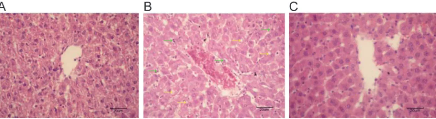
The clear architecture of hepatic lobules, with their regular arrayed hepatic cords, as well as the uniform-sized hepatic cells with round nuclei and clear karyolem-mas were observed in the control group (Figure 1A). In the LPS group, the central vein and its surrounding hepatic sinusoid were dilated and congested. Furthermore, the hepatic cords were disorganized. Hepatic cells showed turbid swelling, steatosis, and cytoplasmic vacuolation. Neutrophil and accidental eosi-nophil neutrophil infiltrations were found in the LPS group (Figure 1B). In the LPS++ENL group, the central vein and surrounding hepatic sinusoid had slight dilations and congestion. The hepatic cords were relatively unimpaired, and the architecture of hepatic lobules was normal. Hepatic cells were of uniform size, with clear karyolem-mas and slight turbid swelling, and neutrophil infiltrations were occasionally observed (Figure 1C).
Effect of ENL on indices of hepatocellular injury in LPS rats
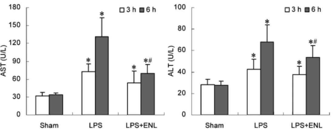
Three and six hours after LPS injection, activities of AST and ALT in plasma of the LPS and LPS++ENL groups were significantly higher than those of the sham group (P,0.05). These two indices in the LPS++ENL group at 6 h were lower than those in the LPS group (P,0.05; Figure 2).
Effect of ENL on W/D of hepatic tissue in LPS rats
The W/D of liver in the LPS (3.56±0.22) and LPS++ENL (3.52±0.10) groups was evidently higher than that of the sham group (3.29±0.07). No significant differences were observed between the LPS and LPS++ENL groups.
Effect of ENL on P-selectin of hepatic tissue in LPS rats
Three hours after LPS injection, P-selectin content of the hepatic homogenate in the LPS group (20.56± 5.88 ng/g protein) was significantly higher than that of the sham group (12.10±3.40 ng/g protein). No significant differences were observed between the LPS++ENL group (15.32±5.46 ng/g protein) and the sham or LPS groups.
Effect of ENL on ICAM-1 of hepatic tissue in LPS rats
Three and six hours after LPS injection, ICAM-1 content of the hepatic homogenate in the LPS group was significantly increased compared with the sham group (P,0.05). However, no significant differences were observed between the sham and LPS++ENL groups (P.0.05). Compared with the LPS group, ICAM-1 content in the LPS++ENL group was lower at 3 h (P,0.05), although no significant differences were observed at 6 h (P.0.05; Figure 3).
Effect of ENL on MPO of hepatic tissue in LPS rats
MPO activity of the hepatic homogenates in the LPS and LPS++ENL groups at 3 and 6 h after LPS adminis-tration was significantly increased, compared with that of the sham group (P,0.05; Figure 4). MPO activity of the LPS++ENL group at 3 h was significantly lower than that of the LPS group (P,0.05).
Figure 1.Effect of exogenous normal lymph (ENL) on hepatic morphology in rats (HE staining, 4006, magnification bar: 20mm).
Effect of ENL on Na+-K+-ATPase of hepatic tissue in LPS rats
Na+-K+-ATPase activity of the hepatic homogenate in the LPS group at 3 and 6 h after LPS administration, as well as that of the LPS++ENL group at 6 h, were obviously decreased, compared with that of the sham group (P,0.05; Figure 5). In contrast, at 3 h after LPS injection, Na+-K+-ATPase activity in the LPS++ENL group was significantly higher than that in the LPS group (P,0.05), but not significantly different from that of the sham group (P.0.05).
Discussion
In this study, we found that administration of ENL could significantly reduce LPS-induced increases in AST and ALT levels in plasma and attenuate the degree of hepatic tissue damage. Liver injury induced by LPS injection was prevented by treatment with ENL. A distinct increase in the W/D ratio of hepatic tissues in the LPS
group of rats was observed, thereby indicating that increased moisture in hepatic tissue was induced by LPS by a mechanism related to vascular endothelial cell (VEC) damage induced by LPS (14). Consequently, vascular hyperpermeability occurred. However, the W/D ratio of hepatic tissues was not changed in the LPS++ENL group. This was probably caused by LPS-induced damage to VECs within 15 min. ENL treatment was unable to restore the damaged VECs within 6 h. Therefore, a longer time period after ENL treatment is recommended for further study.
Experimental investigations show that systemic inflammatory response syndrome (SIRS) is an important indication of organ injury (15). LPS is known to induce SIRS in animal models, and the compound has been widely exploited for the study of SIRS-related conditions and for drug development (16,17). Polymorphonuclear neutrophils (PMNs) contribute to LPS-induced SIRS because PMNs are target cells of LPS attack and effector cells of inflammatory mediator release (18). Therefore, we explored the mechanisms by which ENL attenuates liver
Figure 2.Effect of exogenous normal lymph (ENL) on indices of hepatocellular injury in rats. Data are reported as means±SD (n=10). *P,0.05vsthe sham group;#P
,0.05vsthe lipopolysaccharide (LPS) group (one-way ANOVA). AST: aspartate aminotransferase; ALT: alanine aminotransferase.
Figure 3. Effect of exogenous normal lymph (ENL) on inter-cellular adhesion molecule-1 (ICAM-1) of hepatic homogenate in rats. Data are reported as means±SD (n=10). *P,0.05vsthe sham group; #P
,0.05 vs the lipopolysaccharide (LPS) group (one-way ANOVA).
Figure 4.Effect of exogenous normal lymph (ENL) on myeloper-oxidase (MPO) activity of hepatic tissue in rats. Data are reported as means±SD (n=10). *P,0.05vsthe sham group;#P
injury from PMNs. In general, LPS could activate VECs, and PMNs are generally activated by LPS. An increase in the expression of these cells releases cell adhesion molecules such as P-selectin and ICAM-1, which promote the adhesion, aggregation, and sequestration of PMNs in tissues that may cause SIRS (19). In the current study, the P-selectin level at 3 h was elevated as the ICAM-1 level and MPO activity at 3 and 6 h were increased in hepatic tissues of the LPS group. ENL administration reduced ICAM-1 levels and MPO activity of hepatic tissues at 3 h, but not at 6 h. MPO activity in a specific tissue is significantly correlated with the number of PMNs in affected tissues (20). Therefore, LPS induces PMN adhesion via increased P-selectin and ICAM-1 levels. ENL administration is beneficial for reducing liver injury by decreasing the PMN-mediated inflammatory response. In addition, we found that normal lymph might reduce PMNs attached to the venular wall and improve blood flow for microcirculation, as well as decrease the levels of P-selectin and ICAM-1 in plasma (21). This result is an important supplement to the findings of the current study. ATPase is a membrane-bound protein found in all cells, and it maintains a chemical and electrical gradient across the cell membrane (22,23). The degree of cellular injury is closely related to the activity of ATPase. Any factors that may alter ATPase activity of the cytomem-brane will suppress the transportation of ions into and from these cells (24). A change in the electrochemical gradient may lead to a wide array of cellular errors that ultimately aggravate the dysfunction and morphological damage of organs (25). ATPase in membranes includes Na+-K+-ATPase, Na+-H+-ATPase, Ca2++-ATPase, Ca2++-Mg2++-ATPase, etc. Among them, Na+-K+ -ATPase is the most important, and it is involved in the formation of cell edema. Therefore, we examined Na+ -K+-ATPase activity as an indicator of membrane pump
function of hepatocytes. The results of this study showed that Na+-K+-ATPase activity in hepatic tissues was obviously decreased at 3 and 6 h after LPS injection,
and a low dose of ENL improved Na+-K+-ATPase activity. The present data implied that improvement of membrane pump activity is an important mechanism of ENL in alleviating acute liver injury induced by LPS.
In addition, a previous study in our laboratory (26) showed that ENL administration could increase hepatic microregional blood perfusion at 270-360 min after LPS injection, which might be one of the mechanisms of ENL alleviating LPS-induced liver injury. Meanwhile, the results also showed that ENL administration markedly prolonged survival time of the endotoxically shocked rats from 7.21±1.33 to 11.80±2.67 h, which further con-firmed the beneficial effect of ENL intervention on endotoxic shock.
Several limitations were observed in the present study. First, the model of LPS-induced liver injury does not completely reflect all types of acute liver injury in the human body. Second, although LPS injections were slowly administered for 10 min in the animal model to imitate sepsis in clinical practice, several differences between animal models and human patients were apparent. Third, the long-term effects of ENL treatment of LPS-induced liver injury have to be clarified. Nonetheless, the current results are clinically significant for the treatment of septic shock. Finally, ENL adminis-tration was performed in this study at 15 min after LPS injection. However, whether ENL administration after 15 min has a beneficial effect is still not clear, which is one limitation of the current study, and the effect of ENL administration on endotoxic shock at multiple time points after LPS injection should be investigated in the future.
The amount of lymph used in this experiment was only one-fifteenth of whole blood volume, and an equal volume of normal saline was administered to the LPS group. Consequently, the interventions of normal lymph are difficult to explain by supplementary blood volume and hemodilution. Treatment with a low dose of normal lymph may lessen reperfusion injury induced by fluid resuscita-tion, thereby allowing for a wider range of prospective clinical applications. It should be pointed out that, although the composition of normal lymph is similar to normal plasma, there are differences between them, such as hypoviscosity, hypoproteins, hyperlipoproteins, and so on (27,28), which were involved in the normal lymph ameliorating microcirculation or energy metabolism and, as a result, alleviating organ injury. In addition, Interewicz et al. (29) reported that unknown proteins of low molecular weight were found in lymph but not plasma, using the ProteinChip SELDI MS system. We believe that these unknown proteins may partly explain the observed beneficial effect of normal lymph, alleviating liver injury. However, further research is needed. Meanwhile, ENL obtained from healthy dogs had no immunological rejection in our previous studies (7-9,21) or in the current study. Therefore, healthy dogs may be safely used as donors of ENL. Normal lymph can also be obtained from
Figure 5.Effect of exogenous normal lymph (ENL) on Na+-K+ -ATPase activity of hepatic homogenate in rats. Data are reported as means±SD (n=10). *P,0.05vsthe sham group;#P
large animals such as pigs and sheep. This study provided experimental evidence of lymph development and a novel target for the treatment of septic shock.
We conclude that ENL can alleviate LPS-induced liver injury. The mechanism involved is related to a decrease in neutrophil adhesions and improvement of Na+-K+ -ATPase activity. These data present an alternative therapeutic strategy for the treatment of liver injury accompanied by severe infection or sepsis.
Acknowledgments
Research supported by scientific research projects of the Education Department in Hebei Province (#2005311,
#2007407) and the Foundation of Hundred Innovative Talents in Universities of Hebei Province, China (#CPRC047,#CPRCII026).
References
1. Korish AA, Arafa MM. Propolis derivatives inhibit the systemic inflammatory response and protect hepatic and neuronal cells in acute septic shock.Braz J Infect Dis2011; 15: 332-338.
2. Hozumi H, Tada R, Murakami T, Adachi Y, Ohno N. Comparative analysis of hepatic CD14 expression between two different endotoxin shock model mice: relation between hepatic injury and CD14 expression.PLoS One 2013; 8: e53692, doi: 10.1371/journal.pone.0053692.
3. Marzocco S, Di Paola R, Ribecco MT, Sorrentino R, Domenico B, Genesio M, et al. Effect of methylguanidine in a model of septic shock induced by LPS.Free Radic Res 2004; 38: 1143-1153, doi: 10.1080/107157604100017 25517.
4. Szabo C, Modis K. Pathophysiological roles of peroxynitrite in circulatory shock.Shock2010; 34 (Suppl 1): 4-14, doi: 10.1097/SHK.0b013e3181e7e9ba.
5. Gonzalez-Teran B, Cortes JR, Manieri E, Matesanz N, Verdugo A, Rodriguez ME, et al. Eukaryotic elongation factor 2 controls TNF-alpha translation in LPS-induced hepatitis.J Clin Invest2013; 123: 164-178, doi: 10.1172/ JCI65124.
6. Niu CY, Zhang J, Yao T, Fan G. The antagonistic effect of lymph on shock in rat.Lymphology1998; 31 (Suppl): 159-162.
7. Niu CY, Zhao ZG, Zhang YP, Hou YL, Li JJ, Jiang H, et al. Exogenous normal lymph alleviates microcirculation dis-turbances and abnormal hemorheological properties in rats with disseminated intravascular coagulation. Braz J Med Biol Res2013; 46: 138-147, doi: 10.1590/1414-431X2012 2378.
8. Fan G, Zhao ZG, Niu CY, Yang YQ, Zhang J. [Effects of normal lymph on myeloperoxidase activity and adenosine triphosphatase in rats with endotoxic shock].Zhongguo Wei Zhong Bing Ji Jiu Yi Xue2006; 18: 721-723.
9. Zhao ZG, Niu CY, Zhang LL, Zhang J, Han R, Zhang YP, et al. Exogenous normal lymph alleviates lipopolysacchar-ide-induced acute kidney injury in rats.Ren Fail2013; 35: 806-811, doi: 10.3109/0886022X.2013.794680.
10. Cheng AM, Moore EE, Masuno T, Escobar GA, Sarin EL, Johnson JL, et al. Normal mesenteric lymph blunts the pulmonary inflammatory response to endotoxin.J Surg Res 2006; 136: 166-171, doi: 10.1016/j.jss.2006.05.013. 11. Zhao ZG, Niu CY, Zhang YP, Hou YL, Li JJ, Jiang H.
Exogenous normal lymph alleviating kidney injury by im-proving coagulation function in disseminated intravascular coagulation rats.Ren Fail2012; 34: 221-226.
12. Anderson BO, Brown JM, Shanley PF, Bensard DD, Harken
AH. Marginating neutrophils are reversibly adherent to normal lung endothelium.Surgery1991; 109: 51-61. 13. Fedorova OV, Lakatta EG, Bagrov AY. Endogenous Na,K
pump ligands are differentially regulated during acute NaCl loading of Dahl rats.Circulation2000; 102: 3009-3014, doi: 10.1161/01.CIR.102.24.3009.
14. Dayoub JC, Ortiz F, Lopez LC, Venegas C, Del Pino-Zumaquero A, Roda O, et al. Synergism between melatonin and atorvastatin against endothelial cell damage induced by lipopolysaccharide.J Pineal Res 2011; 51: 324-330, doi: 10.1111/j.1600-079X.2011.00892.x.
15. Zhang Q, Raoof M, Chen Y, Sumi Y, Sursal T, Junger W, et al. Circulating mitochondrial DAMPs cause inflammatory responses to injury.Nature 2010; 464: 104-107, doi: 10. 1038/nature08780.
16. Remick DG, Newcomb DE, Bolgos GL, Call DR. Comparison of the mortality and inflammatory response of two models of sepsis: lipopolysaccharidevscecal ligation and puncture.Shock2000; 13: 110-116, doi: 10.1097/00024 382-200013020-00004.
17. Hollenberg SM, Broussard M, Osman J, Parrillo JE. Increased microvascular reactivity and improved mortality in septic mice lacking inducible nitric oxide synthase.Circ Res2000; 86: 774-778, doi: 10.1161/01.RES.86.7.774. 18. Nakamori Y, Koh T, Ogura H, Tanaka H, Fujimi S, Kasai K,
et al. Enhanced expression of intranuclear NF-kappa B in primed polymorphonuclear leukocytes in systemic inflam-matory response syndrome patients.J Trauma2003; 54: 253-260, doi: 10.1097/01.TA.0000037096.73714.E6. 19. Power C, Wang JH, Sookhai S, Wu QD, Redmond HP.
Proinflammatory effects of bacterial lipoprotein on human neutrophil activation status, function and cytotoxic potential in vitro.Shock2001; 15: 461-466, doi: 10.1097/00024382-200115060-00009.
20. Reino DC, Pisarenko V, Palange D, Doucet D, Bonitz RP, Lu Q, et al. Trauma hemorrhagic shock-induced lung injury involves a gut-lymph-induced TLR4 pathway in mice.PLoS One2011; 6: e14829, doi: 10.1371/journal.pone.0014829. 21. Zhang LL, Zhao ZG, Niu CY, Zhang J. [Interference effects
of lymph plasma on endotoxic shock in rats].Zhongguo Ying Yong Sheng Li Xue Za Zhi2011; 27: 75-80.
22. Murphy E, Eisner DA. Regulation of intracellular and mitochondrial sodium in health and disease.Circ Res2009; 104: 292-303, doi: 10.1161/CIRCRESAHA.108.189050. 23. Billimoria FR, Dave BN, Katyare SS. Neonatal
24. Garces EO, Victorino JA, Veronese FV. [Anticoagulation in continuous renal replacement therapies (CRRT)]. Rev Assoc Med Bras2007; 53: 451-455, doi: 10.1590/S0104-42302007000500023.
25. Berger G, Guetta J, Klorin G, Badarneh R, Braun E, Brod V, et al. Sepsis impairs alveolar epithelial function by down-regulating Na-K-ATPase pump.Am J Physiol Lung Cell Mol Physiol2011; 301: L23-L30, doi: 10.1152/ajplung.00010.2010. 26. Zhang LL, Zhang J, Zhao ZG, Zhang YP, Niu CY. [Intervention role of lymph plasma on renal and hepatic blood perfusion in rats with endotoxic shock].Zhongguo Wei Zhong Bing Ji Jiu Yi Xue2010; 22: 740-743.
27. Zhao ZG, Niu CY, Hdu YL. [Comparation of rheology and ingredient between normal lymph and blood]. Zhongguo Ying Yong Sheng Li Xue Za Zhi2009; 25: 482-484. 28. Nanjee MN, Cooke CJ, Wong JS, Hamilton RL, Olszewski
WL, Miller NE. Composition and ultrastructure of size subclasses of normal human peripheral lymph lipoproteins: quantification of cholesterol uptake by HDL in tissue fluids. J Lipid Res2001; 42: 639-648.