Susceptibility variation of
Malassezia pachydermatis
to antifungal agents
according to isolate source
Caroline Borges Weiler
1, Francielli Pantella Kunz de Jesus
2, Graziela Habib Nardi
3,
Érico Silva Loreto
2, Janio Morais Santurio
2, Selene Dall’Acqua Coutinho
3,
Sydney Hartz Alves
1,21Programa de Pós-graduação em Ciências Farmacêuticas, Universidade Federal de Santa Maria,
Santa Maria, RS, Brasil.
2Departamento de Microbiologia e Parasitologia, Universidade Federal de Santa Maria,
Santa Maria, RS, Brasil.
3Laboratório de Biologia Molecular e Celular, Universidade Paulista, São Paulo, SP, Brasil.
Submitted: August 18, 2011; Approved: July 02, 2012.
Abstract
Malassezia pachydermatisis associated with dermatomycoses and otomycosis in dogs and cats. This study compared the susceptibility ofM. pachydermatisisolates from sick (G1) and healthy (G2) ani-mals to azole and polyene antifungals using the M27-A3 protocol. Isolates from G1 aniani-mals were less sensitive to amphotericin B, nystatin, fluconazole, clotrimazole and miconazole.
Key words:Malassezia pachydermatis, susceptibility, antifungal agents.
Currently, the genus Malassezia includes fourteen species, thirteen of which are lipid-dependent and are most frequently recovered from humans, ruminants and horses (Malassezia furfur, M. globosa, M. obtusa, M. restricta, M.slooffiae, M. sympodialis, M. dermatis, M. nana, M. ja-ponica, M. yamatoensis, M. equina, M. caprae and M.
cuniculi); the non-lipid-dependent species, M.
pachydermatis, is commonly recovered from dogs and cats (Cabañeset al., 2011). The distribution of different species, the colonization prevalence and the population density of Malasseziaspp. varies by carrier and affected sites of the body, which are influenced by the skin lipid composition and the presence of competitive microbiota (Sugitaet al., 2010).
M. pachydermatisis the species most adapted to ani-mals and is often recovered as part of the microbiota of the ear canal and skin of dogs, cats and other species of domes-tic and wild animals. In dogs and cats,M. pachydermatis has been linked to both localized (otitis externa and derma-titis) and systemic disease (Guillot and Bond, 1999). Anti-fungal triazoles are commonly used for treatment, and treatment should be continued until the clinical signs
re-solve and yeast are no longer observed during direct examination (Daigle, 2007).
The aim of this study was to compare the susceptibil-ity ofM. pachydermatisisolates recovered from healthy an-imals and anan-imals with otitis to ketoconazole, fluconazole, itraconazole, voriconazole, clotrimazole, miconazole, nys-tatin and amphotericin B.
We studied two groups ofM. pachydermatisisolates taken from dogs and cats at the Cellular and Molecular Bi-ology Laboratory of the Paulista University (UNIP, São Paulo, Brazil). Group 1 (G1) was comprised of 40 isolates recovered from the ear canals of animals with otitis externa; group 2 (G2) was comprised of 40 isolates recovered from the ear canals of healthy animals. Isolate identification was confirmed by randomly amplified polymorphic DNA using the Mpa-F (CTGCCATACGGATGCGCAAG) and 58S-R (TTCGCTGCGTTCTTCATCGA) primers (Sugita et al., 2003).
Stock solutions of antifungal agents were obtained from the dilution of each antifungal drug in dimethyl sulfo-xide or sterile distilled water for fluconazole. The drugs were serially diluted in RPMI 1640 broth (GIBCOTM) to obtain the following final concentrations: ketoconazole Brazilian Journal of Microbiology 44, 1, 174-178 (2013) Copyright © 2013, Sociedade Brasileira de Microbiologia
ISSN 1678-4405 www.sbmicrobiologia.org.br
Send correspondence to C.B. Weiler. Programa de Pós-graduação em Ciências Farmacêuticas, Universidade Federal de Santa Maria, Santa Maria, RS, Brasil. E-mail: [email protected].
(16 mg/mL-0.007 mg/mL) (Janssen Beerse), itraconazole
(16mg/mL-0.007 mg/mL) (Janssen Beerse), clotrimazole
(64mg/mL-0.125mg/mL) (Bayer), voriconazole (16m
g/mL-0.007 mg/mL) (Pfizer), miconazole (64 m
g/mL-0.125 mg/mL) (Labware), nystatin (64 m
g/mL-0.125mg/mL) (Bristol-Myers), amphotericin B (16m
g/mL-0.007 mg/mL) (Bristol-Myers) and fluconazole
(64mg/mL-0.125mg/mL) (Pfizer). Inocula were obtained
from 48-h pure Dixon agar cultures and consisted of micro-organism suspensions in sterile saline (0.85%) plus Triton X-100 (0.05%) (Merck), whose turbidity was adjusted to 0.5 on the McFarland scale. Inocula were diluted 1:50 in sterile distilled water and then at 1:20 in RPMI 1640 broth. For each isolate, microdilution plates containing 10mLof
antifungals in RPMI 1640 diluted in different concentra-tions were inoculated with 10mLof the standardized
ino-cula. As positive control, the standardized inocula were cultured alone; the negative control was the antifungal alone diluted in RPMI 1640 broth. Culture plates were in-cubated at 37 °C for 48 h. Minimum inhibitory concentra-tions (MICs) were recorded following the M27-A3 protocol (CLSI, 2007). All tests were performed in dupli-cate. The Mann-Whitney test was used to compare the two groups of isolates to determine whether they had similar susceptibility patterns to the antifungal agents tested.
Because no standardized susceptibility testing proce-dures exist forM. pachydermatis, the present study was based on the M27-A3 protocol (CLSI, 2007); in the absence
of specific breakpoints for Malassezia spp., we utilized breakpoints described forCandidaspp.
In this work, the lowest MICs were observed with ketoconazole, itraconazole and voriconazole (Table 1). Voriconazole showed the smallest variations of MICs with the MICs for G1 isolates ranging from 0.01-0.25mg/mL,
while the MICs for G2 isolates ranged from 0.01 to 0.125mg/mL. These data are consistent with findings from
Guptaet al.(2000) that reported MICs ranging from 0.03 to 0.25 mg/mL. Hammer et al. (2000) reported the lowest
MICs with ketoconazole, with MIC50 and MIC90 values similar to values we recorded for G2 isolates (Table 2). Al-though the susceptibility testing for itraconazole, fluco-nazole and amphotericin B showed that 100% of the isolates were susceptible to these antifungals, the MIC range was higher in G1 isolates compared to G2 isolates (Table 2). Nakamuraet al.(2000) tested sevenMalassezia species and all were susceptible to itraconazole; however, M. pachydermatisexhibited the highest MIC range among the species evaluated.
Statistical analysis revealed significant differences in susceptibility between G1 and G2 isolates. In the suscepti-bility tests with amphotericin B, nystatin, fluconazole, miconazole and clotrimazole, G1 isolates had significantly higher MICs compared to G2 isolates (p < 0.05). The exis-tence of a different susceptibility profile between the two groups revealed higher antifungal resistance in the isolates obtained from infected animals (G1). The study by
176 Weileret al.
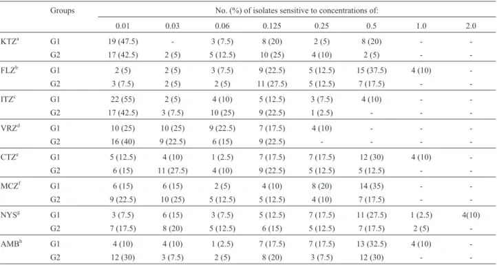
Table 1- Distribution of MICs (mg/mL) forM. pachydermatisisolated from ear canals of animals with and without otitis.
Groups No. (%) of isolates sensitive to concentrations of:
0.01 0.03 0.06 0.125 0.25 0.5 1.0 2.0
KTZa G1 19 (47.5) - 3 (7.5) 8 (20) 2 (5) 8 (20) -
-G2 17 (42.5) 2 (5) 5 (12.5) 10 (25) 4 (10) 2 (5) -
-FLZb G1 2 (5) 2 (5) 3 (7.5) 9 (22.5) 5 (12.5) 15 (37.5) 4 (10)
-G2 3 (7.5) 2 (5) 2 (5) 11 (27.5) 5 (12.5) 7 (17.5) -
-ITZc G1 22 (55) 2 (5) 4 (10) 5 (12.5) 3 (7.5) 4 (10) -
-G2 17 (42.5) 3 (7.5) 10 (25) 9 (22.5) 1 (2.5) - -
-VRZd G1 10 (25) 10 (25) 9 (22.5) 7 (17.5) 4 (10) - -
-G2 16 (40) 9 (22.5) 6 (15) 9 (22.5) - - -
-CTZe G1 5 (12.5) 4 (10) 1 (2.5) 7 (17.5) 7 (17.5) 12 (30) 4 (10)
-G2 6 (15) 11 (27.5) 4 (10) 9 (22.5) 5 (12.5) 5 (12.5) -
-MCZf G1 6 (15) 6 (15) 2 (5) 4 (10) 8 (20) 14 (35) -
-G2 9 (22.5) 10 (25) 5 (12.5) 5 (12.5) 4 (10) 7 (17.5) -
-NYSg G1 3 (7.5) 6 (15) 3 (7.5) 5 (12.5) 7 (17.5) 11 (27.5) 1 (2.5) 4(10)
G2 7 (17.5) 8 (20) 5 (12.5) 6 (15) 5 (12.5) 7 (17.5) 2 (5)
-AMBh G1 4 (10) 4 (10) 1 (2.5) 7 (17.5) 7 (17.5) 13 (32.5) 4 (10)
-G2 12 (30) 3 (7.5) 2 (5) 8 (20) 3 (7.5) 12 (30) -
Lyskovaet al.(2007) usingM. pachydermatisisolates from animals with and without otitis media found that all isolates exhibited high susceptibility to all of the antifungal agents tested in this study, except for fluconazole, to which 4.4% of the isolates were resistant. However, in this same study, significant differences in susceptibility between the two isolate groups were not observed. Fluconazole resistance has been described previously as Eichenberget al.(2003) observed fluconazole resistance in 2.4% of the 82 isolates tested. Further, Jesus et al. (2011) induced resistance to fluconazolein vitro, demonstrating thatM. pachydermatis isolates can become resistant during treatment with this antifungal. Bernardo et al. (1998) found a M. pachydermatis isolate that was resistant to several anti-fungals, including amphotericin B, nystatin and micona-zole.
Malasseziaspecies have a structure resembling a cap-sule around the cell wall that provides protection and is similar in form to the capsule ofCryptococcusspp. (Ashbee and Bond, 2010). In saprophytic strains, this capsule con-tains high levels of lipids, blocking the exposure of fungal antigenic proteins. Factors such as humidity, high tempera-ture and high fat environment favor rapid M. pachydermatis multiplication, which is accompanied by decreased lipid content and exposure of antigenic particles. The reduced susceptibility observed in G1 isolates against azole antifungal agents might be linked to variations in lipid composition. The main mechanism of action of azoles is to inhibit ergosterol synthesis. However, these drugs also act on the cell membrane through direct interaction with its
lipid components (Hitchcocket al., 1986). Thus, during in-fection, the body’s immune cells release substances that promote a lipid imbalance in the constitution of the fungal membrane, affecting membrane permeability (De Kruyffet al., 1973) and interfering with the action of azole antifungal agents. In the case of polyene antifungal agents, the re-duced susceptibility of the G1 isolates may be related to the high activity of fungal intracellular enzymes such as cata-lase and/or superoxide dismutase. In microorganisms, catalase plays an important role in the detoxification of re-active oxygen species that are released by phagocytic cells during the host immune response (Hamptonet al., 1998). Because polyenes act directly on fungal ergosterol causing direct oxidative damage to the cell membrane, the high con-centration of catalase in fungal cells could protect the fun-gal cells from the oxidative action of polyene antifunfun-gal agents, possibly explaining the higher MICs in isolates ob-tained from animals with malasseziosis.
This study demonstrated thatM. pachydermatis iso-lated from animals with otitis are less sensitive to some antifungal agents than yeasts isolated from animals without otitis. This finding may explain because some malassezio-sis treatment failure and emphasizes the importance to evaluate the susceptibility of this pathogenic fungi.
References
Ashbee HR, Bond R (2010)MalasseziaSpecies and Immunity: Host-Pathogen Interactions. In: Boekhout T, Guého-Kellermann E, Mayser P, Velegraki A (eds) Malassezia and
Susceptibility ofM. pachydermatisto antifungal agents 177
Table 2-In vitrosusceptibility ofM. pachydermatisisolates to antifungal agents.
Antifungal Groups MIC rangea(
mg/mL) MIC50b(mg/mL) MIC90c(mg/mL) GMd(mg/mL)
Ketoconazole G1 0.01-0.5 0.06 0.5 0.048
G2 0.01-0.5 0.06 0.25 0.041
Fluconazole G1* 0.01-1.0 0.25 0.5 0.219
G2** 0.01-0.5 0.125 0.5 0.068
Itraconazole G1 0.01-0.5 0.01 0.25 0.032
G2 0.01-0.25 0.03 0.125 0.032
Voriconazole G1 0.01-0.25 0.03 0.125 0.042
G2 0.01-0.125 0.03 0.125 0.029
Clotrimazole G1* 0.01-1.0 0.25 0.5 0.163
G2** 0.01-0.5 0.06 0.5 0.069
Miconazole G1* 0.01-0.5 0.25 0.5 0.124
G2** 0.01-0.5 0.06 0.5 0.061
Nystatin G1* 0.01-2.0 0.25 1.0 0.181
G2** 0.01-1.0 0.06 0.5 0.084
Amphotericin B G1* 0.01-1.0 0.25 0.5 0.180
G2** 0.01-0.5 0.125 0.5 0.081
the Skin - Science and Clincial Practice. Springer, New York, pp 144-146.
Bernardo FM, Martins HM, Martins ML (1998) A survey of mycotic otitis externa of dogs in Lisbon. Rev Iberoam Micol 15:163-165.
Cabañes FJ, Veja S, Castellá G (2011)Malassezia cuniculisp.
nov., a novel yeast species isolated from rabbit skin.Med Mycol 49:40-48.
Clinical and Laboratory Standards Institute (2007) Reference Method for Broth Diluition Antifungal Susceptibility Tes-ting of Yeast: Approved Guideline M27-A3, v. 28.CLSI, Wayne, PA,USA, 1-25.
Daigle JC (2007) Clinical clues, diagnosis and treatment of
Malassezia dermatitis. The North American Veterinary Conference Congress, Orlando, Flórida, 21, pp 317-318. De Kruyff B, De Greef WJ, Eyk RVW, Demel RA, Van Deenen
LLM (1973) The effect of different fatty acid and sterol composition on the erythritol flux through the cell mem-brane ofAcholeplasma laidlawii. Biochimica et Biophysica Acta (BBA) - Biomembranes 298:479-499.
Eichenberg ML, Appelt CE, Berg V, Muschner AC, Nobre MO, Matta D, Alves SH, Ferreiro L (2003) Susceptibility of
Malassezia pachydermatisto azole antifungal agents evalu-ated by a new broth microdilution method. Acta Sci Vet 31:75-80.
Guillot J, Bond R (1999)Malassezia pachydermatis: A review.
Med Mycol 37:295-306.
Gupta AK, Kohli Y, Li A, Summerbell RC (2000)In vitro suscep-tibility of the seven Malasseziaspecies to ketoconazole, voriconazole, itraconazole e terbinafina. Brit J Dermatol 142:758-765.
Hammer KA, Carson CF, Riley TV (2000)In vitroactivities of ketoconazole, econazole, miconazole and Melaleuca
alternifolia (tea tree) oil against Malassezia species.
Antimicrob Agents Ch 44:467-469.
Hampton MB, Kettle AJ, Winterbourn CC (1998) Inside the neutrophil phagosome: Oxidants, myeloperoxidase and bac-terial killing. Blood 92:3007-3017.
Hitchcock C, Barrett-Bee K, Russel N (1986) The lipid composi-tion of azoles-sensitive and azole-resistant strains of
Candida albicans. J Gen Microbiol 132:2421-2431.
Jesus FPK, Lautert C, Zanette RA, Mahl DL, Azevedo MI, Ma-chado MLS, Alves SH, Botton SA, Dutra V, Santurio JM (2011)In vitrosusceptibiliy of fluconazole-susceptible and
-resistent isolates ofMalassezia pachydermatisagainst az-oles. Vet Microbiol 152:161-164.
Lyskova P, Vydrzalova M, Mazurova J (2007) Identification and antimicrobial susceptibility of bacteria and yeasts isolated from healthy dogs and dogs with otitis externa. J Vet Med A 54:559-563.
Nakamura Y, Kano R, Murai T, Watanabe S, Hasegawa A (2000) Susceptibility testing ofMalasseziaspecies using the urea
broth microdilution method. Antimicrob Agents Ch 44:2185-2186.
Sugita T, Boekhout T, Velegraki A, Guillot J, Hadina S, Cabañes J (2010) Epidemiology ofMalassezia- Related skin diseases. In: Boekhout T, Guého E, Mayser P, Velegraki A (eds) Malassezia and the Skin: Science and Clinical Practice. Springer, New York, pp 65-120.
Sugita T, Taqkashima M, Kodama M, Tsubol R, Nishikawa A (2003) Description of a new yeast species,Malassezia ja-ponica, abd its detection in patients with atopic dermatitis and healthy subjects. J Clin Microbiol 41:4695-4699.
All the content of the journal, except where otherwise noted, is licensed under a Creative Commons License CC BY-NC.