Escherichia coli
Biofilms
Stephanie M. Amato, Mark P. Brynildsen*
Department of Chemical and Biological Engineering, Princeton University, Princeton, New Jersey, United States of America
Abstract
Chronic and recurrent infections have been attributed to persisters in biofilms, and despite this importance, the mechanisms of persister formation in biofilms remain unclear. The plethora of biofilm characteristics that could give rise to persisters, including slower growth, quorum signaling, oxidative stress, and nutrient heterogeneity, have complicated efforts to delineate formation pathways that generate persisters during biofilm development. Here we sought to specifically determine whether nutrient transitions, which are a common metabolic stress encountered within surface-attached communities, stimulate persister formation in biofilms and if so, to then identify the pathway. To accomplish this, we established an experimental methodology where nutrient availability to biofilm cells could be controlled exogenously, and then used that method to discover that diauxic carbon source transitions stimulated persister formation inEscherichia coli biofilms. Previously, we found that carbon source transitions stimulate persister formation in planktonic E. colicultures, through a pathway that involved ppGpp and nucleoid-associated proteins, and therefore, tested the functionality of that pathway in biofilms. Biofilm persister formation was also found to be dependent on ppGpp and nucleoid-associated proteins, but the importance of specific proteins and enzymes between biofilm and planktonic lifestyles was significantly different. Data presented here support the increasingly appreciated role of ppGpp as a central mediator of bacterial persistence and demonstrate that nutrient transitions can be a source of persisters in biofilms.
Citation:Amato SM, Brynildsen MP (2014) Nutrient Transitions Are a Source of Persisters inEscherichia coliBiofilms. PLoS ONE 9(3): e93110. doi:10.1371/journal. pone.0093110
Editor:Christophe Beloin, Institut Pasteur, France
ReceivedOctober 7, 2013;AcceptedMarch 3, 2014;PublishedMarch 25, 2014
Copyright:ß2014 Amato, Brynildsen. This is an open-access article distributed under the terms of the Creative Commons Attribution License, which permits unrestricted use, distribution, and reproduction in any medium, provided the original author and source are credited.
Funding:This work was supported by the National Science Foundation (SMA, Graduate Research Fellowship), the Department of the Army (W81XWH-12-2-0138), and with start-up funds from Princeton University. This research was supported by the National Science Foundation Division of Materials Research through the Princeton University MRSEC (NSF-DMR 0819860). The funders had no role in study design, data collection and analysis, decision to publish, or preparation of the manuscript.
Competing Interests:The authors have declared that no competing interests exist. * E-mail: [email protected]
Introduction
Bacterial persisters are rare, phenotypic variants, whose hallmark characteristic is a transient, yet extraordinary, ability to tolerate antibiotics while their surrounding kin are killed. [1] Biofilms contain persisters, and this phenotypic state has been hypothesized to underlie why biofilm infections often relapse. [2,3] Persisters form during biofilm growth, and despite the identifica-tion of several important mediators, including ppGpp,[4–6] Lon, [4] RecA, [6] and YafQ, [7] the aspects of biofilm development that generate persisters, along with their respective pathways, remain ill-defined. The biofilm life-style includes numerous qualities conducive to persister formation, including slower growth, [8,9] decreased metabolism, [10] quorum signaling, [11,12] oxidative stress, [13,14] and nutrient heterogeneity. [15,16] This suggests that the composition of persister sub-populations in mature biofilms is likely heterogeneous,[10,17–19] consisting of persisters formed from different pathways in response to various signals throughout biofilm growth.
Recently, we identified a persister formation pathway in response to nutrient transitions in planktonicE. coli. [16] Nutrient transitions are abundant in biofilms, as cells at the periphery consume favorable substrates and leave less favorable substrates and waste products available to cells deeper in the film. [5,20] Together, these phenomena suggest that nutrient transitions may
be a source of persisters in biofilms. However, several studies have found that genes important to persistence in one lifestyle, biofilm or planktonic, are dispensable to persistence in the other. [6,7] These observations highlight the necessity to test the functionality of persister formation pathways identified under planktonic conditions in biofilm environments. To date, persister formation cascades, from source of stress to antibiotic tolerance, have mainly been studied in planktonic systems, [12,14,16,21] and the extent to which these pathways operate in biofilms remains an open question. Conversely, specific genes important for biofilm persis-tence have been identified [4–7], but their placement in formation cascades are just beginning to be elucidated.[4–6].
formation in biofilms. These findings provide a more thorough understanding of the importance of ppGpp to persistence in biofilms and point to nutrient transitions as an inherent characteristic of biofilm growth that has the capacity for persister generation.
Materials and Methods
Bacterial Strains, Plasmids, and Biofilm Growth Conditions
E. coliMG1655 was the wild-type strain used in this study. Its genetic mutants and plasmids used in this study are displayed in Table S3. Primers used to construct plasmids are displayed in Table S4. Separate colonies were used for each of three replicate experiments. Biofilm experiments were performed using colony biofilms. [20] For these experiments, cells from280uC stock were grown for 4 h in LB, diluted 1:100 into 2 mL of 10 mM glucose M9 media, and grown overnight for 16 h at 37uC and 250 rpm. The overnight culture was diluted into fresh M9 media containing 15 mM carbon content to an optical density at 600 nm (OD600) of
0.01 and 100mL aliquots were inoculated onto sterile, poly-ethersulfone (PES) membranes (0.2mm pore size, 25 mm diam-eter, Pall Corporation, Ann Arbor, MI) positioned on M9 minimal agar plates containing either 60 mM carbon content of carbon source, no carbon, or LB. Plates were incubated at 37uC. To monitor growth, PES membranes were aseptically removed from the agar, vortexed in 2 mL of sterile PBS for 1 minute, and the OD600 of the resulting cell suspensions were measured. Growth
was reported as a fold change in OD600(FCOD600),which is the
ratio of cells present on the membrane to the cells inoculated onto the membrane.
Carbon Source Transition Assay
Colony biofilms were grown as described by diluting the overnight into fresh 15 mM carbon content media, inoculating onto PES membranes atop the desired agar plates, and incubating at 37uC. At desired time points, membranes containing colony biofilms were aseptically removed from the agar, vortexed in 2 mL of PBS for 1 minute, and the OD600 was measured. OD600was
monitored for over 8 h of growth with specified secondary carbon sources and no carbon. Persister measurements were taken prior to glucose exhaustion (FCOD600,14, Figure 1) at FCOD600= 6 and
after glucose exhaustion at FCOD600= 30.
Microscopy of Colony Biofilms
E. coli MG1655 was modified to contain a chromosomally integratedlacIqpromoter in place of thelacIpromoter [10] and a chromosomally integrated PT5 under the control of two lac
operator sites [24] followed bygfpin place oflacZYAto generate the SA034 strain, which was used to image cells grown on the PES membranes. Cells from280uC stock were grown for 4 h in LB, diluted 1:100 into 2 mL of 10 mM glucose M9 media with 2 mM isopropyl b-D-1-thiogalactopyranoside (IPTG), and grown over-night for 16 h at 37uC and 250 rpm.
For biofilm samples, the overnight culture was diluted into fresh 2.5 mM glucose M9 media with 2 mM IPTG to an OD600of 0.01
and 100mL aliquots were inoculated onto sterile, PES membranes positioned on 10 mM glucose M9 minimal agar plates containing 2 mM IPTG. Plates were incubated at 37uC to FCOD600= 30 (,5
doublings). For planktonic samples, the overnight culture was diluted in 25 mL of fresh 10 mM glucose M9 media with 2 mM IPTG to an OD600 of 0.01. Cells were grown at 37uC and
250 rpm until,5 doublings and then,107cells (approximately
the number of cells present on the biofilm at FCOD600= 30) were
applied to a sterile PES membrane.
Membranes were immobilized on a glass slide and a cover slip was placed over them prior to imaging. Imaging was performed using a Nikon Ti-E microscope (Nikon, Melville, NY), a 20X Plan Fluor Nikon objective (0.45 NA), a Chroma 89014 filter set (Chroma, Bellows Falls, VT) with an ET490/20x excitation filter and an ET535/50 m emission filter, a Prior Lumen 200 Pro fluorescence illuminator, and an Andor Clara camera.
Transcriptional Reporters
MG1655 possessing pSA03 [16] was used as a cAMP transcriptional reporter as indicated. Kanamycin (50mg/mL) was present during growth for plasmid retention. Cells were prepared as described above. At FCOD600= 6 (before glucose
exhaustion) and at FCOD600= 30 (after glucose exhaustion),
membranes were aseptically removed from the agar and vortexed in 2 mL of PBS.
All strains including controls were analyzed by LSRII (BD Biosciences, San Jose, CA) flow cytometer. Microorganisms were determined using forward and side scatter parameters (FSC and SSC) and MG1655 carrying pUA66 as a control. The bacteria were assayed with a laser emitting at 488 nm for GFP, and green fluorescence was collected by 525/50 bandpass filter. Data were acquired and analyzed using FACSDiVa software (BD Bioscienc-es, San Jose, CA).
Glucose Measurements
Cells were prepared as described, and at FCOD600= 6 (before
glucose exhaustion) and FCOD600= 30 (after glucose exhaustion),
membranes were aseptically removed from the agar and vortexed in 1 mL of PBS. For the no carbon control, samples were taken at FCOD600= 6 and 2 h post-glucose exhaustion. Glucose was
quantified using the Amplex Red Glucose/Glucose Oxidase Kit (Invitrogen, Eugene, OR).
Persistence Measurements
Persisters were enumerated by determining the number of colony forming units (CFU) after exposure to 10mg/mL ofloxacin or 750mg/mL ampicillin for 5 h. Five hours was sufficient to provide CFU measurements within the second phase of a biphasic, time-dependent kill curve, which is required for persister measurements.[25–27] The antibiotic concentrations used were selected from those that lie on the second phase of a concentration-dependent kill curve (Figure S1), and thus were able to provide concentration-independent persister measurements.[25–27] At specified FCOD600, colony biofilms on membranes were treated
with 200mL of antibiotic solution applied to the top of the membrane and incubated at 37uC. At designated time points post antibiotic treatment, membranes were aseptically removed from the agar, vortexed in 2 mL of PBS, washed and serially diluted in PBS, and 10mL was spotted onto LB agar. For ampicillin-treated cells, the whole sample was plated on LB agar to increase the limit of detection. Cells in PBS were stored at 4uC prior to plating as necessary, and it was confirmed that such storage did not affect CFU measurements when compared to samples plated immedi-ately. Plates were incubated for 16 h at 37uC and CFU were measured to determine persister counts. 10–100 colonies were counted for each data point. [28].
Statistical Analysis
antibiotic treatment CFU measurements were statistically ana-lyzed. We previously confirmed that the data from persister assays, CFU measurements after 5 h antibiotic treatment, can be treated as near-normally distributed with a larger sample dataset. [16] The threshold for significance was set to p-values,0.05.
Results and Discussion
Establishment of a Method to Exogenously Control Carbon Source Availability in Colony Biofilms
Bacteria can exhibit either diauxic or non-diauxic growth when grown in the presence of two carbon sources. [29] During diauxic growth, the preferential carbon source is consumed in the first growth phase, whereas the less favorable secondary carbon source supports growth during the second growth phase. The two growth phases are separated by a lag period associated with physiological changes required for growth on the secondary carbon source. [30] Non-diauxic growth can exhibit preferential carbon source consumption, but lacks the intermediate lag period. [31] We have previously demonstrated that diauxic carbon source transitions stimulate persister formation in planktonic E. coli cultures. [16] Here, we sought to investigate whether carbon source transitions
stimulate persister formation during biofilm growth, and if so, identify the formation pathway responsible.
To study persister formation from carbon source transitions in biofilms, we required an experimental system where only biofilm cells were present and nutrient availability could be controlled exogenously. Biofilms are often grown in the presence of planktonic cells, [32,33] but results from these systems are complicated by biofilm dispersal, and when considering rare events such as persisters, uncertainty arises as to whether a cell originated from the biofilm or planktonic sub-population. Given these considerations, we used the colony biofilm culturing method, where cells are grown on nutrient-permeable membranes positioned atop agar plates. [5,20,34] In this method, which has been used in previous persistence studies, [4,5] all bacteria are surface-attached biofilm cells and planktonic cells are absent. This culturing method also allowed exogenous control of nutrient availability to biofilm cells (Figure 1A). Glucose, the primary carbon source, was delivered with cells to the top of PES membranes and secondary carbon sources or controls (glucose and no carbon) were provided in the agar. Young biofilms were investigated to avoid the in-film nutrient gradients that are present in mature colony biofilms, which would complicate the analysis of a single carbon source transition and its role in persister formation.
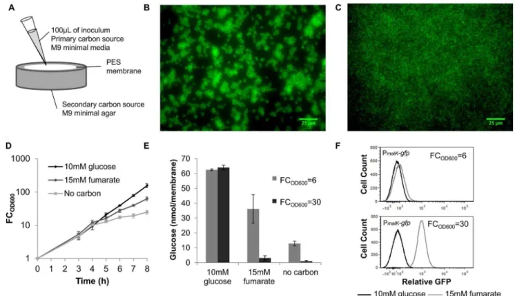
Figure 1. Experimental approach to control carbon source transitions in biofilms.(A) A schematic of our experimental setup where cells and the primary carbon source are applied to a PES membrane set atop agar containing the secondary carbon source, glucose, or no carbon. (B) Biofilms expressing GFP were grown to FCOD600= 30. Membranes were aseptically removed from the agar and analyzed using fluorescence microscopy. (C) Cells expressing GFP were inoculated into 25 mL of 10 mM glucose at 0.01 OD600and after,5 doublings,,107CFU were inoculated
onto a sterilized PES membrane and analyzed using fluorescence microscopy. (D) PES membranes atop agar containing 10 mM glucose, 15 mM fumarate, and no carbon were inoculated with wild-type cells at 0.01 OD600in 2.5 mM glucose and incubated at 37uC. The OD600was measured at specified time intervals and FCOD600was determined. One exponential growth phase was observed for glucose samples. Two regimes of exponential growth were observed for glucose-fumarate samples and no carbon sample exhibited limited growth after glucose exhaustion. (E) Glucose concentration measurements were taken at each persister sampling (FCOD600= 6 and FCOD600= 30) for glucose and glucose-fumarate samples and at FCOD600= 6 and 2 h post glucose exhaustion for the no carbon sample. (F) PmalK-gfpGFP distribution at FCOD600= 6 and FCOD600= 30 in glucose-fumarate and glucose samples. Data are averages of $3 independent experiments for (D) and (E) and data are representative samples of 3 independent experiments for (B), (C), and (F) and error bars indicate standard deviation.
[15,20] To demonstrate that the colony biofilm method produced young biofilms on the timescale of our experiments, we imaged colony biofilms at FCOD600= 30 using fluorescence microscopy
and a strain producing GFP (Figure 1B and 1C). The presence of cell clusters in the biofilm sample compared to a sample where an equal number of planktonic cells were seeded onto the membrane just prior to imaging demonstrates that these young films are immobilized cells growing in surface-attached communities.
To demonstrate that exogenous control of nutrient availability was achieved with the colony biofilm method, we monitored growth, quantified the glucose concentration in membranes, and utilized a transcriptional reporter of glucose exhaustion. Using 2.5 mM glucose in the inoculums, similar growth for films grown on agar containing glucose, fumarate, and no carbon was observed, indicating a common period of glucose consumption (Figure 1D). Fumarate was used here as a representative diauxic carbon source, and analogous measurements with additional secondary carbon sources are presented in the Supporting Information (Figure S2). At FCOD600,14, growth rates of the
fumarate and no carbon samples decreased, suggesting a transition away from glucose-replete conditions. Residual growth in the no carbon sample was due to trace glucose levels, whereas growth in the fumarate sample, which far exceeded that of the no carbon sample, signified fumarate catabolism. This was confirmed by quantifying glucose in the membranes (Figure 1E). Since glucose exhaustion triggers the production of cAMP [35] resulting in cAMP-CRP transcriptional activity, we used a cAMP-CRP transcriptional reporter [16] to demonstrate that cAMP-CRP is comparable between glucose and fumarate samples during the common period of glucose consumption, and cAMP-CRP activity increased in the fumarate sample after glucose depletion (Figure 1F). These flow cytometry data, which consist of only a single, distinct peak for each sample, also suggest that the biofilm cells were exposed to similar nutrient environments. Together these data demonstrate that our experimental setup was successful in achieving well-controlled carbon source transitions in biofilms.
Carbon Source Transitions Stimulate Persister Formation
After establishing a functional system for analyzing carbon source transitions in biofilms, we examined if transitions stimulate persister formation. E. coli persister levels were measured using ofloxacin throughout growth using glucose as the primary carbon source and a panel of secondary carbon sources that are diauxic or non-diauxic with glucose. The antibiotic treatment was delivered to the biofilms as 200mL of 10mg/mL ofloxacin evenly distributed atop the membrane to ensure full treatment of the biofilm. It was observed that diauxic media exhibited an increase in persisters to ofloxacin upon glucose exhaustion compared to the sole glucose control (Figure 2A). The non-diauxic secondary carbon sources, fructose [36] and gluconate [37] did not exhibit an increase in persisters to ofloxacin after glucose exhaustion. These results suggested that diauxic transitions stimulated formation of persisters to ofloxacin in biofilms. In addition, we tested whether carbon source transitions stimulate formation of persisters to additional antibiotics. When the aminoglycoside gentamicin was tested we observed that all cells were killed (Figure S1), a result that was expected given that glucose potentiates aminoglycoside activity in persisters. [38,39] However, we found that carbon transitions also stimulated formation of persisters to theb-lactam ampicillin. Using glucose as the primary carbon source and a panel of secondary carbon sources that are diauxic or non-diauxic with glucose and treating with 200mL of 750mg/mL ampicillin, we observed no persister formation prior to the transition at FCOD600= 6, but observed persister formation for fumarate and
succinate after the transition at FCOD600= 30 (Figure S3A). These
results demonstrate that persister formation in biofilms in response to carbon source transitions is not specific to fluoroquinolones. In addition, these data suggest the existence of distinct pathways, since different sets of secondary carbon sources stimulate ofloxacin and ampicillin persister formation (Figure S3B).
To identify the underlying formation pathway for ofloxacin persisters, we focused on glucose-fumarate transitions, since they elicited the strongest persister formation response and were previously studied in planktonic culture. [16] For comparison, glucose-fructose transitions, which are non-diauxic, were investi-gated. Persister measurements were taken during growth on glucose (FCOD600= 6) and after glucose exhaustion
(FCOD600= 30). During exponential growth on glucose, all
persister levels were the same independent of the secondary
Figure 2. Diauxic shifts stimulate persister formation in biofilms.(A)E. coli were grown on glucose as the primary carbon source and a panel of secondary carbon sources. At hourly time points, biofilms were challenged with 200ml of 10mg/mL ofloxacin for 5 h, aseptically removed from the agar, vortexed in 2 mL PBS for 1 minute, washed, and plated to measure CFUs. To construct the color plot as a function of FCOD600, as needed values plotted were interpolated from two adjacent measurements. Raw values are presented in Table S2. (B) Diauxic growth (glucose-fumarate) results in significant persister formation (p,0.05), whereas non-diauxic growth does not (p.0.05) (glucose and glucose-fructose) when comparing persister levels 5 h post-antibiotic treatment. Time on the x-axis represents time after antibiotic treatment. (C) Growth on fumarate is not responsible for persister formation in glucose-fumarate samples, as evidence by sole fumarate control, which contained fumarate as the only carbon source both in the inoculum and agar. Data are averages of$3 independent experiments, error bars indicate standard deviation, and significance was assessed using the null hypothesis that the mean CFU levels in two sample sets were equal.
carbon source present (Figure 2B). Persister measurements during exponential growth on the secondary carbon source exhibited a statistically significant 13-fold increase in persisters for the glucose-fumarate samples and an insignificant 1.4-fold increase for the glucose-fructose samples when compared to persister levels in the glucose-only samples at the same FCOD600.
To establish that the increase in persisters was due to the transition from glucose to fumarate and not from the slower growth on fumarate, persister measurements were taken at the same film densities during growth on fumarate as the sole carbon source (fumarate provided in the inoculum and agar) (Figure 2C). The persister levels for the culture grown in the fumarate-only samples were 1.4-fold higher and not statistically different from persister levels for glucose-only samples, but were 9-fold lower and statistically different from those for the glucose-fumarate samples, which suggests that the increase in persisters was a result of the carbon source transition and not slower growth on fumarate.
Persisters Form through a RelA-dependent Mechanism
We next sought to determine the molecular mechanism responsible for persister formation from carbon source transitions in biofilms. Upon glucose limitation, the stringent response is activated and the metabolites pppGpp and ppGpp, collectively termed ppGpp for subsequent discussion, are synthesized by both RelA and SpoT. [40,41] RelA is a ribosome-associated ppGpp synthase, whereas SpoT has both synthase and hydrolase activity. [41] We have shown previously that persister formation from carbon source transitions in planktonic cultures is dependent on the ppGpp biochemical network. [16] Therefore, we tested whether ppGpp is also essential for persister formation due to carbon source transitions in biofilms.
We measured persisters prior to and after glucose exhaustion in
DrelA (Figure 3, Table S1). Deletion of the ribosome-dependent ppGpp synthase RelA resulted in an insignificant 1.6-fold increase in persisters when comparing glucose-fumarate to glucose-only samples. The complemented DrelA strain restored the persister formation phenotype confirming that RelA is required for persister formation from carbon source transitions in biofilms (Figure S4). Interestingly, under planktonic conditions,DrelAwas only found to reduce the quantity of persisters formed, whereas deletion of both ppGpp synthases (DrelADspoT)was required to eliminate persister formation. [16] Although the SpoT synthase has been shown to be activated in response to carbon source starvation [42,43] and may experience increased activity during the transition, DspoTis not viable in arelA+background [44] andDrelADspoTis auxotrophic for several amino acids. [45] Since directly assessing the role of SpoT in the carbon source transition model would have required amino acid supplementation, and complete elimination of persister formation was achieved withDrelA, the involvement of SpoT was not further explored.
To validate that E. coli experiences a ppGpp-dependent stringent response during carbon source transitions in biofilms, we monitored levels of stable ribosomal RNA (rRNA) in wild-type and its DrelA derivative during the transition using quantitative PCR (qPCR). The seven rRNA operons are regulated by the stringent response, and, when levels of ppGpp are high, transcription of the rRNA is repressed. [41] However, in DrelA
this inhibition does not occur and levels of stable rRNA have been shown to be ,2- to 10-fold higher than that of arelA+strain in
similar conditions.[46–50] Within colony biofilms, we found that both 23 S and 16 S RNA expression was,2-fold higher inDrelA
than wild-type during carbon source transitions at FCOD600= 14
(Figure S4, Table S5). These data confirm that ppGpp inDrelA,
where persister formation was eliminated, was lower than in
wild-type where persister formation was observed, suggesting that increased ppGpp levels are required for persister formation from carbon source transitions.
ppGpp-dependent Persister Formation in Biofilms Requires NAPs
We next sought to determine how ppGpp in biofilms increases tolerance to ofloxacin, an antibiotic that targets DNA gyrase. [51] Previously, high levels of ppGpp have been observed to lead to relaxation of the chromosome, an indicator of reduced DNA gyrase activity. [52] Though the mechanism underlying this phenomenon remains ill-defined, it is known that DNA gyrase, topoisomerases I, III, and IV, and NAPs work together to control the degree of (2) supercoiling of the chromosome. [52,53] Given the role of NAPs in (2) supercoiling, the knowledge that NAPs are under stringent control both directly and indirectly through a complex interdependent network of regulation,[53–56] and the discovery that several NAPs were involved in persister formation from carbon source transitions in planktonic cultures, we tested mutants of NAPs FIS, HNS, HU, IHF, and SeqA.[57–59].
We found that Dfis, DhupA, DhupB, and DseqA all removed persister formation from carbon source transitions in biofilms, whereas DihfA, DihfB, and Dhns did not eliminate persister formation (Figures 4, S5, and S6). ForDfis, DhupA, andDhupB,
complementation with genes expressed from their native promot-ers on low-copy plasmids restored ppromot-ersister formation (Figure S7). However,DseqAcould not be complemented to a significant level when expression was driven by the putative promoter 86 base pairs upstream of the SeqA start codon on the same low-copy plasmid (Figure S6). Although the possibility of additional mutations in the three DseqA colonies cannot be ruled out, the
seqApromoter is putative and has been shown to be regulated by HU from an undefined location. [56] Given these uncertainties, complementation may be achieved with a different expression system. Interestingly, IHF was previously found to be an important mediator of persister formation in planktonic conditions, but here was not found to be a mediator in the biofilm state. Interactions of NAPs with chromosomal DNA and one another are strongly influenced by growth rate and phase, [53,60,61] so we reason that differences in these interactions between biofilm and planktonic lifestyles underlie why IHF was only found to be important for planktonic persister formation. Together, these results demon-strate that FIS and HU are required for persister generation in response to carbon source transitions in biofilms.
Given the direct connection between NAPs and the primary target of ofloxacin, we tested whether the same pathway was responsible for ampicillin persister formation from carbon source transitions in biofilms. When treating wild-type andDhupB with ampicillin before and after the carbon source transition, we observed no difference in persister formation between the two strains (Figure S8) suggesting that the formation pathway was specific to fluoroquinolones.
To determine whether mediators of the pathway contribute to persister formation in environments more complex than diauxic conditions, we performed analogous assays atop LB agar with mature biofilms, and found thatDrelAandDhupBhad statistically significant fewer persisters than the wild-type,,6-fold (Figure 4).
Conclusions
The clinical importance of persisters has been attributed to their presence in biofilm infections. [3,63] Despite this significance, the means by which persisters form in this bacterial life-style remain obscure. This is in part due to the environmental heterogeneity inherent to biofilm growth. Here we were able to establish an experimental protocol to study, in isolation, the effect of nutrient transitions on persistence in biofilms. Identification that diauxic carbon source transitions generate persisters to ofloxacin in biofilms led us to identify RelA and NAPs as critical mediators of the process. ppGpp has been increasingly identified as a major mediator of persistence.[4–6,16,64] Both Nguyen and colleagues
and Maisonneuve and colleagues established that ppGpp was important for persister formation in matureE. colibiofilms, [4,5] though the aspect of biofilm physiology that was responsible for the significance of ppGpp in persistence was undetermined. In addition, Bernier and colleagues found that ofloxacin persister formation from leucine starvation inE. colibiofilms was partially dependent on ppGpp. [6] Here we identified carbon source transitions as a biofilm property that generates persisters through a ppGpp-dependent mechanism. Further, we found that modulators of chromosomal (2) supercoiling were required for ppGpp to form persisters in response to carbon source transitions, providing a direct connection to the primary target of fluoroquinolones, DNA gyrase. Interestingly, Maisonneuve and colleagues found that the
Figure 3. Genes required for persister formation from carbon source transitions in biofilms.Cells were challenged with 200mL of 10mg/ mL ofloxacin at FCOD600= 6 and FCOD600= 30, representing growth on glucose and growth after glucose exhaustion, respectively (except for glucose-only sample). Carbon source transitions resulted in significant persister formation for (A) wild-type. (B)DrelAeliminated persister formation compared to wild-type (p,0.05). Components of 2 NAPs (C)DhupA,(D)DhupB,and (E)Dfiseliminated persister formation compared to wild-type (p,0.05). Time on the x-axis represents time after antibiotic treatment. Data are averages of 3 independent experiments, error bars indicate standard deviation, and significance was assessed using the null hypothesis that the mutant mean fold-change in persisters was equal to the wild-type fold-change in persisters.
Lon protease was required for the ppGpp-dependent persister formation they observed. [4] We tested Dlon and found that removal of this protease did not eliminate persister formation in response to carbon source transitions (Figure S9). However, we note that Dlon did give rise to fewer persisters than wild-type, supporting its importance to persistence in E. coli biofilms. Increasingly, persistence has been found to depend on ppGpp, suggesting that the alarmone may be a common node for diverse formation mechanisms and thus an attractive candidate for anti-persister therapies. [65].
Supporting Information
Figure S1 Antibiotic killing at various concentrations.
PES membranes placed on 10 mM glucose M9 minimal media plates were inoculated with 100ml of overnightE. coli MG1655 culture that had been diluted to an 0.01 OD600 in 2.5 mM
glucose. After 6 hours of incubation at 37uC, 200ml of (A) ofloxacin, (B) ampicillin, and (C) gentamicin solution at the indicated concentrations were placed on the membranes. Biofilms were treated with antibiotic for 5 h, separated from membranes by vortexing in PBS, washed with PBS, and plated on LB agar to measure CFUs.
(TIF)
Figure S2 Growth of colony biofilms on glucose and a panel of secondary carbon sources. PES membranes atop agar containing specified secondary carbon sources were inocu-lated with wild-type cells at 0.01 OD600in 2.5 mM glucose and
incubated at 37uC. The OD600 was measured at specified time
intervals and FCOD600was determined. Data are averages of$3
independent experiments and error bars indicate standard deviation.
(TIF)
Figure S3 Ampicillin persister formation during carbon source transition.PES membranes placed atop agar containing the specified secondary carbon sources were inoculated with 100ml of overnightE. coliMG1655 culture that had been diluted to 0.01 OD600in 2.5 mM glucose. (A) Cells were challenged with
200mL of 750mg/mL ampicillin at FCOD600= 6 and
FCOD600= 30, treated for 5 h with antibiotic, aseptically removed
from the agar, washed in PBS, and plated on LB agar to measure CFUs. (B) The ratio persisters enumerated after 5 h of antibiotic treatment on the specified secondary carbon to sole glucose at the noted FCOD600 is compared between ofloxacin and ampicillin
treated films at FCOD600= 6 and FCOD600= 30. The persister
formation after ampicillin treatment is distinct from that after ofloxacin treatment.
(TIF)
Figure S4 RelA and the stringent response are impor-tant for persister formation in biofilms.Complementation of RelA was carried out in MG1655DrelA.(A) MG1655 with the pUA66 promoterless vector showed a significant increase in persisters during the carbon source transition. (B) DrelApUA66 eliminated persister formation due to the carbon source transition, while (C) DrelA pUA66-relA complemented strain exhibited a statistically significant increase in persisters due to carbon source transitions restoring the wild-type phenotype. Significance was assessed using the null hypothesis that the mean fold-change in persisters for the complemented strain was equal to the mean fold-change in persisters for the deletion strain carrying the pUA66 vector. (D) RNA from wild-type and DrelA at the transition (FCOD600= 14) was purified, converted to cDNA, and analyzed
using qPCR to determine stringently controlled rRNA expression.
DrelA rRNA showed a statistically significant ,2-fold higher
expression than wild-type for both 16 S and 23 S. Significance was assessed using the null hypothesis that the mean fold-change of
DrelAexpression to wild-type expression was equal to 1. Data are averages of$3 independent experiments and error bars indicate standard deviation.
(TIF)
Figure S5 IHF and HNS are not involved in persister formation from carbon source transitions in biofilms.
Cells were challenged with 200mL of 10mg/mL ofloxacin at FCOD600= 6 and FCOD600= 30, representing growth on glucose
and growth after glucose exhaustion, respectively (except for glucose-only sample). (A)DihfA,(B)DihfB,and (C)Dhnsproduced fold-change increases in persisters (glucose-fumarate persisters/ glucose-only persisters) that were not significantly reduced compared to wild-type. Data are averages of 3 independent experiments, error bars indicate standard deviation, and signifi-cance was assessed using the null hypothesis that the mutant mean fold-change in persisters was equal to the wild-type fold-change in persisters.
(TIF)
Figure S6 Complementation of FIS and HU. Cells were challenged with 200mL of 10mg/mL ofloxacin at FCOD600= 6
and FCOD600= 30, representing growth on glucose and growth
after glucose exhaustion, respectively (except for glucose-only sample). (A)DfispUA66 eliminated persister formation, while (B)
DfispUA66-dusB-fisrestored persister formation. Analogous results were obtained for (C) DhupA pUA66 compared to (D) DhupA
pUA66-hupAand for (E) DhupB pUA66 compared to (F) DhupB
pUA66-hupB. Data are averages of 3 independent experiments, error bars indicate standard deviation, and significance was assessed using the null hypothesis that the mean fold-change in persisters for the complemented strain was equal to the mean fold-change in persisters for the deletion strain carrying the pUA66 vector.
(TIF)
Figure S7 Involvement of SeqA in persister formation from carbon source transitions.Cells were challenged with 200mL of 10mg/mL ofloxacin at FCOD600= 6 and FCOD600= 30,
Figure 4. Mediators of persister formation during carbon source transitions play significant role during growth on complex media.E. coliin 2.5 mM glucose M9 media were inoculated onto PES membranes atop LB agar. At a FCOD600= 1000, cells were challenged with 200mL of 10mg/mL ofloxacin. (A) MG1655 demon-strated a statistically significant 6-fold increase in persisters compared to (B) DrelA, and (C) DhupB (p-value,0.05). Time on the x-axis represents time after antibiotic treatment. Data are averages of 3 independent experiments, error bars indicate standard deviation, and significance was assessed using the null hypothesis that the mean CFU levels in two sample sets were equal.
representing growth on glucose and growth after glucose exhaustion, respectively (except for glucose-only sample). (A)
DseqAeliminated persister formation compared to wild-type (p, 0.05). (B) DseqA pUA66 also eliminated persister formation compared to the wild-type, but complementation of DseqAwith (C) DseqA pUA66-seqA did not give a statistically significant increase in persisters compared to theDseqApUA66 control. Data are averages of 3 independent experiments, error bars indicate standard deviation, and significance was assessed using the null hypothesis that the mean fold-change in persisters for the complemented strain was equal to the mean fold-change in persisters for the deletion strain carrying the pUA66 vector. (TIF)
Figure S8 Ampicillin persister formation from carbon source transitions does not depend on HU. Cells were challenged with 200mL of 750mg/mL ampicillin at FCOD600= 6
and FCOD600= 30, representing growth on glucose and growth
after glucose exhaustion, respectively (except for glucose-only sample). (A) Wild-type and (B) DhupB resulted in statistically significant 5.4-fold and 6.7-fold increase in persisters, respectively. Data are averages of 3 independent experiments, error bars indicate standard deviation, and significance was assessed using the null hypothesis that the mean CFU levels in two sample sets were equal.
(TIF)
Figure S9 Involvement of Lon in persister formation from carbon source transitions in biofilms. Cells were challenged with 200mL of 10mg/mL ofloxacin at FCOD600= 6
and FCOD600= 30, representing growth on glucose and growth
after glucose exhaustion, respectively (except for glucose-only sample).Dlondid not eliminate persister formation due to a carbon source transition in biofilms. The limit of detection was 1 CFU/ membrane. Data are averages of 3 independent experiments and error bars indicate standard deviation.
(TIF)
Table S1 Glucose concentrations (nmol/membrane).
Glucose concentration measurements were taken at each
FCOD600= 6 and = 30 for all strains in both 10 mM glucose
and 15 mM fumarate samples. Measurements were made using an Amplex Red Glucose/Glucose Oxidase Kit (Invitrogen). Three replicates were conducted for each mutant and condition and error indicates standard deviation.
(DOC)
Table S2 Raw values for Figure 2A. FCOD600 prior to
antibiotic treatment and persister measurements after 5 hours of treatment with 200mL of 10mg/mL ofloxacin were taken at the specified time points for a panel of secondary carbon sources using glucose as a primary carbon source. These data were used to linearly interpolate values to generate the heat map for Figure 2A. (DOC)
Table S3 Bacterial strains and plasmids.
(DOC)
Table S4 DNA primers for plasmid construction.
(DOC)
Table S5 DNA primers for qPCR.
(DOC)
Materials and Methods S1. (DOCX)
Acknowledgments
We thank Christina DeCoste for her technical support with flow cytometry experiments, Professor Zemer Gitai and Albert Siryaporn for their technical support with fluorescence microscopy, and Mehmet Orman for construction of SA034 used for fluorescence imaging. We thank the Princeton Microarray Core Facility at the Lewis-Sigler Institute for Integrative Genomics for their assistance with qPCR. We thank the National BioResource Project (NIG, Japan) for their support of the distribution of the Keio collection.
Author Contributions
Conceived and designed the experiments: SMA MPB. Performed the experiments: SMA. Wrote the paper: SMA MPB.
References
1. Kint CI, Verstraeten N, Fauvart M, Michiels J (2012) New-found fundamentals of bacterial persistence. Trends Microbiol 20: 577–585.
2. Lewis K (2007) Persister cells, dormancy and infectious disease. Nat Rev Microbiol 5: 48–56.
3. Fauvart M, De Groote VN, Michiels J (2011) Role of persister cells in chronic infections: clinical relevance and perspectives on anti-persister therapies. J Med Microbiol 60: 699–709.
4. Maisonneuve E, Castro-Camargo M, Gerdes K (2013) (p)ppGpp Controls Bacterial Persistence by Stochastic Induction of Toxin-Antitoxin Activity. Cell 154: 1140–1150.
5. Nguyen D, Joshi-Datar A, Lepine F, Bauerle E, Olakanmi O, et al. (2011) Active starvation responses mediate antibiotic tolerance in biofilms and nutrient-limited bacteria. Science 334: 982–986.
6. Bernier SP, Lebeaux D, DeFrancesco AS, Valomon A, Soubigou G, et al. (2013) Starvation, together with the SOS response, mediates high biofilm-specific tolerance to the fluoroquinolone ofloxacin. PLoS Genet 9: e1003144. 7. Harrison JJ, Wade WD, Akierman S, Vacchi-Suzzi C, Stremick CA, et al. (2009)
The chromosomal toxin gene yafQ is a determinant of multidrug tolerance for Escherichia coli growing in a biofilm. Antimicrob Agents Chemother 53: 2253– 2258.
8. Fasani RA, Savageau MA (2013) Molecular mechanisms of multiple toxin-antitoxin systems are coordinated to govern the persister phenotype. Proc Natl Acad Sci U S A 110: E2528–2537.
9. Klumpp S, Zhang Z, Hwa T (2009) Growth rate-dependent global effects on gene expression in bacteria. Cell 139: 1366–1375.
10. Orman MA, Brynildsen MP (2013) Dormancy is not necessary or sufficient for bacterial persistence. Antimicrob Agents Chemother 57: 3230–3239. 11. Moker N, Dean CR, Tao J (2010) Pseudomonas aeruginosa increases formation
of multidrug-tolerant persister cells in response to quorum-sensing signaling molecules. J Bacteriol 192: 1946–1955.
12. Vega NM, Allison KR, Khalil AS, Collins JJ (2012) Signaling-mediated bacterial persister formation. Nat Chem Biol 8: 431–433.
13. Boles BR, Singh PK (2008) Endogenous oxidative stress produces diversity and adaptability in biofilm communities. Proc Natl Acad Sci U S A 105: 12503– 12508.
14. Wu Y, Vulic M, Keren I, Lewis K (2012) Role of Oxidative Stress in Persister Tolerance. Antimicrob Agents Chemother.
15. Walters MC, 3rd, Roe F, Bugnicourt A, Franklin MJ, Stewart PS (2003) Contributions of antibiotic penetration, oxygen limitation, and low metabolic activity to tolerance of Pseudomonas aeruginosa biofilms to ciprofloxacin and tobramycin. Antimicrob Agents Chemother 47: 317–323.
16. Amato Stephanie M, Orman Mehmet A, Brynildsen Mark P (2013) Metabolic Control of Persister Formation in Escherichia coli. Mol Cell 50: 475–487. 17. Allison KR, Brynildsen MP, Collins JJ (2011) Heterogeneous bacterial persisters
and engineering approaches to eliminate them. Curr Opin Microbiol 14: 593– 598.
18. Li Y, Zhang Y (2007) PhoU is a persistence switch involved in persister formation and tolerance to multiple antibiotics and stresses in Escherichia coli. Antimicrob Agents Chemother 51: 2092–2099.
19. Ma C, Sim S, Shi W, Du L, Xing D, et al. (2010) Energy production genes sucB and ubiF are involved in persister survival and tolerance to multiple antibiotics and stresses in Escherichia coli. FEMS Microbiol Lett 303: 33–40.
20. Williamson KS, Richards LA, Perez-Osorio AC, Pitts B, McInnerney K, et al. (2012) Heterogeneity in Pseudomonas aeruginosa biofilms includes expression of ribosome hibernation factors in the antibiotic-tolerant subpopulation and hypoxia-induced stress response in the metabolically active population. J Bacteriol 194: 2062–2073.
22. Dame RT, Kalmykowa OJ, Grainger DC (2011) Chromosomal macrodomains and associated proteins: implications for DNA organization and replication in gram negative bacteria. PLoS Genet 7: e1002123.
23. Dillon SC, Dorman CJ (2010) Bacterial nucleoid-associated proteins, nucleoid structure and gene expression. Nat Rev Microbiol 8: 185–195.
24. Bujard H, Gentz R, Lanzer M, Stueber D, Mueller M, et al. (1987) A T5 promoter-based transcription-translation system for the analysis of proteins in vitro and in vivo. Methods Enzymol 155: 416–433.
25. Lewis K (2010) Persister cells. Annu Rev Microbiol 64: 357–372.
26. Dhar N, McKinney JD (2007) Microbial phenotypic heterogeneity and antibiotic tolerance. Curr Opin Microbiol 10: 30–38.
27. Balaban NQ, Merrin J, Chait R, Kowalik L, Leibler S (2004) Bacterial persistence as a phenotypic switch. Science 305: 1622–1625.
28. Kohanski MA, Dwyer DJ, Hayete B, Lawrence CA, Collins JJ (2007) A common mechanism of cellular death induced by bactericidal antibiotics. Cell 130: 797– 810.
29. Monod J (1942) Recherches sur la croissance des cultures bacteriennes. 2nd Ed.. 30. Roseman S, Meadow ND (1990) Signal transduction by the bacterial phosphotransferase system. Diauxie and the crr gene (J. Monod revisited). J Biol Chem 265: 2993–2996.
31. Narang A, Pilyugin SS (2007) Bacterial gene regulation in diauxic and non-diauxic growth. J Theor Biol 244: 326–348.
32. Coenye T, Nelis HJ (2010) In vitro and in vivo model systems to study microbial biofilm formation. J Microbiol Methods 83: 89–105.
33. Harrison JJ, Turner RJ, Ceri H (2005) High-throughput metal susceptibility testing of microbial biofilms. BMC Microbiol 5: 53.
34. Zuroff TR, Bernstein H, Lloyd-Randolfi J, Jimenez-Taracido L, Stewart PS, et al. (2010) Robustness analysis of culturing perturbations on Escherichia coli colony biofilm beta-lactam and aminoglycoside antibiotic tolerance. BMC Microbiol 10: 185.
35. Saier MH, Ramseier T.M., Reizer J. (1996) Regulation of Carbon Utilization. Escherichia coli and Salmonella: cellular and molecular biology, 2nd Ed. Washington D.C.: ASM Press.
36. Clark B, Holms WH (1976) Control of the sequential utilization of glucose and fructose by Escherichia coli. J Gen Microbiol 96: 191–201.
37. Bachi B, Kornberg HL (1975) Utilization of gluconate by Escherichia coli. A role of adenosine 3’:5’-cyclic monophosphate in the induction of gluconate catabolism. Biochem J 150: 123–128.
38. Allison KR, Brynildsen MP, Collins JJ (2011) Metabolite-enabled eradication of bacterial persisters by aminoglycosides. Nature 473: 216–220.
39. Orman MA, Brynildsen MP (2013) Establishment of a method to rapidly assay bacterial persister metabolism. Antimicrob Agents Chemother 57: 4398–4409. 40. Traxler MF, Chang DE, Conway T (2006) Guanosine 3’,5’-bispyrophosphate
coordinates global gene expression during glucose-lactose diauxie in Escherichia coli. Proc Natl Acad Sci U S A 103: 2374–2379.
41. Potrykus K, Cashel M (2008) (p)ppGpp: still magical? Annu Rev Microbiol 62: 35–51.
42. Xiao H, Kalman M, Ikehara K, Zemel S, Glaser G, et al. (1991) Residual guanosine 3’,5’-bispyrophosphate synthetic activity of relA null mutants can be eliminated by spoT null mutations. J Biol Chem 266: 5980–5990.
43. Dalebroux ZD, Swanson MS (2012) ppGpp: magic beyond RNA polymerase. Nat Rev Microbiol 10: 203–212.
44. An G, Justesen J, Watson RJ, Friesen JD (1979) Cloning the spoT gene of Escherichia coli: identification of the spoT gene product. J Bacteriol 137: 1100– 1110.
45. Potrykus K, Murphy H, Philippe N, Cashel M (2011) ppGpp is the major source of growth rate control in E. coli. Environ Microbiol 13: 563–575.
46. Kolmsee T, Delic D, Agyenim T, Calles C, Wagner R (2011) Differential stringent control of Escherichia coli rRNA promoters: effects of ppGpp, DksA and the initiating nucleotides. Microbiology 157: 2871–2879.
47. Baracchini E, Bremer H (1988) Stringent and growth control of rRNA synthesis in Escherichia coli are both mediated by ppGpp. J Biol Chem 263: 2597–2602. 48. Ryals J, Little R, Bremer H (1982) Control of rRNA and tRNA syntheses in
Escherichia coli by guanosine tetraphosphate. J Bacteriol 151: 1261–1268. 49. Durfee T, Hansen AM, Zhi H, Blattner FR, Jin DJ (2008) Transcription
profiling of the stringent response in Escherichia coli. J Bacteriol 190: 1084– 1096.
50. Ferullo DJ, Lovett ST (2008) The stringent response and cell cycle arrest in Escherichia coli. PLoS Genet 4: e1000300.
51. Maxwell A (1997) DNA gyrase as a drug target. Trends Microbiol 5: 102–109. 52. Ferullo DJ, Lovett S.T. (2008) The Stringent Response and Cell Cycle Arrest in
Escherichia coli. PLoS Genetics: 1–15.
53. Travers A, Muskhelishvili G (2005) DNA supercoiling - a global transcriptional regulator for enterobacterial growth? Nat Rev Microbiol 3: 157–169. 54. Ninnemann O, Koch C, Kahmann R (1992) The E.coli fis promoter is subject to
stringent control and autoregulation. EMBO J 11: 1075–1083.
55. Claret L, Rouviere-Yaniv J (1996) Regulation of HU alpha and HU beta by CRP and FIS in Escherichia coli. J Mol Biol 263: 126–139.
56. Lee H, Kim HK, Kang S, Hong CB, Yim J, et al. (2001) Expression of the seqA gene is negatively modulated by the HU protein in Escherichia coli. Mol Gen Genet 264: 931–935.
57. Mott ML, Berger JM (2007) DNA replication initiation: mechanisms and regulation in bacteria. Nat Rev Microbiol 5: 343–354.
58. Ali Azam T, Iwata A, Nishimura A, Ueda S, Ishihama A (1999) Growth phase-dependent variation in protein composition of the Escherichia coli nucleoid. J Bacteriol 181: 6361–6370.
59. Kang S, Han JS, Park JH, Skarstad K, Hwang DS (2003) SeqA protein stimulates the relaxing and decatenating activities of topoisomerase IV. J Biol Chem 278: 48779–48785.
60. Dorman CJ (1991) DNA supercoiling and environmental regulation of gene expression in pathogenic bacteria. Infect Immun 59: 745–749.
61. Dorman CJ, Barr GC, Ni Bhriain N, Higgins CF (1988) DNA supercoiling and the anaerobic and growth phase regulation of tonB gene expression. J Bacteriol 170: 2816–2826.
62. Pruss BM, Nelms JM, Park C, Wolfe AJ (1994) Mutations in NADH:ubiquinone oxidoreductase of Escherichia coli affect growth on mixed amino acids. J Bacteriol 176: 2143–2150.
63. Lewis K (2008) Multidrug tolerance of biofilms and persister cells. Curr Top Microbiol Immunol 322: 107–131.
64. Korch SB, Henderson TA, Hill TM (2003) Characterization of the hipA7 allele of Escherichia coli and evidence that high persistence is governed by (p)ppGpp synthesis. Mol Microbiol 50: 1199–1213.