Dehydrogenase 1 in
Staphylococcus aureus
Adrianne K. Crooke.¤, James R. Fuller., Markus W. Obrist, Sarah E. Tomkovich, Nicholas P. Vitko,
Anthony R. Richardson*
Department of Microbiology and Immunology, University of North Carolina at Chapel Hill, Chapel Hill, North Carolina, United States of America
Abstract
Lactate Dehydrogenase 1 (Ldh1) is a key enzyme involved inStaphylococcus aureus NO?-resistance. Full ldh1-induction requires the presence of glucose, and mutants lacking the Carbon-Catabolite Protein (CcpA) exhibit decreased ldh1 transcription and diminished Ldh1 activity. The redox-regulator Rex repressesldh1 directly by binding to Rex-sites within theldh1 promoter (Pldh1). In the absence of Rex, neither glucose nor CcpA affectldh1 expression implying that glucose/ CcpA-mediated activation requires Rex activity. Rex-mediated repression ofldh1 depends on cellular redox status and is maximal when NADH levels are low. However, compared to WT cells, theDccpAmutant exhibited impaired redox balance with relatively high NADH levels, yet ldh1 was still poorly expressed. Furthermore, CcpA did not drastically alter Rex transcript levels, nor did glucose or CcpA affect the expression of other Rex-regulated genes indicating that the glucose/ CcpA effect is specific for Pldh1. A putative catabolite response element (CRE) is located,30 bp upstream of the promoter-distal Rex-binding site in Pldh1. However, CcpA had no affinity for Pldh1in vitroand a genomic mutation of CRE upstream of Pldh1inS. aureushad no affect on Ldh1 expressionin vivo.In contrast to WT,DccpA S. aureuspreferentially consumes non-glycolytic carbon sources. However when grown in defined medium with glucose as the primary carbon source,DccpA mutants express high levels of Ldh1 compared to growth in media devoid of glucose. Thus, the actual consumption of glucose stimulates Ldh1 expression rather than direct CcpA interaction at Pldh1.
Citation:Crooke AK, Fuller JR, Obrist MW, Tomkovich SE, Vitko NP, et al. (2013) CcpA-Independent Glucose Regulation of Lactate Dehydrogenase 1 in
Staphylococcus aureus. PLoS ONE 8(1): e54293. doi:10.1371/journal.pone.0054293
Editor:Tarek Msadek, Institut Pasteur, France
ReceivedOctober 30, 2012;AcceptedDecember 10, 2012;PublishedJanuary 14, 2013
Copyright:ß2013 Crooke et al. This is an open-access article distributed under the terms of the Creative Commons Attribution License, which permits
unrestricted use, distribution, and reproduction in any medium, provided the original author and source are credited.
Funding:This work was supported by funding from the National Institutes of Health (NIH) National Institute of Allergy and Infectious Diseases (NIAID) (AI093613 to ARR) and the Pew Charitable Trusts (Pew Scholars Program in the Biomedical Sciences). The funders had no role in the study design, data collection and analysis, decision to publish, or preparation of the manuscript.
Competing Interests:The authors have declared that no competing interests exist.
* E-mail: anthony_richardson@med.unc.edu
.These authors contributed equally to this work.
¤ Current address: Department of Microbiology, University of Iowa, Iowa City, Iowa, United States of America
Introduction
Staphylococcus aureus is an important human pathogen causing disease ranging from mild skin and soft tissue infections to severe invasive sepsis, pneumonia, osteomyelitis and endocarditis [1]. The prevalence of multi drug-resistant strains, particularly those resistant to Methicillin (MRSA), complicates effective treatment in many cases [2]. While drug-resistance limits treatment options, the high virulence potential ofS. aureusrenders even immunocompe-tent hosts exceedingly susceptible to this pathogen. This is, in part, due to the ability ofS. aureus to resist nearly every facet of host immunity. Indeed,S. aureus encodes many factors that limit the efficacy of opsonophagocytosis, cationic peptides, complement and reactive oxygen species [3]. Consistent with this,S. aureusis also remarkably resistant to nitric oxide (NO?), a lipophilic radical that serves as one of the most broad-spectrum antimicrobial effectors of the innate immune system [4,5]. NO?-resistance distinguishesS. aureusfrom most other bacterial species, including closely related but less pathogenic members of the Staphylococci [6].
S. aureusNO?-resistance hinges upon the ability of this organism to metabolically adapt to the cytotoxic effects of host NO? [6]. NO? attacks many metabolic enzymes by targeting active sites
composed of iron-sulfur clusters, redox-active thiols and heme motifs [7,8]. Consequently, NO?is a potent antagonist of aerobic respiration in the host as well as in invading microbes [6,9]. In response to NO?, S. aureus shifts into a metabolic state heavily reliant on fermentative metabolism to combat the negative effects of NO? on respiration [4,5]. A key enzyme in this response is a lactate dehydrogenase encoded byldh1, a S. aureus-specific allele not found in other species of Staphylococci [6]. Ldh1 catalyzes the reduction of pyruvate to L-lactate with the concomitant oxidation of NADH to NAD+
for the purpose of restoring redox balance, an essential reaction for cells unable to respire. Consistent with its role in redox-balance,ldh1 is normally repressed by the redox-sensing regulator Rex [10]. Rex binds to the ldh1 promoter (Pldh1) and
limits its expression until the level of NADH rises. The binding of NADH to Rex diminishes DNA binding affinity, effectively leading to the derepression ofldh1 as well as many other redox-balancing fermentative enzymes [10,11]. However, during both NO?-stress and anaerobic fermentation, Ldh1-catalyzed L-lactate production serves as the primary source of redox balance forS. aureus[6].
Glucose-Table 1.Strains, Plasmids and Primers.
Strain Description Reference
RN4220 Methicillin Sensitive Restriction DeficientS. aureus [22]
Newman Methicillin Sensitive Clinical Isolate [23]
COL Methicillin Resistant Clinical Isolate [24,25]
AR0168 S. aureusCOLDldh2::KmR [4]
AR0172 S. aureusNewmanDldh2::KmR [4]
AR0212 E. coliDH10B+pCN52 (GFP fusion vector) This Study
AR0352 S. aureusCOLDrex::KmR This Study
AR0407 S. aureusCOLDccpA::SpR This Study
AR0413 S. aureusRN4220+pJF115 (pldh1::GFP fusion 1) This Study
AR0414 S. aureusRN4220+pJF116 (pldh1::GFP fusion 2) This Study
AR0415 S. aureusRN4220+pJF117 (pldh1::GFP fusion 3) This Study
AR0416 S. aureusRN4220+pJF118 (pldh1::GFP fusion 4) This Study
AR0438 S. aureusCOLDrex::KmR
DccpA::SpR This Study
AR0450 S. aureusRN4220+pJF120 (prpoD::GFP fusion) This Study
AR0452 S. aureusRN4220+pJF122 (pldh1::GFP fusion) This Study
AR0480 S. aureusRN4220DccpA::SpR
+pJF120 (pldh1::GFP fusion) This Study
AR0482 S. aureusRN4220DccpA::SpR
+pJF122 (prpoD::GFP fusion) This Study
AR0614 S. aureusCOL pldh1-CRE* This Study
AR0616 S. aureusCOLDldh2::KmRp
ldh1-CRE* This Study
AR0717 E. coliXL-1 Blue harboring pMWO-101 (N’ His6-Rex) This Study
AR0719 E. coliXL-1 Blue harboring pMWO-100 (C’ CcpA-His6) This Study
AR08XX S. aureusNewmanDccpA::SpR
Dldh2::KmR This Study
Plasmid Description Reference
pBT2ts E. coli/S. aureusshuttle vector [26]
pBTK 1.5 kbaphA3 (KmR) allele cloned intoSmaI site in pBT2ts [18]
pBTS 1.3 kbaad9 (SpR) allele cloned into
SmaI site in pBT2ts [18]
pMWO-100 COL C’ CcpA-His6fusion inBamHI/Kpn1 of pQE30 This Study
pMWO-101 COL N’ His6-Rex fusion inNdeI/Not1 of pET24b This Study
pJF102 Drex::KmRin Shuttle Vector pBT2ts This Study
pJF103 DccpA::SpRin Shuttle Vector pBT2ts This Study
pJF115 562 bp full pldh1region cloned into pCN52 This Study
pJF116 248 bp pldh1region lacking CRE cloned into pCN52 This Study
pJF117 204 bp pldh1region lacking CRE and distal Rex site in pCN52 This Study
pJF118 150 bp pldh1region lacking CRE and both Rex sites in pCN52 This Study
pJF119 ReplacedApaI/XhoI ErRregion of pCN52 with CAT (CmR) This Study
pJF120 218 bp prpoDregion in pJF119 This Study
pJF122 562 bp full pldh1region cloned into pJF119 This Study
pAC001 pldh1CRE*in Shuttle Vector pBT2ts This Study
Primers Sequence
ccpA.1A 59CAGGTCGGATCCACAGTTACTATATATGATGTAGC 39
ccpA.1B 59GATGGTACCTTATTTTGTAGTTCCTCGG 39
rex.1A 59GGGAATTCCATATGAGTGACCAAGTTAAAATTCC 39 rex.1B 59ATAGTTTAGCGGCCGCTTCACTGTAATTTTTCATAAAG 39
Pldh1.1A 59GTGTAAATAATCACTGGCGAAGTACG 39
Pldh1.1B 59CGCATAACTTAAAAGGTCATGTGTCATCC 39 ProcD2.1A 59GATTAAGTGTAAAATTATCAATTC 39
ProcD2.1B 59CTCCCCTTTTCATCATTTATAAC 39
mediated induction of Ldh activity in bacteria is not uncommon. For instance in many bacteria, including members of Lactococci and Streptococci, glucose consumption results in high intracellular levels of fructose-1,6-bisphosphate (FBP) an allosteric activator of Ldh that is essential for activity [13]. The binding of FBP stabilizes tetramer formation and improves substrate binding. Many other forms of bacterial Ldh enzymes act independently of FBP and it has been suggested that these enzymes possess intrasubunit salt bridges that obviate the need for FBP allosteric interations [14].S. aureusLdh1 is such an enzyme and consequently does not require FBP for activity [6]. However, in Gram-positive bacteria, high FBP levels resulting from glucose consumption can lead to changes in gene expression in addition to modulating enzyme activity. FBP
accumulation drives additional phosphorylation of Histidine-containing phosphocarrier protein (Hpr) on a conserved serine residue (Ser46) through activation of the kinase activity of Hpr-kinase (HprK) [15]. The resulting P,Ser46-HPr can then directly interact with the staphylococcal catabolite control protein A, CcpA [16]. CcpA-P,Ser46-HPr then activates the expression of glycolytic genes and represses TCA cycle and gluconeogenic gene expression allowing for maximal use of available glycolytic carbon sources [17]. In addition, CcpA mediates the effects of glucose on the expression of someS. aureusvirulence factors including Protein A and FnbB [17]. Here we test whether high intracellular FBP levels activate ldh1 transcription by signaling catabolite control through CcpA inS. aureus, rather than through allosteric activation Table 1.Cont.
Primers Sequence
NS_band.1B 59CGTTTAACGCCAAAAGTTAAATGG 39
ccpA-59.1A 59CGACTCTAGAGGATCCTTCGAGTTATTAAAAGAAGCTGGCCG 39 ccpA-59.1B 59TACTTGCTGGGGATCCAGTAACTGTCATAATTTCCTCCTTGTAAACG 39
ccpA-39.1A 59TACCGAGCTCGAATTCGAATTGAATACCGAGGAACTACAAAATAAATTC 39
ccpA-39.1B 59CAGTGCAGCGGAATTCCGCTTTAACTTTAGCAGATCTTATTATGTCAG 39 rex-59.1A 59ATGCCTGCAGGTCGACCGTAATTACCAACATAGCGTTTGACATCAC 39
rex-59.1B 59ATCCTCTAGAGTCGACCATTCGCTATTTCCTCCTTCGTGTTTG 39
rex-39.1A 59TACCGAGCTCGAATTCCAGTGAATAAATGTGATGTTAGCTTTGAATG 39 rex-39.1B 59CAGTGCAGCGGAATTCTTATACCTAAACGGTCATTTCTTCCAGTTG 39
adhE_RT.1A 59AAGTGGCAATTATGATTGATGCTC 39
adhE_RT.1B 59TCATGTGCTAATTTTGCTAGCACC 39 ald1_RT.1A 59GTGAAGGACGTGTAGCTTGC 39
ald1_RT.1B 59TCGTGAGTTACGATCTTAGCG 39
ddh_RT.1A 59CTTACGGTATTAAACAAATTGCAC 39 ddh_RT.1B 59TAGGGCGATAGAAACAGAATACTC 39
ldh1_RT.1A 59AAAACATGCCACACCATATTCTCC 39
ldh1_RT.1B 59TACTAAATCTAAACGTGTTTCTCC 39
pflA_RT.1A 59AAAAATGGAAGATGGAACAGACAC 39 pflA_RT.1B 59TCGATAACTGCATTACTTGTTCCC 39
rex_RT.1A 59CGTAAAAGAAGATGTTATTGGC 39
rex_RT.1B 59CTGGTGTAGTTAGAATCACAAC 39 rpoD_RT.1A 59AACTGAATCCAAGTGATCTTAGTG 39
rpoD_RT.1B 59TCATCACCTTGTTCAATACGTTTG 39
rpoD_gfp.1A 59TACCGAGCTCGAATTTAGCATGTGATTTTAAAGAATAATACGAATAATG 39 rpoD_gfp.1B 59ATTAGTTAACGAATTCTCCCGATTTTAAATATGAACATTCG 39
ldh1_fusion1.1A 59TACCGAGCTCGAATTCTTTAATCTGTATATTTTGATTATTCTACTAAAAATTTC3’
ldh1_fusion2.1A 59TACCGAGCTCGAATTAGGTGTAAAAATTAAAATAAAATGTGAAATAAATCAC 39 ldh1_fusion3.1A 59TACCGAGCTCGAATTTATTGACCCAGTACTTAATGCATG 39
ldh1_fusion4.1A 59TACCGAGCTCGAATTCTTAAAAGCGGATGACACATGAC 39
ldh1_fusions.1B 59ATTAGTTAACGAATTCAAAAACTCCCTTATGATTAATTCACTAAC 39 ldh1_RT2136.1A 5
9GAAATAAATCACAAACTTAAAAGCGGATG 39 ldh1_RT2176.1A 59CCCAGTACTTAATGCATGTTAC 39
ldh1_UTR_RT.1B 59GTTTAATGCTGCTTATTGACGATAC 39
CRE_mut.FOR 59GTACGAAGACTAAAGACATCTAAGACTTGATCGTACCGGAATTAAAAAGGTGTAAAAA 39
CRE_mut.REV 59TTTTTACACCTTTTTAATTCCGGTACGATCAAGTCTTAGATGTCTTTAGTCTTCGTAC 39
hmp_ldh1.59 59CGTTTGTTTGATAATGTCTTTCTCTTGT 39 hmp_ldh1.39 59TACAAAAACTCCCTTATGATTAATTCAC 39
of Ldh1. Indeed, a putative catabolite response element (CRE) site, the high-affinity CcpA-P,Ser46-HPr binding site, can be identified upstream of ldh1. However, our results show that utilization of glucose stimulates ldh1 expression but that this induction is not directly mediated by CcpA.
Materials and Methods
Bacterial Strains and Culture Conditions
Mutant strains used in this study are listed in Table 1. S. aureus was cultivated in Brain Heart Infusion medium or in chemically defined medium (PN medium) in which primary carbon sources could be modified. Briefly, PN is a phosphate-buffered medium composed of a primary carbon source, nucleobases (Adenine, 5 mg/L; Guanine, 5 mg/L; Cytosine, 5 mg/L; Uracil 5 mg/L and Thymine 20 mg/L), free amino acids (Ala, 60 mg/L; Arg, 70 mg/L; Asp, 90 mg/L; Cystine, 20 mg/L; Glu 100 mg/L; Gly, 50 mg/L; His, 30 mg/L; Iso, 30 mg/L; Leu, 90 mg/L; Lys, 70 mg/L; Met, 10 mg/L; Phe, 40 mg/L; Pro, 10 mg/L; Ser, 30 mg/L; Thr, 30 mg/L; Trp, 10 mg/L; Tyr, 50 mg/L; and Val, 80 mg/L), vitamins (thia-mine, 1 mg/L; niacin, 1.2 mg/L; biotin, 5mg/L; and
panto-thenate, 250mg/L), FeCl3at 8 mg/L, MgSO4at 2.5 mg/L and
trace elements (ZnCl, 70mg/L; MnCl, 63mg/L; Boric Acid, 6mg/L; CoCl2, 190mg/L; CuCl2, 2mg/L; NiCl2, 13mg/L and Na2MoO4, 31mg/L). Antibiotic selection inS. aureus(E. coli) was performed using the following concentrations: ampicillin (100mg/ml), chloramphenicol 20mg/ml, kanamycin 50mg/ml (50mg/ml), spectinomycin 100mg/ml (500mg/ml), erythromy-cin 5mg/ml (300mg/ml). Growth was monitored as change in absorbance (660 nm) assessed using a Tecan infinite M200 plate reader in 200ml cultures within a 96 well plate (100ml headspace). Bacterial cultures used for total RNA isolation, NAD+
/NADH ration determination and Ldh enzyme assays were cultivated in 50 ml of BHI medium shaking (250 rpm) in 250 ml Erlenmeyer flasks to an OD660 , 1.0. For lag phase analyses, 5 ml overnight cultures (BHI) of WT andDrex S. aureus
COL were washed twice in PBS, diluted 1:1000 in fresh defined medium and growth was monitored in 96 well plates (200ml
cultures, 100ml headspace). Viable cfu in inocula were
enumerated to ensure equal numbers of live bacteria were seeded.
Cloning, Mutant Construction and Reporter Fusions Promoter::GFP fusions were constructed by directionally cloning PCR fragments into the EcoRI site of pCN52 (promoterless Gfp fusion vector) using InfusionH technology (Clontech). Fusions with pldh1(fusions 1 through 4) as well as the prpoD fusion were generated by amplifying fragments from S.
aureus COL genomic DNA using primers ldh1_fusion1.1A/ ldh1_fusions.1B, ldh1_fusion2.1A/ldh1_fusions.1B, ldh1_fu-sion3.1A/ldh1_fusions.1B, ldh1_fusion4.1A/ldh1_fusions.1B and rpoD_gfp.1A/rpoD_gfp.1B, yielding pJF115, pJF116, pJF117, pJF118 and pJF120, respectively. Promoter fusions for prpoDand
pldh1 (full length promoter fragment) were also generated in pJF119 in which the ApaI/XhoI fragment of pCN52 harboring the ermC gene was replaced with a fragment encoding chloramphenicol acetyltransferase (CAT) yielding pJF120 and pJF122, respectively. CRE* was generated by amplifying S. aureus COL genomic DNA with primers hmp_ldh1.59 /CRE_-mut.1B and CRE_mut.1A/hmp_ldh.39. The two resulting amplimers were combined using overlapping PCR using primers hmp_ldh.59/hmp-ldh.39. The resulting fragment was cloned into pCR BluntII TopoH (Invitrogen) from which the EcoRI
fragment harboring CRE* was moved into pBT2ts. Allelic replacement was performed as described previously [18]. Drex
and DccpA were generated by cloning 59- and 39-homology regions for each gene on either side of a KmR (rex) or SpR(ccpA) cassette in the pBTK and pBTS yielding pJF102 and pJF103, respectively. Mutants were made using an allelic replacement scheme previously described [18].
Quantitative Reverse Transcriptase Real Time PCR (Q RT-PCR)
Cells were grown to OD660= 0.5 and either treated with NO? (2 mM DEA-NO, AG Scientific) for 15 minutes or left untreated. Twenty-five ml of culture was added to an equal volume of ice cold ethanol:acetone (1:1) and incubated at280uC until further use. All frozen cell suspensions were thawed at room temperature, pelleted by centrifugation and resuspended in 500ml TE for
mechanical disruption using Lysing Matrix B (MP Biomedicals, Solon, OH) in a standard cell disruptor. One-hundredml of lysates
were used for RNA isolation using an RNAEasyH Mini Kit (Qiagen, Valencia, CA) per manufacturer instructions. RNA was spectrophotometrically quantified and 50 ng of total RNA analyzed per reaction using the SensimixTMSYBR & Fluorescein One-Step kit (Bioline). Reaction conditions were as specified by Bioline and performed on a MyIQ Single color Real-Time PCR Detection System (BioRad). Primers used for analysis are listed in Table 1. All transcript levels were normalized to those ofrpoD, which exhibited little variation across the growth conditions described here. Transcriptional start site for pldh1 was tested using RT-PCR by amplifying either cDNA or genomic DNA with primers ldh1_RT2136.1A/ldh1_UTR_RT.1B and ldh1_RT2176.1A/ldh1_UTR_RT.1B (Table 1). Statistical signif-icance was determined using Student’s t-test (2-tailed).
Electromobility Shift Assays (EMSAs)
CcpA and Rex were purified as N’-terminal and C’-terminal His6 tagged fusions, respectively. CcpA was amplified from S.
at 75 V) 8.0% Polyacrylamide gel (1X TBE) and run ice-cooled at 125 V. Rex::DNA mixtures were similarly run through 7.0% polyacrylamide gels. Gels were stained 1:10,000 with GelRed (Biotum) for 5 min, destained with deionized H2O and then imaged via UV-exposure on a GelDoc system (BioRad).
Ldh1 Enzyme Activities
Cultures ofS. aureusDldh2 strains were grown in 1 L volumes in BHI medium to OD660= 1.0 then aliquoted into 250 ml sealed Sorvall centrifuge tubes and incubated at 37uC for 2 h. Cells were pelleted, disrupted mechanically using Lysing Matrix B (MP Biomedicals, Solon, OH) and then cell debris was removed via centrifugation. Protein concentration for each lysate was deter-mined using a BCA method (Pierce, Rockford, IL). Ldh1 reaction mixtures contained 1 mg total protein from cell-free extracts, 100 mM Tris?Cl pH8.5, and 3 mM NAD+. Reactions were
initiated by the addition of 13.9 mM L-lactate. Ldh1 activity was defined as the L-lactate-dependent reduction of NAD+
, which was monitored spectrophotometrically at 340 nm (mM j340 for NADH = 6.2). Significance was determined using Student’s t-test (2-tailed).
Metabolite Analyses
Extracellular ammonia and glucose levels in culture superna-tants were determined enzymatically using commercially available kits (R-Biopharm). Culture samples were heat inactivated at 55uC for 5 min, cells were pelleted and supernatants were used for analyses. NAD+
/NADH ratios were also determined using commercially available kits (BioVision). Cells were cultured in PN defined medium with glucose and Casamino Acid carbon sources (0.5% each). Aerated cultures (50 ml in 250 ml Erlen-meyer flasks at 250 rpm) were grown to an OD660= 1.0 then immediately filtered through a 0.45mm nylon filter (GE Health).
Alternatively, NO?-treated cultures were exposed to 2 mM DETA-NO for 20 minutes prior to filtering. Cells were harvested from the nylon filter and resuspended in 600ml of cold NAD
Extraction Buffer (BioVision), flash frozen in a dry ice/EtOH bath, thawed and then mechanically disrupted via lysing matrix B (MP Biomedicals). Cell debris was removed via centrifugation and supernatants were assayed for NAD+
and NADH levels as per manufacturer instructions (BioVision). Statistical significance was determined using Student’s t-test (2-tailed).
Results
NO?-mediated Induction of Ldh1 Activity Relies on the Presence of Glucose
Exposing S. aureus strain COL to NO? in a chemically defined medium with casamino acids as the primary carbon source did not result in detectible induction of a Pldh1::GFP
fusion (Figure 1A). However, supplementation of this defined medium with 0.5% glucose restored robust Pldh1::GFP
expres-sion upon NO?-exposure. In contrast, the stimulatory effect of glucose was not observed in the isogenic DccpA mutant (Figure 1A). Using quantitative real-time PCR to assess transcript levels from chromosomal ldh1, a .3-log induction inldh1 transcript levels upon NO?-stimulation in the presence of glucose was observed relative to rpoD (Figure 1B). This induction was diminished by more than 13-fold when S. aureus
COL was exposed to NO? in media devoid of glucose. Again, similar to what was observed with the GFP promoter fusions, the positive effect of glucose on ldh1 transcript levels was not apparent in the DccpA mutant (Figure 1B). It should be noted, that in aerobic environments in the absence of NO?, ldh1
transcript is barely detectible resulting in large apparent induction ratios upon NO?-exposure. Thus, in the absence of glucose and/or CcpA,ldh1 transcript levels were still effectively induced ,200-fold by NO? (Figure 1B). However, in compar-ison to WT cells grown in the presence of glucose, the .1-log reduction in overall ldh1 transcript in the absence of glucose and/or CcpA correlates with the absence of detectible GFP signal on a multi-copy promoter fusion (Figure 1A). Likewise, measuring Ldh1 enzyme activity revealed that without CcpA, Ldh1 activity is barely above background implying that without glucose stimulation, S. aureus effectively lacks Ldh1 activity despite detectible increases in transcript (Figure 1C).
Glucose-dependent Stimulation ofldh1 Requires both CcpA and Rex
In response to NO?, the high level induction ofldh1 transcript in the presence of glucose was identical to the basalldh1 transcript level in theDrex mutant (Figure 2A). Furthermore, exposure to NO?did not affectldh1 transcript levels in theDrex background. Additionally, inactivation ofccpAin theDrexbackground did not attenuate the constitutive expression of ldh1 (Figure 1B). These data imply that CcpA does not activateldh1 independently of Rex-mediated repression. Thus, glucose-dependent induction ofldh1 relies on the presence of both Rex and CcpA, but CcpA must affect the ability of Rex to repressldh1 transcription.
Two putative Rex binding sites (TGTGAWWWWWWT-CACA) can be identified 2160 bp and 2218 bp upstream of theldh1 start-codon. Purified Rex bound specifically to both sites
in vitroconsistent with the role of Rex inldh1 repression (Figure 2B). Interestingly, a similar consensus sequence has been proposed for the iron-sulfur cluster containing oxygen sensor ArcR [19]. However, deletion of ArcR had no affect on ldh1 expression under anaerobiosis (data not shown, [10]). Furthermore, theDarcR
mutant exhibited no alterations in the expression of other Rex-regulated genes includingpflA,adhEorddhimplying that the two regulators do not share identical binding sites and that Rex, not ArcR, controlsldh1 expression.
The fact that the putative Rex binding sites were between 160 and 218 bp upstream of theldh1 coding sequence prompted us to map the transcriptional start site ofldh1 using 59-RACE. However, given the apparent secondary structure predicted to occur within the 59UTR of theldh1 transcript, 59RACE was unsuccessful at identifying the start ofldh1 transcription (Figure 2C). By using a combination ofin silicoprediction models combined with truncated GFP-promoter fusions and Reverse-Transcriptase PCR we established that the start ofldh1 transcription was likely within the proximal Rex binding site (Figure 2C). That is, a reverse oriented primer withinldh1 was able to amplify a product (later verified by sequencing) with a forward-oriented primer positioned at 2136 from ldh1 using both genomic and cDNA templates (Figure 2D). In contrast, using a forward-oriented primer positioned at2176 from theldh1 ATG was unable to amplify a product specifically from cDNA. Furthermore, Pldh1::GFP
pro-moter fusions were highly active until the cloned fragment was truncated past2150 relative toldh1 ATG (Figure 2C). Thus, the 26 bp fragment from 2150 to 2176 likely contains the ldh1 transcriptional start site. A putative210 and235 sequence could be identified upstream of this region putting theldh1 transcrip-tional start site at the guanine residue2167 bp upstream of the
The CcpA-dependent Glucose Effect onldh1 Expression does not Involve Altered Rex Activity
The fact that the presence of glucose stimulatesldh1 transcrip-tion in a manner dependent on both Rex and CcpA suggests that CcpA might affect Rex activity thereby modulatingldh1 expres-sion. Indeed,rextranscript levels were a modest 50% increased in the DccpA background (Figure 3A). Higher Rex levels could be predictive of moreldh1-repression as seen in Figure 1B. However, this modest increase in rex transcript did not result in reduced expression of other Rex-regulated genes including ddh, ald1 and
adhE. Alternatively, the DccpA mutant may exhibit less redox-imbalance under NO?-stress thereby increasing the repressive effects of Rex. That is, Rex activity should be more pronounced in conditions of redox balance with relatively low NADH levels (e.g.
high NAD+
/NADH ratios). However, upon exposure to NO?the DccpAmutant exhibited similar redox imbalance compared to WT (same NAD+
/NADH ratios) (Figure 3C). In unexposed cells,DccpA
mutants actually exhibited enhanced redox imbalance (lower NAD+
/NADH ratios). This should act to further relieve Rex-mediated repression, however Ldh1 activity is diminished in a Rex-dependent fashion in this strain (Figures 1C and 2A). Thus, CcpA must affect the ability of Rex to repress Ldh1 in a manner that does not alter Rex protein levels or activity. One hypothesis is that CcpA binding to the CRE site upstream of the distal Rex binding site reduces the ability of Rex to bind DNA and therefore repressldh1 transcription.
CcpA does not Interact Directly with theldh1 Promoter but rather Acts Indirectly by Promoting Glucose Utilization
We sought to show that CcpA binding to the CRE site near Pldh1 could occlude Rex from the promoter and relieveldh1 repression. However, purified CcpA showed no affinity for Pldh1over that of non-specific DNA probe (Figure 4A). In contrast, purified CcpA was able to shift therocD2 promoter, a DNA fragment know to be directly bound by CcpA [16]. The lack of CcpA binding to Pldh1 was surprising given that the putative CRE site upstream of ldh1 was completely consistent with the proposed Rex-consensus sequence fromB. subtilis(Figure 4B). CcpA normally binds with higher affinity to CRE sites when complexed with P,Ser46-HPr, so an attempt to showin vivoCcpA interactions with the CRE of Pldh1was undertaken. Despite altering seven key base pairs of the CRE consensus (CRE*) in the genomic Pldh1, there was no measurable effect of CRE*onldh1 transcription (Figure 4C). Similarly, the chromosomal CRE* mutation had no affect on Ldh1 enzyme activity inS. aureusCOL (data not shown). Thus, CcpA does not directly bind to the predicted CRE site of Pldh1in vitroand the CRE site upstream ofldh1 exerts no measurable effects on Ldh1 expressionin vivo.
Together, these data suggest that the effect of CcpA on ldh1 expression is indirect. We therefore wanted to determine whether the glucose-mediated activation of Ldh1 was dependent on CcpA at all (Figure 1). SinceDccpAmutants grow poorly in chemically defined medium with glucose alone as the primary carbon source, Figure 1. Ldh1 expression inS. aureusis dependent on glucose and CcpA. A.WTS. aureusstrain Newman and an isogenicDccpAmutant harboring Pldh1::GFP promoter fusions were grown in chemically defined medium with 0.5% casamino acids as primary carbon/energy sources. Glucose (0.5%) was added when indicated. Once cultures reached early exponential phase, NO?was administered (1 mM DETA/NO) and fluorescence and optical density were monitored for two hours.B.Quantitative Real-Time Reverse Transcriptase PCR (Q RT-PCR) was used to determineldh1 transcript levels relative torpoDin WTS. aureusstrain COL and an isogenic isogenicDccpAmutant grown in media as described in Figure 1A. NO?was administerd as 2 mM DEA-NO.C.Ldh1 enzyme activity from cell extracts of WT and isogenicDccpA S. aureusstrain COL lackingldh2. Cells were cultured in BHI and stimulated with NO?(2 mM DEA-NO) 15 minutes prior to obtaining lysates.
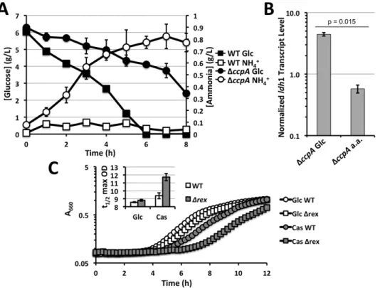
throughout this study WT andDccpAstrains were grown in media with casamino acids as carbon sources supplemented with or without 0.5% glucose (Figure S2). In media supplemented with both casamino acids and glucose, WT cells preferentially consume glucose with little ammonia production indicating limited consumption of amino acids (Figure 5A). In contrast, DccpA
mutants actively produced ammonia and consumed less glucose compared to WT. Thus, a DccpA mutant primarily consumes amino acids in this defined medium whereas WT preferentially performs glycolysis. When ‘‘forcing’’ the DccpAmutant to utilize glucose by growing in defined medium with no casamino acids,
ldh1 was expressed eight-fold higher compared to growth on casamino acids alone (Figure 5B). This increased expression was evident despite poor growth rates ofDccpAon glucose (Figure S2). These results indicate that utilization of glucose promotes Ldh1 expression independently of CcpA. Furthermore, the deficiency in glucose utilization observed in theDccpAmutant can explain the apparent dependency of Ldh1 expression on CcpA.
The Glucose-requirement for Maximal Ldh1 Activation Prevents Carbon Loss during Gluconeogenesis
Lactate production via Ldh1 provides redox balance by re-oxidizing NADH produced during glycolytic conversion of
carbo-hydrates to pyruvate. However, when carbocarbo-hydrates are scarce,S. aureus can utilize amino acids, lactate, pyruvate and other gluconeogenic substrates for carbon and/or energy. Under these conditions, high level Ldh activity would be detrimental since pyruvate pools would need to be converted to phosphoenopyruvate (PEP) via oxaloacetate for gluconeogenesis. High Ldh activity would effectively compete for available pyruvate, diminishing flux through gluconeogenesis. The Drex mutant expresses high Ldh1 activity irrespective of the presence/absence of glucose (Figure 2A). Likewise, Drex mutants have difficulty growing in media with pyruvate or casamino acids as a sole carbon/energy sources (Figure 5C). That is,Drexmutants exhibit an extended lag phase (,2.5 h) only on gluconeogenic carbon sources such as amino acids and pyruvate. Thus, the conserved dependence on glucose catabolism in bacteria for maximal Ldh activity, either transcrip-tionally as inS. aureus or posttranscriptionally as in many other bacteria, represents a fundamental control mechanism ensuring efficient carbon utilization in the absence of abundant carbohy-drates.
Discussion
The ability to resist NO?-mediated toxicity separates S. aureus
coccal species [6]. A major difference in theS. aureusNO?-response compared to other bacteria is the metabolic adaptations mounted by this pathogen against nitrosative stress [4,5]. Glucose has been shown to be essential forS. aureusNO?-resistance [18]. There are several possible explanations as to why glycolysis is the primary central metabolic pathway used byS. aureusunder NO?-stress. For instance, NO?may target key gluconeogenic enzymes limiting use of non-glycolytic carbon/energy sources. Alternatively, even with a full repertoire of active non-glycolytic pathways, gluconeogenic metab-olism may simply be incompatible with the effects of NO?(e.g.elicits excessive redox imbalance). On the other hand, glycolysis may be required for the induction of key metabolic enzymes in S. aureus
during growth in the presence of NO?. Here we demonstrate that, at least in the case of ldh1, glucose stimulates its expression and is required for full Ldh1 activity. Given that Ldh1 is the primary redox-balancing enzyme for S. aureus during periods of diminished respiratory activity, reduced ldh1 transcription in the absence of glucose may partially account for inability ofS. aureusto thrive under NO?-stress on gluconeogenic carbon/energy sources.
S. aureusacquiredldh1 after emerging evolutionarily from other staphylococci given that the allele can only be found inS. aureus
genomes where it seems to be universally present. Transcript levels ofldh1 are kept virtually undetectable in respiring cells by the direct binding of the Rex repressor to two Rex-sites within Pldh1(Figure 2). Thus, Ldh1 activity is only detected in redox-stressed cells (increased NADH levels). Here we show, as others have suggested [12], that maximal Ldh1 activity also relies on the presence of glucose. It is interesting that of all the tested Rex-regulated genes, only ldh1 appears to be under glucose/CcpA control (Figure 1B). This creates
a scenario whereby in the presence of glucose,S. aureuspreferentially balances redox by the production of L-lactate. This is not the case in other staphylococcal species, which produce commensurate levels of D- and L-lactate anaerobically [6]. The ‘‘enantiomer preference’’ of
S. aureusmay stem from the fact that L-lactate alone can be utilized by lactate-quinone oxidoreductase (Lqo) [18]. Thus, L-lactate may represent more of a metabolic intermediate than D-lactate, which is primarily a metabolic endproduct. However, the true selective advantage of L-lactate production over D-lactate inS. aureusis still unknown.
The fact that glucose-stimulation of Ldh1 expression required the presence of Rex implies that glucose somehow modulates that ability of Rex to repressldh1 transcription. While theDccpAmutant produced marginally more rex transcript (,50% increase over WT), the impaired redox balance of the DccpA mutant (high NADH levels) would predict that the excess Rex would be less active in these cells (Figure 3). However, Ldh1 activity is barely detectible in non-glucose grown cells orDccpAmutants grown even in the presence of glucose. Furthermore, given that the entire Rex regulon did not exhibit glucose-dependent induction implies that the glucose affect on Rex is specific for pldh1. This does not exclude
the possibility that the Rex has higher affinity for sites at the promoters forddh,adhE, andald1 and therefore the glucose effect is masked. However, the higher NADH levels in cells cultured on amino acid carbon sources predict that all Rex regulated genes should be derepressed on amino acids. Indeed,ald1 did exhibit a significant.2-fold induction on amino acid media compared to glucose consistent with NADH-mediated Rex inactivation (Figure 3B). The fact thatldh1 expression behaves in an opposing Figure 3. Alteration of Rex levels and/or activity cannot explain the Rex-dependency of ldh1 glucose-induction. A.Q RT-PCR analyses ofrextranscript levels normalized to those ofrpoDin WT versusDccpA S. aureusstrain COL following NO?stimulation.B.Q RT-PCR analyses of other Rex-regulated genes normalized torpoDupon NO?-stimulation. Onlyldh1 exhibits CcpA-dependent activation.C.Redox status depicted as NAD+
/ NADH ratios of WT versusDccpA S. aureusCOL prior to and after stimulation with NO?.
trend (lower expression on amino acids leading to higher NADH-levels) implies a separate form of regulation. The requirement for Rex to observe glucose-stimulated induction also eliminates the role for a ‘‘traditional’’ activator driving ldh1 transcription since that would predict a drop in Ldh1 expression in theDrexmutant grown in the absence of glucose. Rather, the data suggest that a glucose-responsive regulator is acting as an anti-repressor limiting the ability of Rex to blockldh1 transcription (Figure S1). A CRE consensus site ,30 bp upstream of the promoter distal Rex site seemed to be in a prime position to allow CcpA to serve as an effective anti-repressor against Rex. However, despite the com-plete conservation of the Pldh1CRE with the published consensus fromB. subtilis[20], CcpA has no affinity for Pldh1eitherin vitroor
in vivo (Figure 4). This may result from the presence of a T-A basepair at position 7 of the pldh1CRE (Figure 4). This position is never occupied by a T in high-affinity CcpA binding sites and only is present in only 3% of low-affinity sites in B. subtilis[20]. The pldh1CRE is completely conserved among all available sequences (data not shown). Alternatively, the CRE consensus in S. aureus
may be significantly divergent from that of Bacillus spp. More investigation into the sequences of theS. aureusCRE required for CcpA binding will explain these curious results.
The fact that theDccpAmutant exhibits altered carbon source utilization preferentially oxidizing amino acids over glucose (Figure 5), and the indirect reduction ofldh1 transcription in the DccpA background suggest that Pldh1 actually responds to the performance of glycolysis (Figure S1). This glycolysis-stimulated Ldh1 expression theory is further supported by the enhanced transcription ofldh1 in theDccpAmutant grown in glucose relative to amino acids despite the poor growth ofDccpAon glucose alone (Figures 5 and S2). Carbohydrate utilization would result in reduced pH whereas peptide catabolism would raise local pH given the excessive ammonia production. Perhaps Pldh1responds to a drop in intracellular pH thereby being indirectly affected by glycolysis. Alternatively, organic acid production by glycolytic fermentation may triggerldh1 transcription. CidR is a regulator known to respond to the presence of very short-chain organic acids (e.g.acetate and lactate), however theDcidRmutant exhibited no reported alterations inldh1 transcription [21]. Thus the mecha-nism behind enhanced Ldh1 expression in the presence of glucose is still unknown. However, whatever the mechanism, glucose stimulatedldh1 transcription inS. aureus will mimic species that express fructose bisphosphate activated Ldh enzymes in that maximal activity will only be present in cells that are actively Figure 4. CcpA affect at Pldh1is indirect. A.EMSA with His-tagged CcpA using Pldh1(LEFT) or ProcD2(RIGHT) as probes and an internalhmp fragment as a non-specific probe (N.S. band). Only at highest ratios of CcpA::DNA did non-specific shifted bands become evident using Pldh1as a probe (white arrows). 250 fmol of DNA probes were used in all wells.B.Alignment of CRE from Pldh1, ProcD2, theB. subtilisconsensus sequence and the mutated CRE*.C.Q RT-PCR analyses ofldh1 transcript levels normalized to those ofrpoDin WT,DccpAand CRE*derivatives ofS. aureusstrain COL following 15 min. NO?exposure (2 mM DEA-NO).
performing glycolysis. This level of regulation is necessary to conserve carbon during growth in the absence of glucose (Figure 5). Thus it would seem evolutionarily advantageous to limit high Ldh activity when grow under low carbohydrate conditions. Many bacteria have evolved allosteric control of Ldh to achieve this regulation [13].S. aureusappears to have evolved to transcription-ally modulate Ldh activity to achieve the same level of control.
Supporting Information
Figure S1 Model of S. aureus glucose-dependent ldh1 regulation. NO? blocks respiration leading to a buildup of NADH (low NAD+
/NADH ratios), which diminishes Rex DNA binding activity leading to derepression ofldh1. The presence of glucose also diminishes the repressive activity of Rex by an unidentified mechanism. CcpA acts to direct S. aureus to preferentially utilize glycolytic carbon sources therefor maximizing the glycolytic effect on Rex-repression. Utilization of glycolytic
carbon sources leads to increased steady-state levels of fructose 1,6,-bisphosphate (FBP), which signals the phosphorylation of HPr on a conserved Ser residue. HPr-PO4acts as a co-activator with CcpA.
(TIF)
Figure S2 Growth defect ofDccpA S. aureusCOL when ‘‘forced’’ to use glucose as a primary carbon/energy source.Bacteria were cultivated in chemically defined medium with either 0.5% glucose (Glc), 0.5% casamino acid (a.a.) or the combination (both at 0.5%, Glc/a.a.) as primary carbon/energy sources.
(TIF)
Author Contributions
Conceived and designed the experiments: ARR AKC JRF MWO. Performed the experiments: AKC JRF SET NPV MWO. Analyzed the data: ARR JRF AKC MWO. Wrote the paper: ARR.
References
1. Lowy FD (1998) Staphylococcus aureus infections. N Engl J Med 339: 520–532. doi:10.1056/NEJM199808203390806.
2. Lambert M (2011) IDSA guidelines on the treatment of MRSA infections in adults and children. Am Fam Physician 84: 455–463.
3. Foster TJ (2005) Immune evasion by staphylococci. Nat Rev Microbiol 3: 948– 958. doi:10.1038/nrmicro1289.
4. Richardson AR, Dunman PM, Fang FC (2006) The nitrosative stress response of Staphylococcus aureus is required for resistance to innate immunity. Mol Microbiol 61: 927–939. doi:10.1111/j.1365-2958.2006.05290.x.
5. Hochgra¨fe F, Wolf C, Fuchs S, Liebeke M, Lalk M, et al. (2008) Nitric oxide stress induces different responses but mediates comparable protein thiol
protection in Bacillus subtilis and Staphylococcus aureus. J Bacteriol 190: 4997–5008. doi:10.1128/JB.01846-07.
6. Richardson AR, Libby SJ, Fang FC (2008) A nitric oxide-inducible lactate dehydrogenase enables Staphylococcus aureus to resist innate immunity. Science 319: 1672–1676. doi:10.1126/science.1155207.
7. Richardson AR, Payne EC, Younger N, Karlinsey JE, Thomas VC, et al. (2011) Multiple targets of nitric oxide in the tricarboxylic acid cycle of Salmonella enterica serovar typhimurium. Cell Host Microbe 10: 33–43. doi:10.1016/ j.chom.2011.06.004.
8. Toledo JC, Augusto O (2012) Connecting the chemical and biological properties of nitric oxide. Chem Res Toxicol 25: 975–989. doi:10.1021/tx300042g.
Figure 5. Performing glycolysis stimulatesldh1 expression. A.Metabolite analyses of WTS. aureusCOL and an isogenicDccpAmutant grown in defined medium devoid of ammonia with both glucose (0.5%) and casamino acids (0.5%) as carbon sources. Glucose utilization is delayed and ammonia production is enhanced in theDccpAmutant implying altered carbon source preference.B.Q RT-PCR analyses ofldh1 transcript levels normalized to those ofrpoDinDccpA S. aureusCOL following 15 min NO?-exposure (2 mM DEA-NO) cultivated in defined medium with indicated carbon source.C.Representative growth curve demonstrating the defect of aDrexmutant compared to isogenic WTS. aureusCOL when utilizing gluconeogenic carbon sources. Inset: average time toKmaximum optical density as a metric for length of lag phase from four independent curves.
9. Brown GC, McBride AG, Fox EJ, McNaught KS, Borutaite V (1997) Nitric oxide and oxygen metabolism. Biochem Soc Trans 25: 901–904.
10. Pagels M, Fuchs S, Pane´-Farre´ J, Kohler C, Menschner L, et al. (2010) Redox sensing by a Rex-family repressor is involved in the regulation of anaerobic gene expression in Staphylococcus aureus. Mol Microbiol 76: 1142–1161. doi:10.1111/j.1365-2958.2010.07105.x.
11. Wang E, Bauer MC, Rogstam A, Linse S, Logan DT, et al. (2008) Structure and functional properties of the Bacillus subtilis transcriptional repressor Rex. Mol Microbiol 69: 466–478. doi:10.1111/j.1365-2958.2008.06295.x.
12. Seidl K, Stucki M, Ruegg M, Goerke C, Wolz C, et al. (2006) Staphylococcus aureus CcpA affects virulence determinant production and antibiotic resistance. Antimicrob Agents Chemother 50: 1183–1194. doi:10.1128/AAC.50.4.1183-1194.2006.
13. Garvie EI (1980) Bacterial lactate dehydrogenases. Microbiol Rev 44: 106–139. 14. Arai K, Ichikawa J, Nonaka S, Miyanaga A, Uchikoba H, et al. (2011) A molecular design that stabilizes active state in bacterial allosteric L-lactate dehydrogenases. J Biochem 150: 579–591. doi:10.1093/jb/mvr100. 15. Titgemeyer F, Hillen W (2002) Global control of sugar metabolism: a
gram-positive solution. Antonie Van Leeuwenhoek 82: 59–71.
16. Li C, Sun F, Cho H, Yelavarthi V, Sohn C, et al. (2010) CcpA mediates proline auxotrophy and is required for Staphylococcus aureus pathogenesis. J Bacteriol 192: 3883–3892. doi:10.1128/JB.00237-10.
17. Seidl K, Mu¨ller S, Francois P, Kriebitzsch C, Schrenzel J, et al. (2009) Effect of a glucose impulse on the CcpA regulon in Staphylococcus aureus. BMC Microbiol 9: 95. doi:10.1186/1471-2180-9-95.
18. Fuller JR, Vitko NP, Perkowski EF, Scott E, Khatri D, et al. (2011) Identification of a Lactate-Quinone Oxidoreductase in Staphylococcus aureus that is Essential for Virulence. Front Cell Infect Microbiol 1: 19. doi:10.3389/fcimb.2011.00019. 19. Makhlin J, Kofman T, Borovok I, Kohler C, Engelmann S, et al. (2007) Staphylococcus aureus ArcR controls expression of the arginine deiminase operon. J Bacteriol 189: 5976–5986. doi:10.1128/JB.00592-07.
20. Marciniak BC, Pabijaniak M, de Jong A, Du˝hring R, Seidel G, et al. (2012) High- and low-affinity cre boxes for CcpA binding in Bacillus subtilis revealed by genome-wide analysis. BMC Genomics 13: 401. doi:10.1186/1471-2164-13-401.
21. Yang S-J, Dunman PM, Projan SJ, Bayles KW (2006) Characterization of the Staphylococcus aureus CidR regulon: elucidation of a novel role for acetoin metabolism in cell death and lysis. Mol Microbiol 60: 458–468. doi:10.1111/ j.1365-2958.2006.05105.x.
22. Kreiswirth BN, Lo¨fdahl S, Betley MJ, O’Reilly M, Schlievert PM, et al. (1983) The toxic shock syndrome exotoxin structural gene is not detectably transmitted by a prophage. Nature 305: 709–712.
23. Duthie ES, Lorenz LL (1952) Staphylococcal coagulase; mode of action and antigenicity. J Gen Microbiol 6: 95–107.
24. Dyke KG (1969) Penicillinase production and intrinsic resistance to penicillins in methicillin-resistant cultures of Staphylococcus aureus. J Med Microbiol 2: 261– 278.
25. Sabath LD, Wallace SJ, Gerstein DA (1972) Suppression of intrinsic resistance to methicillin and other penicillins in Staphylococcus aureus. Antimicrob Agents Chemother 2: 350–355.