Histophilus somni
Stimulates Expression of
Antiviral Proteins and Inhibits BRSV
Replication in Bovine Respiratory Epithelial
Cells
C. Lin1, J. T. Agnes1, N. Behrens2, M. Shao2†, Y. Tagawa3, L. J. Gershwin2, L. B. Corbeil1,4*
1Department of Pathology, University of California San Diego, San Diego, CA, United States of America,
2Department of Pathology, Microbiology, and Immunology, School of Veterinary Medicine, University of California Davis, Davis, CA, United States of America,3Bacterial and Parasitic Diseases Research Division, National Institute of Animal Health, NARO, Tsukuba, Ibaraki, Japan,4Department of Population Health and Reproduction, School of Veterinary Medicine, University of California Davis, Davis, CA, United States of America
†Deceased. *[email protected]
Abstract
Our previous studies showed that bovine respiratory syncytial virus (BRSV) followed by His-tophilus somnicauses more severe bovine respiratory disease and a more permeable alve-olar barrierin vitrothan either agent alone. However, microarray analysis revealed the treatment of bovine alveolar type 2 (BAT2) epithelial cells withH.somniconcentrated cul-ture supernatant (CCS) stimulated up-regulation of four antiviral protein genes as compared with BRSV infection or dual treatment. This suggested that inhibition of viral infection, rather than synergy, may occur if the bacterial infection occurred before the viral infection. Viperin (or radical S-adenosyl methionine domain containing 2—RSAD2) and ISG15 (IFN-stimu-lated gene 15—ubiquitin-like modifier) were most up-regulated. CCS dose and time course for up-regulation of viperin protein levels were determined in treated bovine turbinate (BT) upper respiratory cells and BAT2 lower respiratory cells by Western blotting. Treatment of BAT2 cells withH.somniculture supernatant before BRSV infection dramatically reduced viral replication as determined by qRT PCR, supporting the hypothesis that the bacterial infection may inhibit viral infection. Studies of the role of the two knownH.somnicytotoxins showed that viperin protein expression was induced by endotoxin (lipooligosaccharide) but not by IbpA, which mediates alveolar permeability andH.somniinvasion. A naturally occur-ring IbpA negative asymptomatic carrier strain ofH.somni(129Pt) does not cause BAT2 cell retraction or permeability of alveolar cell monolayers, so lacks virulencein vitro. To investigate initial steps of pathogenesis, we showed that strain 129Pt attached to BT cells and induced a strong viperin responsein vitro. Thus colonization of the bovine upper respi-ratory tract with an asymptomatic carrier strain lacking virulence may decrease viral infec-tion and the subsequent enhancement of bacterial respiratory infecinfec-tionin vivo.
OPEN ACCESS
Citation:Lin C, Agnes JT, Behrens N, Shao M, Tagawa Y, Gershwin LJ, et al. (2016)Histophilus somniStimulates Expression of Antiviral Proteins and Inhibits BRSV Replication in Bovine Respiratory Epithelial Cells. PLoS ONE 11(2): e0148551. doi:10.1371/journal.pone.0148551
Editor:Yung-Fu Chang, Cornell University, UNITED STATES
Received:May 7, 2015
Accepted:January 20, 2016
Published:February 9, 2016
Copyright:© 2016 Lin et al. This is an open access article distributed under the terms of theCreative Commons Attribution License, which permits unrestricted use, distribution, and reproduction in any medium, provided the original author and source are credited.
Data Availability Statement:All relevant data are within the paper.
Introduction
Human or bovine respiratory disease often has a multifactorial etiology. Interactions between viruses and bacteria are known to cause more severe disease than either one alone even though both may colonize the upper respiratory tract without causing disease [1–3]. Several studies have revealed mechanisms of viral predisposition to bacterial pneumonia [1,3]. We have inves-tigated viral bacterial interactions in bovine respiratory disease (BRD) which is caused by a variety of pathogens including the following bacteria:Mannheimia haemolytica,Pasteurella multocida,Mycoplasma bovisandHistophilus somniand viruses: bovine respiratory syncytial virus (BRSV), bovine viral diarrhea virus (BVDV), bovine parainfluenza 3 (BPIV-3) and bovine herpes virus 1 (BHV-1). Our team has reproduced experimental pneumonia in calves with eitherH.somni[4–6] or BRSV alone [7–10]. Studies of interactions of BRSV andH.
somnishowed that aerosol infection of calves with BRSV 6 days before intrabronchial inocula-tion ofH.somni, resulted in more severe pneumonia of longer duration than either pathogen alone [11]. This was associated with higher increases in specific IgE anti-H.somnilevels in the dual infection than in either single infection [11]. Since BRSV andH.somniinfect both upper respiratory and lower respiratory tract cellsin vivoandin vitro, we have used both bovine tur-binate cells (BT) and bovine alveolar type 2 (BAT2) epithelial cells for further investigation of pathogenic mechanisms. To study bacterial viral synergy, BAT2 cells were infected with BRSV 60 h before treatment withH.somniconcentrated culture supernatant (CCS) for 4 h. This resulted in increased retraction of the BAT2 cells and microarray analysis showed increased BAT2 cell expression of matrix metalloproteinase (MMP)1 and MMP3 over either treatment alone [12]. The dual treatment of BAT2 cells increased passage ofH.somniacross the alveolar cell monolayer and increased digestion of collagen IV, a major component of the alveolar base-ment membrane. Thus, dual infection facilitated invasion by the bacteria [12]. We found that the IbpA was the major factor in CCS which caused retraction of BAT2 cells [13]. IbpA consists of a surface fibrillar network that is released into culture supernatant from all strains isolated from disease and most strains from asymptomatic carriers [14,15]. However, theibpAgene was missing in four serum sensitive strains from asymptomatic preputial carriers (1P, 129Pt, 130Pf and 133P) [16]. The complete genome sequence of one of these IbpA negative asymp-tomatic carrier stains (129Pt) has been reported [17]. IbpA from disease isolates ofH.somni
has two direct repeats (DR1 and DR2), each with a cytotoxic fic motif which adenylylates Rho GTPases interfering with the cytoskeleton [18]. The motivation for the current study came from the observation that treatment of BAT2 cells withH.somniCCS as described above increased mRNA expression of four antiviral proteins over that of either BRSV or dual treated cells. Viperin (virus-inhibitory protein, endoplasmic reticulum associated, IFN-inducible) or RSAD2 (radical S-adenosyl methionine domain containing 2) and ISG15 (IFN-stimulated gene 15—ubiquitin-like modifier) were the most up-regulated antiviral genes. Therefore we hypoth-esized that release ofH.somnifactors on the surface of respiratory epithelial cells before viral infection may inhibit subsequent viral infection, the opposite of synergy. To test this hypothe-sis, we investigated up-regulation of viperin protein in BAT2 cells and BT cells as well as the role ofH.somnitoxins on increasing expression of antiviral proteins. To address antiviral func-tion, we examined the effect of CCS treatment of BAT2 cells on BRSV replication and mecha-nisms of up-regulation of antiviral genes. Lastly, adherence to BT cells was investigated becauseH.somnidoes colonize the bovine upper respiratory tractin vivo[19], does form bio-filmsin vitroas well asin vivoand adherence is a step in biofilm formation [20,21]. Sustained adherence to the epithelial surface would allow continuous release ofH.somnisecreted prod-ucts which stimulate increased expression of antiviral proteins.
Materials and Methods
Bacteria
PathogenicH.somnistrain 2336 and asymptomatic carrierH.somnistrain 129Pt, which have been previously described [4,16,17], were grown on Difco BHI agar (BD Diagnostics, Sparks, MD) plates with 5% bovine blood in Alsever's solution (Lampire Biological Laboratories, Pipersville, PA) in candle jars at 37°C. Strain 2336.A1, with theibpAgene deleted [22], was grown on BHI blood agar supplemented with Kanamycin (145μg/ml). Growth from 18 h plates was suspended in BHI broth containing 0.1% Tris base and 0.01% thiamine monophos-phate (BHITT). For studies of interaction of live bacteria with BT or BAT2 cells, the cfus of bacteria were estimated spectrophotometrically, since 75% light Transmission at 610 nm equals approximately 2 x10⁸cfu/ml. Concentrated culture supernatant (CCS) was prepared by incu-batingH.somnifor 7 h in BHITT, with an estimated starting concentration of 5 x 10⁷cfu/ml. After 7 h of incubation, (approximately late log phase) the culture supernatant was concen-trated 40 times with Amicon Ultra-15 Centrifugal Filter Units (EMD Millipore, Billerica, MA) and filtered through 0.22 um sterile syringe filters (EMD Millipore, Billerica, MA) as previously described [12].
Bovine Respiratory Syncytial Virus preparation
A virulent clinical isolate of BRSV (strain CA-1) was propagated in primary bovine turbinate (BT) cells as previously described (12). A 1ml aliquot was used for measuring the plaque form-ing units (PFU) /ml and the rest stored at -80°C in aliquots of 1ml in BT cell media containform-ing 10% horse serum and transferred to liquid nitrogen after 2–4 hours.
BAT2 and BT cell culture
Microarray analysis
BAT2 cells at 50% confluence were treated with BAT2 media alone or with BRSV at 0.5 MOI for 60 h at which time cells were almost confluent. This was followed by 4 h incubation withH.
somnistrain 2336 CCS at 20X concentration (final concentration in cell culture media) or media alone. RNA was extracted with the RNeasy minikit (Qiagen, Maryland) and gene expression was profiled using Affymetrix GeneChip bovine genome arrays, which were pro-cessed in the UC Davis School of Medicine Microarray Core Facility. The experiment was done 3 times and the data analyzed using dChip (DNA–Chip Analyszer) and DAVID (The Database for Annotation, Visualization and Integrated Discovery).
Lipooligosaccharide (LOS) preparation and quantitation
Strain 2336 LOS (also called endotoxin) was prepared by hot phenol extraction as previously reported [23,24]. The amount of endotoxin in CCS was determined with a Pierce LAL Chro-mogenic Endotoxin Quantitation Kit (Thermo Scientific, Rockford, IL) according to the manu-facturer’s instructions. 1 ug/ml LOS was considered to be equal to 20,000 EU/ml as reported by others [25]. This LOS preparation was used in assays of viperin expression by BT cells to inves-tigate the role of thisH.somnitoxin in stimulation of antiviral proteins as described in the Results section.
Western blotting
H.somniCCS, live bacteria or BT/BAT2 cell lysates boiled in SDS loading buffer (Biorad, Her-cules, CA) were loaded onto 10% SDS PAGE gels, run at 180V for 65 min, and transferred onto nitrocellulose membranes in a cold room at 30V overnight and 70V for an additional 1 h the next morning. Membranes were blocked with TBSTG (TBS + 0.05% Tween-20 + 0.3% gelatin) for 1 h. To detect viperin and GAPDH, membranes were incubated with primary antibodies to viperin, 1:100 (MAP-VIP, a generous gift from Dr. Peter Cresswell, Yale University) or mouse anti GAPDH, 1:4,000 (Life Technologies, Grand Island, NY) for 2 h at room temperature. Bac-terial antigens were detected with convalescent phase serum (calves E5/E7, 1:1, at 1:1000) or rabbit (405) antibody to IbpA DR2 at 1:1000 [13]. After washing, membranes were incubated with alkaline phosphatase labeled goat anti mouse IgG (H+L) at 1:10,000, goat anti bovine IgG (H+L) at 1:16,000 or goat anti rabbit IgG (H+L) at 1:8,000 (Kirkegaard & Perry Laboratories, KPL, Gaithersburg, MA) for 1 h and developed in NBT/BCIP (Thermo Scientific Pierce, Rock-ford, IL) for 10 min.
Bacterial adherence assay
ml by spectrophotometry and 100 ul of this bacterial suspension was added to each well of BT cells at 300 MOI. After 1.5 h incubation at 37°C, wells were washed 4 times with PBS, and fixed with 5% formalin (Sigma, St. Louis, MO) in PBS. ELISA plates were blocked with 3% gelatin in PBS with 0.02% sodium azide at 37°C overnight, incubated with convalescent phase bovine serum (calves E5+E7, 1:1) at 1:1000 dilution, followed by peroxidase labeled goat anti bovine IgG (KPL, Gaithersburg, MA) and then developed with TMB sure blue (KPL, Gaithersburg, MA). The plates were read on a dual wavelength Vmax kinetic microplate reader (Molecular Devices Corp., Menlo Park, CA) at 450/650nm. Morphology ofH.somniadherence was deter-mined by treating BT cells with live bacteria at 100 MOI in 12 well plates as above but with cells grown on coverslips in the wells. Conditions were the same as for ELISA evaluation of adherence. Coverslips were washed in the wells, fixed with 4% paraformaldehyde, stained with 0.5% crystal violet for 5 m, washed and air dried.
BRSV proliferation in BAT2 cells
Confluent BAT2 cells in 12 well plates coated with 0.1% gelatin were treated with 1X CCS final concentration in BAT2 media in appropriate wells. Control wells were treated with BAT2 media alone. Six hours post initial CCS treatment, media or CCS treated wells were infected with BRSV at a concentration of 5.0 MOI. Additional wells with and without CCS treatment were not treated with BRSV. Cells in appropriate wells were retreated with 1X CCS at 24-h intervals for all time points longer than 24-h. Cells and supernatant were harvested at 24 h, 36 h, 48 h, and 72 h.
point for the standard curve and this value was then diluted ten fold for 6 dilutions. By using the TCID50 value only live virus is measured. The Ct value for each sample for the housekeep-ing gene, GAPDH, and BRSV was then converted to a copy number ushousekeep-ing the standard curve. The copy number for BRSV was then divided by the copy number for GAPDH, to get the final value for viral shedding.
Statistical analysis
All graphs and statistical analyses by unpaired t tests were made using GraphPad Prism, ver-sion 6. P values of<0.05 () and P<0.01 () were considered significant. Data presented are
the means and standard error of the mean (SEM).
Results
Induction of antiviral genes by BRSV,
H.
somni
or dual treatment of
BAT2 cells
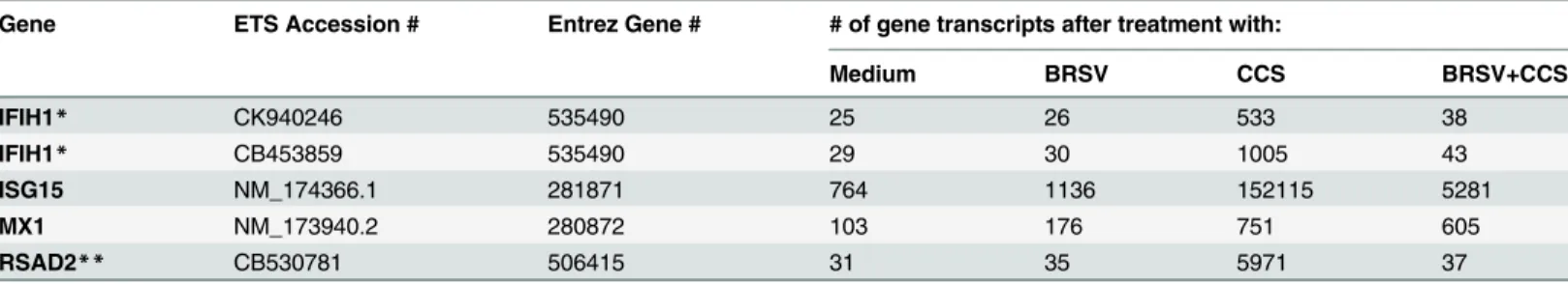
We previously showed that treatment of BAT2 cells with BRSV,H.somniCCS (20X concentra-tion) or BRSV plus CCS caused up-regulation ofmmp1andmmp3genes [12], with the dual treatment causing significantly more up-regulation than BRSV orH.somniCCS alone. There-fore, we investigated other changes in gene expression by microarray analysis of BAT2 cells after exposure to BRSV, CCS or dual treatment. We found that CCS alone induces up-regulation of mRNA transcripts for antiviral genes IFIH1 (interferon induced with helicase C domain 1), ISG15, MX1 (Myxovirus resistance 1), and RSAD2, also called viperin (Table 1). Interestingly, infection with BRSV or the dual treatment of BRSV followed by CCS up-regulated antiviral gene expression much less than CCS alone. These results were of special interest becauseH.somni
may inhibit viral infection, unlike the viral bacterial synergy which is seen when the viral infec-tion precedes the bacterial infecinfec-tion. Thus, we further investigatedH.somniup-regulation of antiviral genes with a view to defining the role of increased antiviral proteins in inhibition of virally induced disease. ISG15 and viperin gene expression were up-regulated the most by treat-ment of BAT2 cells withH.somniCCS (Fig 1). We focused on viperin for studies of up-regula-tion of antiviral protein expression byH.somniand for studies of mechanism of up-regulation.
Up-regulation of viperin protein expression in cells treated with
H.
somni
or its secreted products
The kinetics of viperin induction by CCS was determined for both bovine upper respiratory and lower respiratory cells (bovine turbinate and alveolar epithelial cells) becauseH.somnican
Table 1. Microarray analysis of BAT2 cell gene expression after treatment with BRSV and/orH.somniCCS.
Gene ETS Accession # Entrez Gene # # of gene transcripts after treatment with:
Medium BRSV CCS BRSV+CCS
IFIH1* CK940246 535490 25 26 533 38
IFIH1* CB453859 535490 29 30 1005 43
ISG15 NM_174366.1 281871 764 1136 152115 5281
MX1 NM_173940.2 280872 103 176 751 605
RSAD2** CB530781 506415 31 35 5971 37
*Two different IFIH1 ESTs from the same gene.
**RSAD2 is also called viperin. Data presented equal means calculated from three independent experiments.
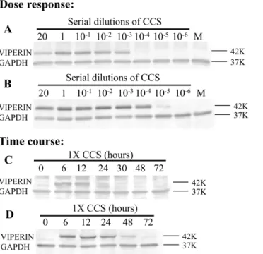
be carried in the upper respiratory tract but is found primarily in the alveolus during experimen-tal pneumonia [4] and because BRSV infects both cell types. Although functional studies and microarray studies with BAT2 cells were done by stimulating cells with CCS concentrated 20 times [12,13], dose response studies showed that this CCS could be diluted at least 1000 times before less viperin protein expression was detected in BT cells or BAT2 cells by Western blotting (Fig 2A and 2B). Therefore, subsequent studies used 1X CCS (equivalent to undiluted culture supernatant, which should be physiologic). Preliminary studies using the trypan blue exclusion method, showed that this concentration did not kill or inhibit proliferation of BT cells. The time course of viperin protein expression was then studied by stimulating cells with 1x concentrated CCS (equivalent to undiluted culture supernatant). Western blots of cell lysates showed that viperin protein expression was detectable in BT cells for at least 12 h (Fig 2C) and in BAT2 cells for at least 48 h after addition of CCS (Fig 2D). Thus,H.somniCCS up-regulates viperin protein expression in a dose dependent and time dependent manner in both BT and BAT2 cells.
Effect of antiviral proteins on BRSV replication
Up-regulation of antiviral proteins in respiratory epithelial cells should result in decreased viral load in cells treated withH.somnior its culture supernatant, if the protein is functional. We tested this hypothesis in by treating BAT2 cells withH.somniCCS before infecting with BRSV since viperin protein expression was increased for longest in that cell type. BRSV was detected by qRT PCR of BAT2 control cells by 36 hours but only at very low levels of BRSV were detected in cells treated withH.somniculture supernatant throughout the 72 h study (Fig 3). So pretreatment of BAT2 cells withH.somnisecreted products greatly depressed BRSV replication.
Fig 1.Histophilus somniCCS up-regulation of mRNA expressions of antiviral proteins.BAT2 cells were treated with medium alone orH.somnistrain 2336 CCS (concentrated 20X) for 6h, and mRNA was purified for microarray analysis. Fold increase was calculated as the mean number of transcripts for cells treated with CCS over that of control cells treated with medium alone. Means were calculated from three independent experiments. The IFIH1 value is the mean of the 2 IFIH1 EST results inTable 1. Note that four antiviral protein genes (IFIH1, ISG15, MX1 and RSAD2 or viperin) were greatly up-regulated in CCS treated cells as compared with BRSV treated cells. Significant differences were analyzed by two tailed unpaired t test (*, P<0.05;**, P<0.01, NS, not significant).
Fig 2. Dose response and time course ofH.somni2336 induction of viperin protein expression.For the dose response: BT (A) or BAT2 (B) cells were incubated with dilutions ofH.somni2336 CCS
(concentrated 20X) or with BHI medium (M). Cells were lysed at indicated time points. For the time course: BT (C) or BAT 2 (D) cells were treated withH.somni2336 1X CCS or medium (M) alone and cell lysates were collected after 6 h. In each case, cell lysates were separated by SDS PAGE and Western Blots reacted with antibody to viperin and to GAPDH. Controls with dilutions of concentrated BHI did not induce viperin expression (data not shown). Molecular weight markers are on the right.
doi:10.1371/journal.pone.0148551.g002
Fig 3. Effect ofH.somniculture supernatant on BRSV replication in BAT2 cells.Cells were treated with 1X CCS or BAT2 control medium at the time of BRSV infection (0 time) and at 24 and 48 hours post BRSV infection based on the time course of viperin expression shown inFig 2D. BRSV proliferation determined by qRT-PCR was depressed after treatment of cells with CCS. Significant differences were analyzed by one-tailed unpaired t test (*, P<0.05;**, P<0.01).
Virulence factors in
H.
somni
CCS
PathogenicH.somnistrain 2336 culture supernatant is potent in stimulating antiviral proteins expression. SinceH.somnireleases two known toxins (LOS or endotoxin and the IbpA cyto-toxin), we hypothesized that one of these factors in CCS may be stimulating viperin expression. BT cells were used in these experiments since inhibition of BRSV or other respiratory virus in the upper respiratory tract could result in fewer virus particles reaching the lung to produce pneumonia. Western blotting against convalescent phase serum showed that CCS contained the high molecular weight“ladder”of immunoglobulin binding proteins (IgBPs) as is charac-teristic of the IbpA cytotoxin as well as immunodominant 78, 60 and 40 K antigens which have not been shown to be cytotoxic (Fig 4A). Antibodies to IbpA DR2 revealed many IbpA bands from above 250 K to below 60 K, perhaps at least partly due to the multiple start codons in the IbpA gene as demonstrated earlier [27,28]. The amount of endotoxin or LOS (the second cyto-toxin) was determined by LAL assay in 3 different CCS preparations (which had been used for cytotoxicity assays in the past). Average levels of LOS in CCS were shown to be about 5.8 ug/ ml (Fig 4B). To determine whether one of the two toxins (IbpA cytotoxin or LOS endotoxin) were involved in viperin induction, we compared BT cells challenged with liveH.somni2336 and the homologous strain withibpAgene deleted (2336.A1) [22] at 10 MOI.Both the wild type (2336) and the knockout (2336.A1) induced viperin protein expression in a similar dose dependent manner, showing that the IbpA gene and protein were not necessary forH.somni
up-regulation of viperin expression (Fig 5A and 5B). To determine whetherH.somniLOS stimulated viperin, BT cells were treated with LOS extracted fromH.somnistrain 2336. Treat-ment of BT cells with serial dilutions of purified LOS from strain 2336 showed that as little as 8 ng/ml LOS induced a detectable amount of viperin as determined by Western blotting (Fig 5C). This is comparable to the amount of endotoxin in the highest dilution of CCS (10−3) which induced viperin expression (Fig 2), or 5.8 ng/ml as calculated from the LAL assay (Fig 4B). Whether other factors in CCS, such asH.somniOMPs, might contribute to up-regulation of viperin expression was not determined.
The ability of a naturally occurringibpAnegative strain 129Pt [17] to stimulate viperin expression in bovine upper respiratory epithelial cells was then determined since this strain was isolated from the epithelium of an asymptomatic carrier bull [29]. AnibpAnegative strain Fig 4. Virulence factors ofH.somnireleased into the culture supernatant.(A) Western blotting of strain 2336 CCS against convalescent phase serum (from calves E5 and E7) or rabbit antibodies to IbpA DR2 showing many high molecular weight IgBPs reacting slightly with the convalescent phase serum but strongly with anti-IbpA as well as immunodominant 78K, 60 K and 40 K antigens. Antigens are labeled on the left and molecular weight markers on the right. (B) Endotoxin levels in 3 batches of strain 2336 CCS (at 1 x
concentration) as determined by LAL assay.
was chosen because it lacks a critical virulence factor, so would not cause disease. LiveH.somni
129Pt at a low MOI (2.5) induced viperin expression, and CCS from strain 129Pt at a 1:10,000 dilution induced as strong a viperin protein expression in BT cells as strain 2336 (Fig 6A and 6Bcompared with Figs2Aand4A). These were similar results to those with the virulent strain 2336. Since colonization of the bovine upper respiratory tract with viperin inducing, non-viru-lentH.somnimay induce antiviral protein expression and attachment is one step in coloniza-tion by biofilm formacoloniza-tion, we examined attachment of strains 129Pt and 2336 to BT cellsin vitro. Both microscopy (Fig 7A, 7B and 7C) and ELISA based assays (Fig 7D) showed thatH.
somnistrain 129Pt adheres to BT cells approximately as well as the pathogenic strain 2336.
Discussion
This study shows thatH.somnilive bacteria and culture supernatant stimulate an antiviral response in the bovine upper and lower respiratory tract epithelial cells. This has implications Fig 5.Histophilus somnitoxins involved in stimulating viperin expression in BT cells.(A) Pathogenic strain 2336 and (B) the IbpA deleted strain (2336 A1) live bacteria induce viperin expression equally as shown by Western Blotting of cell lysates after 6 hrs of treatment. Therefore, IbpA is not necessary to for up-regulation of viperin. (C) PurifiedH.somnistrain 2336 lipooligosaccharide (LOS) induces viperin expression even at very low doses, showing that LOS is sufficient to up-regulate viperin expression. All compared with treatment by medium (M) alone or strain 2336 CCS as negative and positive controls, respectively. Molecular weight markers are on the right.
doi:10.1371/journal.pone.0148551.g005
Fig 6. Viperin expression in BT cells after treatments with the IbpA negative asymptomatic carrier strain ofH.somnistrain129Pt.Both CCS and live strain 129Pt induced viperin expression as determined by Western blotting. Media (M) and strain 2336 CCS were included as negative and positive controls, respectively. Molecular weight markers are on the right. Viperin induction by strain 129Pt CCS or live strain 129Pt was comparable to that of strain 2336 in Figs2and5respectively.
for host resistance since bacterial/viral interactions are critical to the etiology of this polymicro-bial disease, and respiratory epithelial cells are a first line of defense. All or some of the four up-regulated antiviral proteins in this study (IFIH1, ISG15, MX1 and RSAD/viperin) also were shown to be up-regulated in epithelial cells by cigarette smoke, or several viruses [30–34]. In our studies,H.somniup-regulated the antiviral response strongly, but BRSV infection alone up-regulated it only a little, if any. Surprisingly, BRSV, followed byH.somniCCS, down-regu-lated these antiviral genes as compared with CCS alone. This suggests that antiviral responses may be up-regulated if the bacterial infection occurs first but down regulated if the BRSV infec-tion occurs first. Although many viruses and some bacterial products have been shown to induce an antiviral response, there has been little investigation of the effect of bacterial stimula-tion of antiviral proteins on viral infecstimula-tion.
Induction of bacterial inhibition by bacterial respiratory pathogens has been demonstrated previously. We showed that some isolates of bovine nasal flora inhibitin vitrogrowth of bovine bacterial respiratory pathogens, others enhance pathogen growth while still others have no effect [35]. Other investigators found that human bacterial pathogens, such asStreptococcus pneumoniae and Haemophilus influenzae, compete with each otherin vitro and in vivo[2]. Viral predisposition to bacterial infection has been reported by our team and by others [2,11,
12]. However, bacterial influence on viral infection is less well investigated. Bosch et al [1] cite Fig 7. Adherence ofH.somnistrain 2336 and 129Pt to BT cells.Live bacteria were co-incubated with BT cells at 300 MOI in 96-well plates for 2 h, free bacteria were washed off and adherence determined by ELISA withH.somnispecific antibody. The morphology of adherence is shown in Giemsa stained BT cells on coverslips treated with 100 MOIH.somni2336, 129Pt or no bacteria. Arrows indicate adhered bacteria. Although there was variation from field to field, adherence of both strains was similar. Significant differences were analyzed by two-tailed unpaired t test (**, P<0.01, NS, not significant).
several studies that suggest a preceding bacterial infection may increase susceptibility to a sub-sequent viral infection. Our studies indicate thatH.somniinfection of respiratory epithelial cells may have the opposite effect on BRSV infection. With a qRT-PCR assay based on the dose response and time course of viperin protein expression, we showed that the antiviral response induced by secreted factors ofH.somniwas associated with decreased production of BRSV by BAT2 cells. Many studies have shown that antiviral responses initiated by many DNA and RNA viruses do inhibit viral production [36–39]. For example, replication of human RSV virus in respiratory tract cells is inhibited by viperinin vitro and in vivo[34]. Large numbers of inter-feron stimulated genes were screened recently for antiviral activity against a panel of 14 viruses [40]. Infectivity of RSV was inhibited by several ISGs. The roles of ISG15 and RSAD2in vivo
andin vitrohave been reviewed [41]. Most of thein vivoandin vitroISG studies involved sin-gle genes investigated by ectopic expression or gene deletion. Although we did not do combina-torial studies of gene networks, our studies did look at several antiviral proteins expressed together in response to bacterial stimulation. The fact that this increased expression of several antiviral genes occurred when target respiratory epithelial cells from the natural host were treated with shed factors from bacteria involved in the disease complex suggests that the results may be relevant to disease prevention. Others have shown that infection withSalmonella typhi-murium,Listeria monocytogenes,Lactobacillus acidophilusorStreptococcus agalactiaeresulted in up-regulation of viperin expression by macrophages or dendritic cells but the effect on viral replication was not tested and epithelial cells responses were not examined [42–44]. Little information is available on induction of antiviral proteins by bacteria with subsequent inhibi-tion of viral replicainhibi-tionin vitroorin vivo. Therefore, we investigated the mechanisms ofH.
somniinduction of antiviral responses, showing thatH.somniLOS but not IbpA increased viperin protein expression. Both are toxic for bovine respiratory tract epithelial and endothelial cells [13,15,45]. We expected that IbpA may be involved in the antiviral response, since Borde-tella pertussisFHA stimulates human peripheral blood mononuclear cells to up-regulate the ISG15 antiviral protein gene [46] andH.somniIbpA and FHA are quite homologous in the N terminal region. This was not the case. The C terminal portion ofH.somniIbpA, with the two fic toxic domains, has no homology to FHA and was also not associated with the antiviral response. On the other hand, very low amounts ofH.somniendotoxin (LOS) stimulated strong viperin responses in a dose dependent manner. This is consistent with other studies showing that endotoxin or lipopolysaccharide is a strong inducer of antiviral proteins, including IFIH1, ISG15, MX1 and viperin [47,48] but this has not been extended to studies ofin vitroorin vivo
inhibition of viral replication.
perhaps other respiratory viral infections. This may occur naturally in cattle carryingH.somni
in the upper respiratory tract and may partially explain individual animal differences in suscep-tibility to respiratory infection. Although biofilm formation byH.somnistrain 129Pt in the upper respiratory tract of cattle has not been tested experimentally, thein vitroresults provide hope that it may be possible to introduce a bovine asymptomatic carrier isolate ofH.somni
(like strain 129Pt) to the upper respiratory microbiome of calves by intranasal inoculation to stimulate antiviral responses and decrease susceptibility to respiratory disease. Lactobacilli are often used as probiotics in the gut, andLactobacillus acidophilusinduces strong antiviral pro-tein gene up-regulation [43]. Maybe a similar approach could be used prophylactically in the upper respiratory tract.
Acknowledgments
We thank Lauren Crum for technical assistance, Riccardo Rosenbusch for originally supplying the BAT2 cells and Peter Cresswell for the monoclonal antibodies to viperin (MAP-VIP)
Author Contributions
Conceived and designed the experiments: LBC LJG. Performed the experiments: CL JTA NB MS YT. Analyzed the data: CL JTA NB MS. Contributed reagents/materials/analysis tools: YT. Wrote the paper: CL LBC.
References
1. Bosch AA, Biesbroek G, Trzcinski K, Sanders EA, Bogaert D. Viral and bacterial interactions in the upper respiratory tract. PLoS Pathog. 2013; 9(1):e1003057. doi:10.1371/journal.ppat.1003057PMID:
23326226
2. Murphy TF, Bakaletz LO, Smeesters PR. Microbial interactions in the respiratory tract. Pediatr Infect Dis J. 2009; 28(10 Suppl):S121–6. doi:10.1097/INF.0b013e3181b6d7ecPMID:19918134 3. Caswell JL. Failure of respiratory defenses in the pathogenesis of bacterial pneumonia of cattle. Vet
Pathol. 2014; 51(2):393–409. doi:10.1177/0300985813502821PMID:24021557
4. Gogolewski RP, Leathers CW, Liggitt HD, Corbeil LB. ExperimentalHaemophilus somnuspneumonia in calves and immunoperoxidase localization of bacteria. Vet Pathol. 1987; 24(3):250–6. PMID:
3300006
5. Gogolewski RP, Schaefer DC, Wasson SK, Corbeil RR, Corbeil LB. Pulmonary persistence of Haemo-philus somnusin the presence of specific antibody. J Clin Microbiol. 1989; 27(8):1767–74. PMID:
2768464
6. Geertsema RS, Zekarias B, La Franco Scheuch L, Worby C, Russo R, Gershwin LJ, et al. IbpA DR2 subunit immunization protects calves againstHistophilus somnipneumonia. Vaccine. 2011; 29(29– 30):4805–12. doi:10.1016/j.vaccine.2011.04.075PMID:21557979
7. Kalina WV, Wollums AR, Berghaus RD, Gershwin LJ. Formalin-inactivated bovine RSV vaccine enhances a Th2 mediated immune response in infected cattle. Vaccine. 2004; 22(11–12):1465–74. PMID:15063570
8. Kalina WV, Woolums AR, Gershwin LJ. Formalin-inactivated bovine RSV vaccine influences antibody levels in bronchoalveolar lavage fluid and disease outcome in experimentally infected calves. Vaccine. 2005; 23(37):4625–30. PMID:15967545
9. Woolums AR, Gunther RA, McArthur-Vaughan K, Anderson ML, Omlor A, Boyle GA, et al. Cytotoxic T lymphocyte activity and cytokine expression in calves vaccinated with formalin-inactivated bovine respiratory syncytial virus prior to challenge. Comp Immunol Microbiol Infect Dis. 2004; 27(1):57–74. PMID:14656542
10. Schmidt U, Beyer J, Polster U, Gershwin LJ, Buchholz UJ. Mucosal immunization with live recombinant bovine respiratory syncytial virus (BRSV) and recombinant BRSV lacking the envelope glycoprotein G protects against challenge with wild-type BRSV. J Virol. 2002; 76(23):12355–9. PMID:12414977 11. Gershwin LJ, Berghaus LJ, Arnold K, Anderson ML, Corbeil LB. Immune mechanisms of pathogenetic
12. Agnes JT, Zekarias B, Shao M, Anderson ML, Gershwin LJ, Corbeil LB. Bovine respiratory syncytial virus andHistophilus somniinteraction at the alveolar barrier. Infect Immun. 2013; 81(7):2592–7. doi:
10.1128/IAI.00108-13PMID:23649093
13. Zekarias B, Mattoo S, Worby C, Lehmann J, Rosenbusch RF, Corbeil LB.Histophilus somniIbpA DR2/ Fic in virulence and immunoprotection at the natural host alveolar epithelial barrier. Infect Immun. 2010; 78(5):1850–8. doi:10.1128/IAI.01277-09PMID:20176790
14. Corbeil LB, Bastida-Corcuera FD, Beveridge TJ.Haemophilus somnusimmunoglobulin binding pro-teins and surface fibrils. Infect Immun. 1997; 65(10):4250–7. PMID:9317034
15. Zekarias B, O'Toole D, Lehmann J, Corbeil LB.Histophilus somniIbpA Fic cytotoxin is conserved in disease strains and most carrier strains from cattle, sheep and bison. Vet Microbiol. 2011; 149(1– 2):177–85. doi:10.1016/j.vetmic.2010.10.012PMID:21112704
16. Cole SP, Guiney DG, Corbeil LB. Two linked genes for outer membrane proteins are absent in four non-disease strains ofHaemophilus somnus. Mol Microbiol. 1992; 6(14):1895–902. PMID:1508038 17. Challacombe JF, Duncan AJ, Brettin TS, Bruce D, Chertkov O, Detter JC, et al. Complete genome
sequence ofHaemophilus somnus(Histophilus somni) strain 129Pt and comparison to Haemophilus ducreyi 35000HP and Haemophilus influenzae Rd. J Bacteriol. 2007; 189(5):1890–8. PMID:17172329 18. Worby CA, Mattoo S, Kruger RP, Corbeil LB, Koller A, Mendez JC, et al. The fic domain: regulation of
cell signaling by adenylylation. Mol Cell. 2009; 34(1):93–103. doi:10.1016/j.molcel.2009.03.008PMID:
19362538
19. Humphrey JD, Stephens LR.‘Haemophilus somnus’: A Review. Vet Bull 1983; 53:987–1003.
20. Sandal I, Hong W, Swords WE, Inzana TJ. Characterization and comparison of biofilm development by pathogenic and commensal isolates of Histophilus somni. J Bacteriol. 2007; 189(22):8179–85. PMID:
17644581
21. Sandal I, Shao JQ, Annadata S, Apicella MA, Boye M, Jensen TK, et al.Histophilus somnibiofilm for-mation in cardiopulmonary tissue of the bovine host following respiratory challenge. Microbes Infect. 2009; 11(2):254–63. doi:10.1016/j.micinf.2008.11.011PMID:19095078
22. Hoshinoo K, Sasaki K, Tanaka A, Corbeil LB, Tagawa Y. Virulence attributes ofHistophilus somniwith a deletion mutation in the ibpA gene. Microb Pathog. 2009; 46(5):273–82. doi:10.1016/j.micpath.2009. 02.003PMID:19269314
23. Inzana TJ, Iritani B, Gogolewski RP, Kania SA, Corbeil LB. Purification and characterization of lipooli-gosaccharides from four strains of "Haemophilus somnus". Infect Immun. 1988; 56(11):2830–7. PMID:
3169988
24. Gogolewski RP, Kania SA, Inzana TJ, Widders PR, Liggitt HD, Corbeil LB. Protective ability and speci-ficity of convalescent serum from calves withHaemophilus somnuspneumonia. Infect Immun. 1987; 55 (6):1403–11. PMID:3570472
25. Gu XX, Chen J, Barenkamp SJ, Robbins JB, Tsai CM, Lim DJ, et al. Synthesis and characterization of lipooligosaccharide-based conjugates as vaccine candidates forMoraxella(Branhamella)catarrhalis. Infection and Immunity. 1998; 66(5):1891–7. PMID:9573066
26. Sanders JD, Bastida-Corcuera FD, Arnold KF, Wunderlich AC, Corbeil LB. Genetic manipulation of immunoglobulin binding proteins ofHaemophilus somnus. Microb Pathog. 2003; 34(3):131–9. PMID:
12631474
27. Cole SP, Guiney DG, Corbeil LB. Molecular analysis of a gene encoding a serum-resistance-associ-ated 76 kDa surface antigen ofHaemophilus somnus. J Gen Microbiol. 1993; 139(9):2135–43. PMID:
8245839
28. Tagawa Y, Sanders JD, Uchida I, Bastida-Corcuera FD, Kawashima K, Corbeil LB. Genetic and func-tional analysis ofHaemophilus somnushigh molecular weight-immunoglobulin binding proteins. Microb Pathog. 2005; 39(5–6):159–70. PMID:16169703
29. Corbeil LB, Blau K, Prieur DJ, Ward AC. Serum susceptibility ofHaemophilus somnusfrom bovine clini-cal cases and carriers. J Clin Microbiol. 1985; 22(2):192–8. PMID:4031034
30. Proud D, Hudy MH, Wiehler S, Zaheer RS, Amin MA, Pelikan JB, et al. Cigarette smoke modulates expression of human rhinovirus-induced airway epithelial host defense genes. PLoS One. 2012; 7(7): e40762. doi:10.1371/journal.pone.0040762PMID:22808255
31. Zhao C, Collins MN, Hsiang TY, Krug RM. Interferon-induced ISG15 pathway: an ongoing virus-host battle. Trends in Microbiology. 2013; 21(4):181–6. doi:10.1016/j.tim.2013.01.005PMID:23414970 32. Smirnova NP, Webb BT, Bielefeldt-Ohmann H, Van Campen H, Antoniazzi AQ, Morarie SE, et al.
33. Proud D, Turner RB, Winther B, Wiehler S, Tiesman JP, Reichling TD, et al. Gene Expression Profiles during In Vivo Human Rhinovirus Infection Insights into the Host Response. American Journal of Respi-ratory and Critical Care Medicine. 2008; 178(9):962–8. doi:10.1164/rccm.200805-670OCPMID:
18658112
34. McGillivary G, Jordan ZB, Peeples ME, Bakaletz LO. Replication of respiratory syncytial virus is inhib-ited by the host defense molecule viperin. J Innate Immun. 2013; 5(1):60–71. doi:10.1159/000342473
PMID:23018837
35. Corbeil LB, Woodward W, Ward ACS, Mickelsen WD, Paisley L. Bacterial interactions in bovine respi-ratory and reproductive infections. Journal of Clinical Microbiology. 1985; 21(5):803–7. PMID:3998115 36. Seo JY, Yaneva R, Hinson ER, Cresswell P. Human cytomegalovirus directly induces the antiviral
pro-tein viperin to enhance infectivity. Science. 2011; 332(6033):1093–7. doi:10.1126/science.1202007
PMID:21527675
37. Morales DJ, Lenschow DJ. The antiviral activities of ISG15. J Mol Biol. 2013; 425(24):4995–5008. doi:
10.1016/j.jmb.2013.09.041PMID:24095857
38. Sadler AJ, Williams BRG. Interferon-inducible antiviral effectors. Nature Reviews Immunology. 2008; 8 (7):559–68. doi:10.1038/nri2314PMID:18575461
39. Verhelst J, Hulpiau P, Saelens X. Mx Proteins: Antiviral Gatekeepers That Restrain the Uninvited. Microbiology and Molecular Biology Reviews. 2013; 77(4):551–66. doi:10.1128/MMBR.00024-13
PMID:24296571
40. Schoggins JW, MacDuff DA, Imanaka N, Gainey MD, Shrestha B, Eitson JL, et al. Pan-viral specificity of IFN-induced genes reveals new roles for cGAS in innate immunity. Nature. 2014; 505(7485):691–5. doi:10.1038/nature12862PMID:24284630
41. Schoggins JW. Interferon-stimulated genes: roles in viral pathogenesis. Curr Opin Virol. 2014; 6:40–6. doi:10.1016/j.coviro.2014.03.006PMID:24713352
42. Saitoh T, Satoh T, Yamamoto N, Uematsu S, Takeuchi O, Kawai T, et al. Antiviral protein Viperin pro-motes Toll-like receptor 7- and Toll-like receptor 9-mediated type I interferon production in plasmacytoid dendritic cells. Immunity. 2011; 34(3):352–63. doi:10.1016/j.immuni.2011.03.010PMID:21435586 43. Weiss G, Rasmussen S, Zeuthen LH, Nielsen BN, Jarmer H, Jespersen L, et al.Lactobacillus
acidoph-ilusinduces virus immune defence genes in murine dendritic cells by a Toll-like receptor-2-dependent mechanism. Immunology. 2010; 131(2):268–81. doi:10.1111/j.1365-2567.2010.03301.xPMID:
20545783
44. Charrel-Dennis M, Latz E, Halmen KA, Trieu-Cuot P, Fitzgerald KA, Kasper DL, et al. TLR-Independent Type I Interferon Induction in Response to an Extracellular Bacterial Pathogen via Intracellular Recog-nition of Its DNA. Cell Host & Microbe. 2008; 4(6):543–54.
45. Sylte MJ, Corbeil LB, Inzana TJ, Czuprynski CJ.Haemophilus somnusinduces apoptosis in bovine endothelial cells in vitro. Infect Immun. 2001; 69(3):1650–60. PMID:11179340
46. Dieterich C, Relman DA. Modulation of the host interferon response and ISGylation pathway byB. per-tussisfilamentous hemagglutinin. PLoS One. 2011; 6(11):e27535. doi:10.1371/journal.pone.0027535
PMID:22140447
47. Severa M, Coccia EM, Fitzgerald KA. Toll-like receptor-dependent and -independent viperin gene expression and counter-regulation by PRDI-binding factor-1/BLIMP1. J Biol Chem. 2006; 281 (36):26188–95. PMID:16849320
48. Kandasamy S, Kerr DE. Genomic analysis of between-cow variation in dermal fibroblast response to lipopolysaccharide. Journal of Dairy Science. 2012; 95(7):3852–64. doi:10.3168/jds.2011-5251PMID: