This content has been downloaded from IOPscience. Please scroll down to see the full text.
Download details:
IP Address: 177.20.130.27
This content was downloaded on 14/06/2016 at 14:04
Please note that terms and conditions apply.
Degree of conversion of different composite resins photo-activated with light-emitting diode
and argon ion laser
View the table of contents for this issue, or go to the journal homepage for more
1. Introduction
Frequently, light-cured composite resins are prepared by the mixing of organic resin matrix with inorganic fillers. Different types of fillers such as silicon dioxide (silica, SiO2),
zirco-nium dioxide (zirconia, ZrO2) and aluminum trioxide
(alu-mina, Al2O3) of micron or submicron particle size are usually
used [1]. The organic matrix is often composed of methac-rylate resins, such as 2, 2-bis[4-(3-methacryloxy-2-hydroxy-propoxy) phenyl] propane (Bis-GMA) and triethylene glycol dimethacrylate (TEGDMA). Bis-GMA is the primary organic compound in nearly every commercial restorative composite
resin [2, 3]. Bis-GMA has become a vital monomer for den-tal restorative composites, due to its superior mechanical strength, less shrinkage, high modulus and reduced toxicity because of its lower volatility and diffusivity into the tissue. Because of the very high viscosity of Bis-GMA, TEGDMA is added to the composition in order to reduce its viscosity and to enhance filler loading and as a result, physical and mechanical properties [4, 5].
Composite resins have been classified in different ways, depending on their composition, to make it easier for dentists to identify and to use them for therapeutic purposes. A usual and very popular classification is based on filler particle size.
Degree of conversion of different composite
resins photo-activated with light-emitting
diode and argon ion laser
A M Messias1, M R Galvão1, J M C Boaventura1, D P Jacomassi2, M I B Bernardi3, V S Bagnato2, A N S Rastelli1,2 and M F Andrade1 1 University Estadual Paulista—UNESP, Araraquara, School of Dentistry, Department of Restorative
Dentistry, Araraquara, SP, Brazil
2 University of São Paulo, São Carlos Physics Institute, Optical Group, Biophotonics Lab., São Carlos,
SP, Brazil
3 University of São Paulo, São Carlos Physics Institute, Crystal Growth and Ceramic Materials Group,
São Carlos, SP, Brazil
E-mail: [email protected]
Received 24 April 2014, revised 16 November 2014 Accepted for publication 16 November 2014 Published 24 December 2014
Abstract
This study evaluated the degree of conversion (DC%) of one experimental and different brands of composite resins light-cured by two light sources (one LED and one argon laser). The percentage of unreacted C = C was determined from the ratio of absorbance intensities of aliphatic C = C (peak at 1637 cm−1) against internal standards before and after curing:
aromatic C–C (peak at 1610 cm−1) except for P90, where %C = C bonds was given for C–O–C
(883 cm−1) and C–C (1257 cm−1). ANOVA and Tukey’s test revealed no statistically significant
difference among Z350 (67.17), Z250 (69.52) and experimental (66.61 ± 2.03) with LED, just among them and Evolu-X (75.51) and P90 (32.05) that showed higher and lower DC%, respectively. For the argon laser, there were no differences among Z250 (70.67), Z350 (69.60), experimental (65.66) and Evolu-X (73, 37), however a significant difference was observed for P90 (36.80), which showed lowest DC%. The light sources showed similar DC%, however the main difference was observed regarding the composite resins. The lowest DC% was observed for the argon laser. P90 showed the lowest DC% for both light-curing sources.
Keywords: composite resins, argon ion laser, LED, photopolymerization
(Some figures may appear in colour only in the online journal)
A M Messias et al
2
The composite resins are divided into macro filler compos-ites (particles from 0.1 to 100 µm), micro filler composites (0.04 µm particles) and hybrid composites (fillers of differ-ent sizes) [6]. However, more recently, to create an universal composite resin used for both anterior and posterior teeth, a new kind of composite resin based on nanotechnology with filler particle size ranging between 5–75 nm was introduced in the market.
Nanotechnology is known as the production and manipu-lation of materials and structures with sizes ranging from approximately 0.1 to 100 nm. Much interest has been shown in research with composite resins. With the reduced size and distribution of particles, much more can be incorpo-rated with a consequent reduction of polymerization shrink-age and increase in mechanical properties such as tensile strength, compression and fracture and an adequate clinical performance. These properties appear to be similar to those of hybrid composites and microhybrids and significantly higher than the microfilled composite [7–12].
Apart from the material’s characteristics, light-curing units (LCUs) significantly influence the degree of polymerization of light-activated composite resins [13–17].
In this sense, LCUs are one part of the daily practice of restorative dentistry. Quartz–tungsten–halogen (QTH), plasma-arc (PAC), argon ion laser and light-emitting diodes (LED) are currently commercially available and may also influence the final physical properties of composite resins [18,
19]. Today, the most common LCU used to start the polym-erization process is based on blue LEDs and has the advan-tage of a narrower spectral range than the QTH light and a better match of light emitted with the absorption spectrum of the photoinitiator camphorquinone [20, 21]. Additionally, LED units do not need filters, which are required with halo-gen units for wavelength selection. Thus, LED units repre-sent an improvement over halogen lamps [22–25]. According to Aravamudhan et al [26] and Calixto et al [27], in general, there were no differences between the halogen and LED LCUs with the same parameters.
As an alternative but expensive and complex technology, argon ion lasers, have been used [28–30]. The main advantage of this LCU is the high-power density of the emitted radia-tion, which reduces the polymerization time. Additionally, the argon ion laser has a narrow wavelength band that is optimally correlated to the absorption peak for initiating the polymeriza-tion of composite resins with camphorquinone in their com-position, coherency, collimation, low beam divergence and fiber delivery capability. They have been considered a suitable light source for the polymerization of composite resins, which effectively can provide a greater degree of conversion (DC) of monomers, reduce curing time and enhance physical proper-ties of cured composites [31, 32].
The most important features associated with the effec-tiveness of light-curing seem to be the power density i.e. mW cm−2 of the light emitted, the spectral output of the light
source and the curing mode. In this way, different LCUs have been evaluated regarding their effectiveness on light-curing composite resins, but there is still some controversy in the literature [15].
The degree of conversion is one important tool to verify the polymerization efficacy and measure the percentage con-version of carbon–carbon double bonds monomeric carbonic to carbonic simple polymer [33, 34]. This process results from the replacement of power connections of Van-der-Waals, pre-existing by covalent bonds [35]. According to Araújo et al [36], different techniques can be used to assess the degree of conversion of composite resins, such as FT-IR (Fourier transform infrared spectroscopy), micro-Raman and hardness.
The literature is still unclear about the influence of the nature of the LCU used to cure different composite resins. Then, the aim of this study was to evaluate the degree of conversion (DC%) of one experimental and different brands of composite resins light-cured by one light-emitting diode (LED) and one argon ion laser light-curing source.
2. Materials and methods
2.1. Composite resins
Four brands of composite resins, FiltekTM Z250, FiltekTM
Z350 and FiltekTM P90 (3 M Espe, Dental Products, St Paul,
MN, USA) and Evolu-X® (Dentsply DeTrey, Konstanz,
Germany) at color A2 (table 1) and one experimental
nano-filled composite resin were used in this study. The main com-position can be seen in table 1.
2.2. Light-curing units (LCUs)
One blue LED (LED D-2000® DMC, São Carlos, SP, Brazil,
serial number: 002041) at 430–490 nm and one argon ion laser (Coherent, Innova 200–20 serial number 3240, USA) at 488 nm were used with a power density of 1100 mW cm−2.
First, the power output was measured using a Fieldmaster powermeter (Fieldmaster Power to Put, Coherent-model no. FM, set no. WX65, part no. 33–0506, USA) and then, the power density (mW cm−2) was calculated.
2.3. Sample preparation
The samples (n = 50) were made in a metallic mould with central orifice (4 mm in diameter and 2 mm in thickness) according to ISO 4049 [34]. The metallic mould was posi-tioned in a 10 mm thickness glass plate. The composite resin was packed in a single increment and the top and base surfaces were covered by a mylar strip. A 1 mm thickness glass sheet was positioned on the top surface and then a 1 kg weight was used to pack the composite resin. The photo-activation was performed by positioning the light tip on the top surface of the composite resin samples. The samples were irradiated for 40 s. Before making the samples with the experimental composite resin, their components were weighed on a precision balance (model BG Ltd Gehaka 440). The organic matrix was prepared by mixing bisphenol A glycol dimethacrylate (Bis-GMA) and triethylene glycol dimethacrylate (TEGDMA) that were obtained from Sigma-Aldrich Chemie GmbH, 82018 Taufkirchen, Germany)
et al
and the inorganic fillers were based on crystalline zirconia nanoparticles (Zr2O3) at a ratio of 70/30%, respectively. The
initiator system used in this study was the visible light-initi-ating system of camphorquinone (CQ) (0.5 wt%) and N, N′ -dimethyl amino-ethyl methacrylate (DMAEMA, 0.5 wt%) (6 × 10⁻⁶ molg−1) Sigma-Aldrich Chemie GmbH, 82018
Taufkirchen, Germany).
After photo-activation, the samples were stored in a dry mean at 37 °C (±1 °C) for 24 h.
2.4. Degree of conversion measurements (%DC)
After 24h, the composite resin was pulverized into fine pow-der. The pulverized composite resin was maintained in a dark room until the moment of the FT-IR analyses. Five milligrams of the ground powder was thoroughly mixed with one hundred milligrams of KBr powder salt. This mixture was placed into a pelleting device and then pressed in a press with a load of 10 tons for 1min to obtain a pellet.
To measure the degree of conversion, the pellet was then placed into a holder attachment into the spectrophotometer Nexus-470 FT-IR (Thermo Nicolet, Vernon Hills, Illinois, USA) The Fourier transform infrared spectroscopy (FTIR) spectra for both uncured and cured samples were analyzed using an accessory of the diffuse reflectance. The measure-ments were recorded in the absorbance operating under the following conditions: 32 scans, a 4 cm−1 resolution and a 300
to 4000 cm−1 wavelength. The percentage of unreacted
car-bon–carbon double bonds (%C = C) was determined from the ratio of the absorbance intensities of aliphatic C = C (peak at 1637 cm−1) against an internal standard before and after
the photoactivation of the specimen: aromatic C–C (peak at 1610 cm−1). This experiment was carried out in triplicate.
The degree of conversion was determined by subtracting the % C = C from 100%, according to the formula:
= −
− −
− −
DC (%) 1 (1637 cm / 1610 cm ) (1637 cm / 1610 cm ) .
1 1
cured
1 1
uncured
For the resin-based silorane, the percentage of unreacted carbon–carbon double bonds (%C = C) was determined from the ratio of absorbance intensities of connections between C–O–C in 883 cm−1 compared with an internal standard peak
at 1257 cm−1 [37]. The corresponding degree of conversion
was calculated by the formula:
= −
− −
− −
DC (%) 1 (883 cm / 1257 cm ) (883 cm / 1257cm ) .
1 1
cured
1 1
uncured
2.5. Statistical analysis
As data presented normal distribution and homogeneity, they were submitted to a factorial ANOVA and Tukey’s Test at a significance level of 5% (p < 0.05) considering the light-cur-ing sources and composite resins used.
The Shapiro–Wilks test was used to test the data for normality. The homogeneity of variance was tested by the Levene test.
3. Results
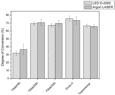
Regarding the factors evaluated in this study (composite resins and light-curing sources), composite resins showed a statistically significant effect (p < 0.001) on the degree of conversion. The two light-curing sources used did not show a statistically significant effect on the degree of conversion (p = 0.227).
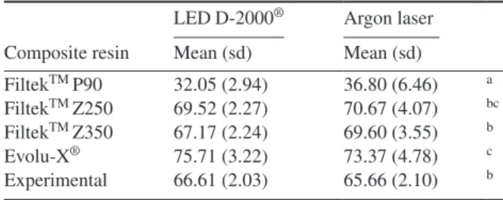
Table 2 shows the mean values and standard deviations for the degree of conversion (DC%).
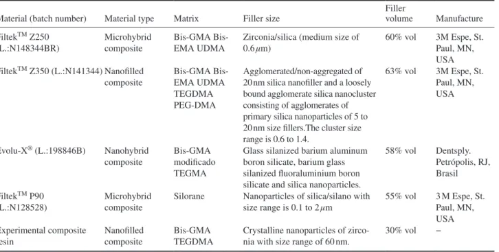
Regardless of light-curing sources (LCUs) evaluated in this study, there was a significant reduction for degree of Table 1. The main compositions of the composite resins used in this study (manufacturers’ data).
Material (batch number) Material type Matrix Filler size
Filler
volume Manufacture
FiltekTM Z250
(L.:N148344BR)
Microhybrid composite
GMA Bis-EMA UDMA
Zirconia/silica (medium size of 0.6 μm)
60% vol 3M Espe, St. Paul, MN, USA FiltekTM Z350 (L.:N141344) Nanofilled
composite GMA Bis-EMA UDMA TEGDMA PEG-DMA Agglomerated/non-aggregated of 20 nm silica nanofiller and a loosely bound agglomerate silica nanocluster consisting of agglomerates of primary silica nanoparticles of 5 to 20 nm size fillers.The cluster size range is 0.6 to 1.4.
63% vol 3M Espe, St. Paul, MN, USA
Evolu-X® (L.:198846B) Nanohybrid
composite
Bis-GMA modificado TEGMA
Glass silanized barium aluminum boron silicate, barium glass silanized fluoraluminium boron silicate and silica nanoparticles.
58% vol Dentsply.
Petrópolis, RJ, Brasil
FiltekTM P90
(L.:N128528)
Microhybrid composite
Silorane Nanoparticles of silica/silano with size range is 0.1 to 2 µm
55% vol 3 M Espe, St. Paul, MN, USA Experimental composite resin Nanofilled composite Bis-GMA TEGDMA
Crystalline nanoparticles of zirco-nia with size range of 60 nm.
30% vol −
A M Messias et al
4
conversion mean values mainly for FiltekTM P90. For this
composite resin the lowest mean values were observed, while for Evolu-X® the highest mean values were observed. These
results are displayed in figure 1.
4. Discussion
In the dental profession, there has been an increase in the use of light-cured restorative materials and hence a corresponding increase in research into the light-curing sources used to pro-mote adequate polymerization of composite resins [38]. This
in vitro study was conducted in order to compare the effective-ness of one LED and argon ion laser on the polymerization of composite resins with different filler loading and size by means of degree of conversion.
The two major components of dental composites are the polymer matrix and the filler particles. Changes in composi-tion and chemistry of the constituent monomers and filler can change their physical properties [39].
Degree of conversion, defined as the percentage of aliphatic C = C bonds converted dimethacrylate monomer present in their polymeric matrices is critical for the optimization of physical and mechanical properties [40], clinical performance, longevity and biocompatibility in order not to cause cytotoxic effects in pulp tissue, an effect attributed to the unconverted monomers that are released uncured matrix [18, 41, 42].
Ideally, the degree of conversion during the polymeriza-tion reacpolymeriza-tion, should achieve a high percentage, which would imply a full conversion of monomers into polymers [19]. However, due to residual unsaturation at the end of the reac-tion, the conversion has a final average of between 43 and 75% [18, 42–45].
Factors such as the filler particle size and refraction index, restorative material thickness, nature of polymeric matrix and the radiant exposure generated by the light polymerization mode, can influence the DC of dental composites [46].
In this sense, according to the results presented in table 2
and figure 1 there was no statistical difference in the DC (%) values between the two light-curing sources and composite resins considered, except to FiltekTM Z250. For the composite
resins based on methacrylate (FiltekTM Z250, FiltekTM Z350,
Evolu-X® and experimental) the DC% mean values ranged
from 65.66% to 75.71%. Just FiltekTM P90 did not show an
adequate degree of conversion according to other studies
previously published in the literature [18, 42–45]. FiltekTM
P90 showed the low DC% mean values for both, LED and argon ion laser LCUs used.
For an experimental nanoparticulated dental composite based on dioxide zirconia it was possible to show the arith-metic mean of the degree of conversion when photo-activa-tion with LED of 66.61 (± 2.03) and with argon ion laser of 65.66 (± 2.10), which was not statistically significant and for nanoparticulated resin FiltekTM Z350 also used in this study.
This fact can be explained by the organic composition of such resins as well as the size, volume and type of particle, which according to Knezevic et al [47], interferes with the depth of cure and scattering of incident light.
The generation of radical species for methacrylate curing is produced using a two component system consisting of cam-phoroquinone, which is the actual photoinitiator and a tertiary amine, responsible for the hydrogen transfer reaction [48]. This system decomposes immediately due to exposure of light with a wavelength between 410 and 500 nm, generating the radical species to start the polymerization process [49]. The develop-ment of a photo-activated silorane-based composite occurs with a three component initiating system comprised of camphorqui-none, iodonium salt and an electron donor. In this reaction path, the electron donor acts in a redox process and decomposes the iodonium salt into an acidic cation, which starts the ring open-ing polymerization process (1). It is beneficial to use non-coor-dinative counter-anions A—such as SbF6 or B[(C6F5)4]—to
enhance the reactivity. The 3-component system provides the optimal balance between high polymerization reactivity and light stability [48]. In the present study, all composite resins presented camphorquinone as photoinitiator in their composi-tion, except FiltekTM P90 which does not present
camphorqui-none. It is possible that the low degree of conversion mean values obtained for FiltekTM P90 can be explained by the
dif-ferences on radical species generated systems used during the polymerization process as previously related.
The resin-based silorane (FiltekTM P90) showed a lower
degree of conversion, getting around 32.05 (± 2.94%) when photo-activated with an LED and 36.80 (± 6.46%) when photo-activated with an argon laser. The differences between them were not statistically significant. These results are in agreement with Kusgoz et al [50].
Another factor to consider is described by Weinmann
et al [48]. When based methacrylate is compared to resin-based silorane, the polymerization process begins with an acid cation, which opens the oxirane ring and generates a new carbocation. Subsequently, the current spread of crosslinking, the polymerization continues. However, during this process, the acidic Si-OH groups on the particles’ released inorganic quartz can potentially result in an undesired initiation of cati-onic polymerization process. This unwanted process could increase the total amount of unreacted monomer oxirane, causing a lower degree of conversion, which can explain the results found in this study. The lower degree of conversion for this process described above also implies lower mechanical properties of the material analyzed, as shown in the results of Lien et al [51] who observed these characteristics in FiltekTM P90 composite resin.
Table 2. Mean values and standard deviations (sd) for degree of conversion, according to composite resins and the light-curing units used.
Composite resin
LED D-2000® Argon laser
Mean (sd) Mean (sd)
FiltekTM P90 32.05 (2.94) 36.80 (6.46) a
FiltekTM Z250 69.52 (2.27) 70.67 (4.07) bc
FiltekTM Z350 67.17 (2.24) 69.60 (3.55) b
Evolu-X® 75.71 (3.22) 73.37 (4.78) c
Experimental 66.61 (2.03) 65.66 (2.10) b
* Means followed by different lowercase letters indicate statistical signifi-cant difference (p < 0.05).
et al
Another paper published by Xiong et al [52] showed that the degree of conversion for FiltekTM P90 composite resin was
the lowest among the other resins based on Bis-GMA and can be explained by the reaction described above.
Regarding the LCUs used in this study, the LED light-cur-ing unit showed similar results for metahacrylate composite resins used in this study. When the argon ion laser was used, the differences among methacrylate based composite resins were just observed to FiltekTM Z250.
Some factors related to LCUs can affect the polymeriza-tion of composite resins and then the degree of conversion. The total energy delivered by LCUs remarkably influences the degree of polymerization of composite resins. However, in this study, the LCUs were used with the same final power density.
The argon ion laser has been described as a promising source for light-curing, as its wavelength is expected to be highly absorbed by the initiator present in the composition of the most of composite resins [15].
Some authors have reported that the argon ion laser can promote a greater depth and degree of polymerization inducing enhancement of the physical properties of com-posite resins after polymerization [15, 53–59]. However, the absorption peak of camphoroquinone is at approximately 470 nm and the argon ion laser works at a wavelength of 488 nm and this distance between them can make the laser activation inefficient [60–62]. This fact can explain the results obtained in our study, where the blue LED showed similar degree of conversion for all composite resins used, except for FiltekTM Z250.
Regarding the use of LEDs for composite resin curing, the technology appears to be interesting, because the internal com-ponents are very small and consequently, allow the equipment to be carried to and from the clinical office and mainly because it produces a low increase in temperature during its use [63, 64].
Under clinical conditions, it may be necessary to increase the exposure time in silorane-based composites, or use LCUs with greater irradiance than that of the LED and argon ion laser used in the present study (1100 mW cm−2) to obtain the
best results. The irradiance must be sufficient to form free radicals and form polymers in both silorane and methacrylate-based composites. In summary, silorane methacrylate-based composites are not as well polymerized as methacrylate-based composites.
5. Conclusion
Although this study was performed in vitro and thus has some limitations, the following conclusions can be drawn.
(1) The different light-curing sources promoted similar DC% values in methacrylate-based resins, however there was a great difference between them and silorane-based composites.
(2) The different composite resins showed different DC% mean values and this fact can be explained by the differ-ences in chemical composition.
Acknowledgment
This study was supported by FAPESP–Process number: 2010/08998-2 and FUNDUNESP–Process number: 00832/11.
References
[1] Chen M, Chen C, Hsu S, Sun S H and Su W 2006 Dent. Mater.22138–45
[2] Bowen R L 1962 US Patent 3066112
A M Messias et al
6 [3] Bowen R L 1962 J. Dent. Res.581493–503
[4] Sankarapandian M and Shobha H 1997 J. Mater. Sci. Mater. Med.8465–8
[5] Khosroshahi M E, Atai M and Nourbakhsh M S 2008 Lasers Med. Sci.23399–406
[6] Lutz F and Phillips R W 1983 J. Prosthet. Dent.50480–8
[7] Beun S, Glorieux T, Devaux J, Vreven J and Leloup G 2007 J. Dent. Mater.23 51–9
[8] Kroschwitz J and Howe-Grant M (ed) 1991 Kirk–Othmer Ency-clopedia of Chemical Technology 4th ed (New York: Wiley) [9] Moszner N and Klapdohr S 2004 Int. J. Nanotechnol.
1 130–56
[10] Moszner N and Salz U 2001 Prog. Polym. Sci.26535–76
[11] Mitra S D, Wu D and Holmer B N 2003 J. Am. Dent. Assoc. 1341382–90
[12] Terry D A 2004 Pract. Proc. Aest. Dent.16 417–22 [13] Rahiotis C, Kakaboura A, Loukidis M and Vougiouklakis G
2004 Eur. J. Oral Sci.11289–94
[14] Knobloch L A, Kerby R E, Clelland N and Lee J 2004 Oper. Dent.29 642–9
[15] Powell G L and Blankenau R J 2000 Dent. Clin. North Am. 44 923–30
[16] Vandewalle K S, Roberts H W, Tiba A and Charlton D G 2005 Oper. Dent.30 257–64
[17] Peris A R, Mitsui F H O, Amaral C M, Ambrosano G M B and Pimenta L A F 2005 Oper. Dent.30 649–54
[18] Costa S X S, Martins L M, Franscisconi P A S, Bagnato V S, Saad J R C, Rastelli A N S and Andrade M F 2009 Laser Phys.192210–8
[19] Rastelli A N S, Jacomassi D P and Bagnato V S 2008 Laser Phys.181570–5
[20] Leonard D L, Charlton D G, Roberts H W and Cohen M E 2002 J. Esthet. Restor. Dent.14286–95
[21] Nomura Y, Teshima W, Tanaka N, Yoshida Y, Nahara Y and Okazaki M 2002 J. Biomed. Mater. Res.63209–13
[22] Mills R W, Uhl A, Blackwell G B and Jandt K D 2002 Biomaterials232955–63
[23] Uhl A, Mills R W, Rzanny A E and Jandt K D 2005 Dent. Mater.21278–86
[24] Vandewalle K S, Roberts H W, Andrus J L and Dunn W J 2005 J. Esthet. Restor. Dent.17244–54
[25] Leonard D L 2007 J. Esthet. Restor. Dent.1956–62
[26] Aravamudhan K, Floyd C J, Rakowski D, Flaim G, Dickens S H, Eichmiller F C and Fan P L 2006 J. Am. Dent. Assoc. 137213–23
[27] Calixto L R, Lima D M, Queiroz R S, Rastelli A N S, Bagnato V S and Andrade M F 2008 Laser Phys.181365–9
[28] Vargas M A, Cobb D S and Rundle T 1998 Oper. Dent.23 87–93
[29] Blankenau R J, Kelsey W P, Powell G L, Shearer G O, Barkmeier W W and Cavel T 1991 Am. J. Dent.4 40–2 [30] Cobb D S, Vargas M A and Rundle T 1996 Am. J. Dent.
9 199–2
[31] Fleming M and Mailet W 1999 Clin. Pract.65 447–50 [32] Conrado L, Munin E and Zangaro R 2004 Lasers Med. Sci.
1995–9
[33] Ruyter I E and Svedsen S A 1979 Acta Odontol. Scand. 3675–82
[34] Lovell L G, Elliott J E, Stansburry J W and Bowman C N 2001 Dent. Mater.17504–11
[35] Saade E G, Bandeca M C, Rastelli A N S, Bagnato V S and Porto-Neto S T 2009 Laser Phys.191276–81
[36] Araújo C S A, Schein M T, Znchi C H, Rodrigues S A Jr and Demarco F F 2008 J. Contemp. Dent. Pract.9 43–50 [37] Ilie N and Hickel R 2006 Dent. Mater.25445–54
[38] Rode K M, Freitas P M, Lloret P R, Powell L G and Turbino M L 2009 Laser Med. Sci.24 87–92
[39] Mirsasaani S S, Atai M M and Hasani-Sadrabadi M M 2011 Lasers Med. Sci.26553–61
[40] Ferracane J L 1985 Dent. Mater.111–4
[41] Cook W D 1992 Polymer33600–9
[42] Emami N and Soderholm K J 2005 J. Mater. Sci. Mater. Med. 1647–52
[43] Ferracane J L and Greener E H 1986 J. Biomed. Mater. Res. 20121–31
[44] Halvorson R H, Erckson R L and Davidson C L 2003 Dent. Mater.19327–33
[45] Moraes L G, Rocha R S, Menegazzo L M, Araujo E B, Yukimito K and Moraes J C 2008 J. Appl. Oral Sci. 16145–9
[46] Silva E M, Poskus L T, Guimarães J G A, Barcellos A A L and Fellows C E 2008 J. Mater. Sci. Mater. Med.191027–32
[47] Knezević A, Tarle Z, Meniga A, Sutalo J, Pichler G and Ristić M 2001 J. Oral Rehabil.28586–91
[48] Weinmann W, Thalacher C and Guggenberger R 2005 Dent. Mater.2168–8
[49] Cook W D 1982 J. Dent. Res.611436–8
[50] Kusgoz A, Ülker M, Yesilyurt C, Yoldas O H, Ozil M and Tanriver M 2011 J. Esthet. Restor. Dent.23324–35
[51] Lien W and Vandewalle K S 2010 Dent. Mater.26337–44
[52] Xiong J, Sun X, Li Y and Chen J 2011 J. Appl. Polym. Sci. 1221882–8
[53] Blankenau R J, Kelsey W P, Powell G L, Shearer G O, Barkmeier W W and Cavel W T 1991 Am. J. Dent.4 40–2 [54] Peutzfeldt A, Sahafi A and Asmussen E 2000 Dent. Mater.
16330–6
[55] Frentzen M and Koort H J 1990 Int. Dent. J.40 323–31 [56] Docktor M H 1994 J. Esthet. Dent.677–82
[57] Cobb D S, Vargas M A and Rundle T 1996 Am. J. Dent. 9 199–202
[58] Vargas M A, Cobb D S and Schmit J L 1998 Oper. Dent. 23 87–93
[59] Verheyen P J 2001 J. Oral Laser Appl.1 49–54
[60] Rode K M, Kawano Y and Turbino M L 2007 Oper. Dent. 32571–8
[61] Burgess J O, Walker R S, Porche C J and Rappold A J 2002 Compendium23 889–906
[62] Hammesfahr P D, O’Connor M T and Wang X 2002 Comp. Contin. Educ. Dent.23 18–24
[63] Cunha L G, Sinhoreti M A C, Consani S and Correr-Sobrinho L 2003 Oper. Dent.28 155–59 [64] Dunn W J and Bush A C 2002 J. Am. Dent. Assoc.