See discussions, stats, and author profiles for this publication at: https://www.researchgate.net/publication/232724141
Compressive Strength of Dental Composite
Resins Photo-Activated with Different Light
Tips
Article in Laser Physics · April 2013
Impact Factor: 1.03 · DOI: 10.1088/1054-660X/23/4/045604
READS
326
12 authors, including:
Sergei Rabelo Caldas
Universidade Federal do Rio Grande do Norte
20PUBLICATIONS 24CITATIONS
SEE PROFILE
Saturnino Calabrez-Filho
São Paulo State University, UNIUBE
14PUBLICATIONS 19CITATIONS
SEE PROFILE
Edson Alves Campos
São Paulo State University
96PUBLICATIONS 472CITATIONS
SEE PROFILE
Marcelo Ferrarezi de Andrade
São Paulo State University
103PUBLICATIONS 423CITATIONS
SEE PROFILE
L P
Laser Phys.23(2013) 045604 (5pp) doi:10.1088/1054-660X/23/4/045604
Compressive strength of dental
composites photo-activated with different
light tips
M R Galv˜ao
1, S G F R Caldas
2, S Calabrez-Filho
3, E A Campos
1,
V S Bagnato
4, A N S Rastelli
1,4,5and M F Andrade
11Department of Restorative Dentistry, Araraquara School of Dentistry, Univ. Estadual Paulista-UNESP
Araraquara, SP, Brazil
2Department of Pediatric Dentistry, Araraquara School of Dentistry, Univ. Estadual Paulista-UNESP,
Araraquara, SP, Brazil
3Department of Dental Materials and Restorative Dentistry, University of Uberaba, Uberaba, MG, Brazil 4S˜ao Carlos Physics Institute, Optical Group, Biophotonics Lab., University of S˜ao Paulo,
S˜ao Carlos-SP, Brazil
E-mail:[email protected]
Received 31 March 2012, in final form 9 October 2012 Accepted for publication 18 October 2012
Published 21 February 2013
Online atstacks.iop.org/LP/23/045604
Abstract
The aim of this study was to evaluate the compressive strength of microhybrid
(FiltekTMZ250) and nanofilled (FiltekTMSupreme XT) composite resins photo-activated with two different light guide tips, fiber optic and polymer, coupled with one LED. The power density was 653 mW cm−2when using the fiber optic light tip and 596 mW cm−2with the polymer. After storage in distilled water at 37±2◦C for seven days, the samples were subjected to mechanical testing of compressive strength in an EMIC universal mechanical testing machine with a load cell of 5 kN and speed of 0.5 mm min−1. The statistical analysis was performed using ANOVA with a confidence interval of 95% and Tamhane’s test. The results showed that the mean values of compressive strength were not influenced by the different light tips (p>0.05). However, a statistical difference was observed (p<0.001) between the microhybrid composite resin photo-activated with the fiber optic light tip and the nanofilled composite resin. Based on these results, it can be concluded that microhybrid composite resin photo-activated with the fiber optic light tip showed better results than nanofilled, regardless of the tip used, and the type of the light tip did not influence the compressive strength of either composite. Thus, the presented results suggest that both the fiber optic and polymer light guide tips provide adequate compressive strength to be used to make restorations. However, the fiber optic light tip associated with microhybrid composite resin may be an interesting option for restorations mainly in posterior teeth.
(Some figures may appear in colour only in the online journal)
1. Introduction
Dental composite was developed by Bowen in the 1960s. Since then, this material has suffered various transformations
5 Address for correspondence: Araraquara School of Dentistry, Department of Restorative Dentistry, Univ. Estadual Paulista-UNESP, Araraquara, SP, Brazil. Humait´a St. 1680, Araraquara, SP, 14.801-903, Brazil.
to improve its physical and mechanical properties, making it increasingly acceptable for dental restorations in the posterior teeth [1,2].
Laser Phys.23(2013) 045604 M R Galv˜aoet al
Table 1. Characteristics of composite resins used in the study (manufacturers’ data).
Material Manufacturer Shade Material type Matrix Filler size
Filler
volume Lot
FiltekTMSupreme XT
3M Espe A2 Nanofilled
composite
Bis-GMA TEGDMA UDMA and Bis-EMA
Agglomerated/non-aggregated of 20 nm silica nanofiller and a loosely bound agglomeratic silica nanocluster consisting of agglomerates of primary silica nanoparticles of 20 nm size fillers.
59.5% 8BK
FiltekTMZ250 3M Espe A
2 Microhybrid
composite
Bis-GMA TEGDMA UDMA and Bis-EMA
Zirconia/silica (medium size of 0.6µm)
60% 9KK
human eye and limitations of curing depth [3]. QTH lamps are composed of a quartz tungsten thread found in the bulb, enclosed by an inert gas filter, refrigerating system and optic fibers for light conduction. Besides the heat production, another inconvenience is that the lamp, reflector and filter can degrade over time due to high operating temperatures. This effect leads to decreased effectiveness of polymerization, promoting inadequate physical properties and increased risk of premature failure of restorations [4,5].
Different light-curing units (LCUs) have been developed, with newer types of light-curing source using other curing methods such as lasers and xenon arcs. Laser and xenon arc curing units have the advantage of reduced curing times; however, these LCUs have a larger and more complicated construction, and are more costly than halogen. The use of lasers is currently more concerned with the suppression of dental hypersensitivity, soft tissue surgeries, intracanal disinfection, caries removal, and cavity preparation [6–11].
More recently, to overcome the problems inherent to halogen light, light emitting diodes (LEDs) have been used for curing resinous materials. LED units have some advantages over QTH lamps as they have lifetimes of over 10,000 h and no need for cooling systems or filters, and the thermal emission is significantly lower than that of halogen lamps with little wasted energy and minimum heat generation [4,12–14]. Technologies have been developed that enable production of the appropriate amount of light for the efficient conversion of composite resins [15–18], resulting in improved physical and chemical properties, which can be analyzed and studied by several methods, such as hardness testing and analysis of the degree of conversion and compressive strength [19–23]. The compressive strength indicates the ability demonstrated by a material to withstand vertical stress. It is known that, during the act of chewing, the forces that are transmitted to the restorations can break them or promote tooth fracture [24–27]. Some factors can influence the polymerization of composite resins such as the different LCUs, power density, and wavelength and irradiation times. Another factor affecting the polymerization process is the light guide tip used for light transmission [28–30]. Nowadays, a wide variety of commercially available light guide tips claim to fit different operative procedures related to various clinical situations. Another problem that should be pointed out is that the light
guide tips which are available for LED LCUs have a variety of diameters and are made from different materials. The light conductor system of such devices is based on a rigid tube that contains optical fibers with a vitreous inclusion, usually covered with amber glass, metal, fiber optic, or polymer. This coating is important to prevent the passage of light, especially on the lateral surface of the tip, and decrease the light scattering. Some studies have shown that the polymer tip scatters the guided light, thus reducing the power density at the end of the tip, which would have direct repercussions in the polymerization process of the composite [17,31,32].
Therefore, it is believed that the material covering the tips of the LCU can influence the values of final power density due to light scattering over its route. In this way, this study evaluated the influence of the light guide tips used in the photo-activation on the compressive strength of dental composite resins.
2. Material and methods
In this experiment two different composites were used: the universal microhybrid FiltekTM Z250 (3M ESPE Dental Products Division, St Paul, MN, USA), and the nanofilled FiltekTM Supreme XT (3M ESPE Dental Products Division, St Paul, MN, USA) (table1).
A blue LED LCU (Ultrablue IS, DMC, S˜ao Carlos, SP, Brazil, serial number 002041) with two different light guide tips (fiber optic and polymer) was used in this study. The power output was measured using a Fieldmaster power meter (Fieldmaster Power to Put, Coherent model no FM, set no WX65, part no 33-0506, USA). The values of power density (mW cm−2) were computed as the ratio of the output power and the area of the tip with the following formula:
I=P/A
wherePis the power in milliwatts,Ais the area of the light tip in squared centimeters and I is the power density. The LED LCU produced 653 mW cm−2coupled with the metal light guide tip and 596 mW cm−2with the polymeric tip. The characteristics of the light guide tips are shown in table2.
Cylindrical specimens, 4 mm in diameter and 8 mm in height, were prepared by composite insertion into a stainless steel split mold. The specimens were photo-activated for
Table 2. Characteristics of the light guide tip used in the study.
LCU
Light guide tip
Diameter entry (mm)
Diameter
exit (mm) Geometry
Ultrablue IS Fiber optic 11 8 Turbo
Polymer 10 8 Turbo
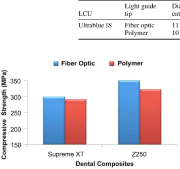
Figure 1. Mean values of compressive strength depending on the light guide tip and dental composites.
20 s (n=8). The photo-activation was carried out at every increment of 2 mm. The specimens were removed from the split mold and were photo-activated for a further 20 s on the bottom and on the four lateral surfaces. The specimens were then stored in distilled water at 37±2◦C for 7 days [33].
Following the storage period, the compressive strength test was performed employing an EMIC mechanical test machine (model DL2000, S˜ao Jos´e dos Pinhais, Brazil), with a load cell of 5 kN and programmed speed of 0.5 mm min−1. Data recording and processing for compressive strength values in megapascals were performed by the computer program Tesc.
The data were statistically analyzed by analysis of variance (ANOVA) using a confidence interval of 95% and Tamhane’s test.
3. Results
Figure 1 shows the mean values of compressive strength obtained with the different light guide tips and dental composites. The statistical test showed that the compressive strength was not influenced by the light guide tips (p >
0.05); however, some differences were observed for dental composites.
By observing the results of the nanofilled composite resin photo-activated with different tips, it was found that there were no statistically significant differences. The same can be observed for microhybrid composite resin, which was not influenced by the different tips.
Tamhane’s test indicates that significant differences were found between the microhybrid composite resin photo-activated with the fiber optic light guide and the nanofilled composite resin photo-activated with both light guide tips. In absolute values, the nanofilled composite resins
associated with the polymer tip showed the lowest values for compressive strength. However, they were statistically equivalent to the microhybrid composite resin photo-activated using the polymer light tip.
4. Discussion
The purpose of this study was to evaluate whether the type of material used in the light guide tip might have an influence on the resulting compressive strength of resin composites. For this purpose, two light guide tips (fiber optic and polymer) were used coupled to one LED LCU, and two composites (microhybrid and nanofilled).
Adequate polymerization is a crucial factor to obtain optimal physical properties and clinical performance of composite resins. Inadequate polymerization has been associated with poor physical properties, high solubility, low retention, adverse pulpal responses and low biocompatibility, which may affect the clinical performance of restorative procedures [13,34].
To compare the ability of different light guide tips coupled to an LED to cure dental composite material, suitable tests had to be chosen. Although many methods for testing the physical properties of dental composites are known, most are orientated towards comparing the properties of the different materials rather than the LCUs [35–37]. Compressive strength tests have previously been used to compare different LCUs but specifically in the present study this test was employed to compare different light guide tips [38,39].
Compressive strength has a particularly important role in the mastication process since most of the masticatory forces are of compressive nature. The maximum resistance to compression is calculated by the original cross-sectional area of the test specimen and the maximum force applied [4,40].
A clinically relevant compressive strength value may be based on the compressive strength values of natural mineralized tissues. The compressive strength of enamel has been measured to be 384 MPa. The fracture strength of natural molars however is around 305 MPa while other teeth have generally lower fracture strengths. The latter value may offer a good mechanical standard to select the optimal strength of composite resins used in posterior teeth [20,41].
Laser Phys.23(2013) 045604 M R Galv˜aoet al
Simply considering each material, no statistically significant differences in compressive strength were found between groups light cured with fiber optic or polymer light guide tips, although there was a tendency to higher compressive strength values for the fiber optic light guide tip. This can be explained by the dispersion of light in the route of the tip, which have direct impact on the final power density, and then on mechanical properties [28].
The power density from the LCU, also referred as light intensity, is the number of photons per second (watts, W) emitted by the light source per unit area (W cm−2). It has been reported that a minimum power density of 300–400 mW cm−2 is required to adequately cure one increment of 1.5–2 mm of composite resin at the manufacturers’ recommended curing time [42]. In a more effective LCU, more photons will be available for absorption by the photosensitizers. With more photons, more camphoroquinone molecules are raised to the excited state, react with the amine and form free radicals for polymerization [43–45]. This is in agreement with our study, because the power density using the fiber optic tip was 653 mW cm−2and that using the polymer was 596 mW cm−2, and as a final result the compressive strength of composite resins photo-activated with the fiber optic light tip was greater than that obtained with the polymer tip.
In the present study, the dental composites (microhybrid and nanofilled) provided an important role in polymerization. The microhybrid composite resin presented higher absolutes values of compressive strength than the nanofilled one (figure1). The literature has shown that chemical composition can influence mechanical properties [46–49]. According to Yearn [50] and Swartzet al[51], factors related to composites include shade, translucency and filler particle size, load and distribution. Mitra et al [2] believe that the composite of nanoparticles has good light transmission and presents physical and mechanical properties equivalent to those of microhybrid resin.
The resin matrix composites are an important group of materials in restorative dentistry [52,53]. Their development and formulation are based on the fact that the addition of inert fillers to acrylic and dimethacrylate resins can significantly improve certain properties. The effect of filler depends on the type, shape, size and amount used and on the existence of efficient coupling between filler and matrix resin [54–56]. Many properties (e.g. compressive strength) are improved as the filler content is increased.
Another interesting observation is that the variability of the compressive strength of composite resins photo-activated with the polymer tip was greater than with the fiber optic, as shown by the standard deviation (±SD). This can be considered an important characteristic of the tip, as it was shown to be less predictable than the fiber optic tip, which probably resulted in lower values of compressive strength.
5. Conclusions
The results obtained for this study indicate that the light guide tips did not influence the compressive strength of the dental composites. The microhybrid Filtek Z250 composite
photo-activated with a fiber optic tip showed better results than the nanofilled composite resin, mainly when the fiber optic tip was used.
Based on the results of this study it may be suggested that the fiber optic tip associated with microhybrid composite resin may be an interesting option for restorations in the posterior teeth.
Acknowledgments
This study was supported by CAPES Brazil. The authors would like thank the Physics Institute of S˜ao Carlos, University of S˜ao Paulo, S˜ao Carlos, SP, Brazil, Department of Dental Materials and the Laboratory of Mechanics Faculty of Dentistry of Araraquara Universidade Estadual Paulista ‘J´ulio de Mesquita Filho’—UNESP, Araraquara School of Dentistry, University of S˜ao Paulo, SP, Brazil, for the use of the EMIC mechanical test machine.
References
[1] St-Georges A J, Swift E J, Thompson J Y and Heymann H O 2003Dent. Mater. J.19406
[2] Mitra S B, Wu D and Holmes B N 2003J. Am. Dent. Assoc.
1341382
[3] Saade E G, Band´eca M C, Rastelli A N S, Bagnato V S and Porto-Neto S T 2009Laser Phys.191276
[4] Silva C M and Dias K R H C 2009Braz. Dent. J.2054 [5] Rueggeberg F A 1999Compend. Cont. Educ. Dent.204 [6] Jel´ınkov´a H, Dost´alov´a T, Nemec M, Koranda P, Miyagi M,
Iwai K, Shi W-Y and Matsuura Y 2007Laser Phys. Lett.
4835
[7] Malta D A M P, Kreidler M A M, Villa G E, Andrade M F, Fontana C R and Lizarelli R F Z 2007Laser Phys. Lett.
4153
[8] Marraccini T M, Bachmann L, Wigdor H A, Walsh J T Jr, Turbino M L, Stabholtz A and Zezell D M 2006Laser Phys. Lett.396
[9] Marraccini T M, Bachmann L, Wigdor H A, Walsh J T Jr, Stabholtz A and Zezell D M 2005Laser Phys. Lett.2551 [10] Clavijo V R G, Band´eca M C, Calixto L R, Nadalin M R,
Saade E G, Oliveira-junior O B and Andrade M F 2009
Laser Phys.191920
[11] Rossato D M, Band´eca M C, Saade E G, Lizarelli R F Z, Bagnato V S and Saad J R C 2009Laser Phys.192144 [12] Nomoto R 1997Dent. Mater. J.1660
[13] Rastelli A N S, Jacomassi D P and Bagnato V S 2008Laser Phys.181570
[14] Rastelli A N S, Jacomassi D P and Bagnato V S 2008Laser Phys.181003
[15] Rode K M, Kawano Y and Turbino M L 2007Oper. Dent.
32571
[16] Hammesfahr P D, O’Connor M T and Wang X 2002
Compend. Cont. Educ. Dent.2318
[17] Galv˜ao M R, Costa S X S, Victorino K R, Ribeiro A A, Menezes F C H, Rastelli A N S, Bagnato V S and Andrade M F 2010Laser Phys.201
[18] Calixto L R, Band´eca M C, Silva F B, Rastelli A N S, Porto-Neto S T and Andrade M F 2009Laser Phys.191867 [19] Aravamudhan K, Floyd C J, Rakowski D, Flaim G,
Dickens S H, Eichmiller F C and Fan P L 2006J. Am. Dent. Assoc.137213
[20] Jandt K D, Mills R W, Blackwell G B and Ashworth S H 2000
Dent. Mater. J.1641
[21] Aguiar T C, Lima D M, Calixto L R, Saad J R C,
Bandeca M C, Pinto S C S and Silva M A S 2011Polymers
3998
[22] Rastelli A N S, Jacomassi D P and Bagnato V S 2008Laser Phys.181074
[23] Band´eca M C, El-Mowafy O, Saade E G, Rastelli A N S, Bagnato V S and Porto-Neto S T 2009Laser Phys.191050 [24] Maciel D, Dias A L, Moys´es M R, Ribeiro J C R,
Dias S C and Reis A C 2005Arch. Dent.41235 [25] Baharav H, Abraham D, Cardash H S and Helft M 1988
J. Oral Rehabil.15167
[26] Oliveira F C, Denehy G E and Boyer D B 1987J. Am. Dent. Assoc.11757
[27] Roulet J F 1988J. Dent.16101
[28] Bhamra G S and Fleming G J 2008J. Dent.36643 [29] Kabbach W, Zezell D M, Band´eca M C, Pereira T M and
Andrade M F 2010Laser Phys.201833 [30] Kabbach W, Zezell D M, Band´eca M C and
Andrade M F 2010Laser Phys.201654
[31] Soares L E, Liporoni P C and Martin A A 2007Oper. Dent.
32160
[32] Corciolani G, Vichi A, Davidson C L and Ferrari M 2008
Oper. Dent.33325
[33] Brandao L, Adabo G L, Vaz L G and Saad J R 2005Braz. Oral Res.19272
[34] Queiroz R S, Band´eca M C, Calixto L R, Gaiao U, Cuin A and Porto-Neto S T 2010Laser Phys.201647
[35] Band´eca M C, Kassem A S, El-Mowafy O, Nadalin M R, Queiroz R S, Clavijo V G R and Saad J R C 2010Mater. Res.1325
[36] Queiroz R S, Band´eca M C, Calixto L R, Saade E G,
Nadalin M R, Andrade M F and Porto-Neto S T 2009Laser Phys.11909
[37] Bandeca M C, Pinto S C S, Calixto L R, Saad J R C, Barros E L R D, El-Mowafy O and Porto-Neto S T 2012
Mater. Res.151
[38] Tanoue N, Matsumura H and Atsuta M 1998J. Oral Rehab.
25358
[39] Cobb D S, Vargas M A and Rundle T 1996Am. J. Dent.9199 [40] Rueggeberg F A, Caughman W F and Curtis J W Jr 1994
Oper. Dent.1926
[41] Willems G, Lambrechts P, Braem M and Vanherle G 1993
Quint. Int.24641
[42] Price R B, Felix C A and Andreou P 2005Compend. Contin. Educ. Dent.25336
[43] Vandewalle K S, Ferracane J L, Hilton T J, Erickson R L and Sakaguchi R L 2004Dent. Mater. J.2096
[44] Silva P C G, Porto-Neto S T, Lizarelli R F Z and Bagnato V S 2008Laser Phys. Lett.5220 [45] Valentino T A, Calabrez-Filho S, Menezes F C H,
Cavalcante L M A, Pimenta L F A, Andrade M F, Dantas A A R and Rastelli A N S 2011Laser Phys.211 [46] Koupis N S, Vercruysse C W, Marks L A, Martens L C and
Verbeeck R M 2004Dent. Mater. J.20908
[47] Obici A C, Sinhoreti M A, de Goes M F, Consani S and Sobrinho L C 2002Oper. Dent.27192
[48] Soh M S, Yap A U and Siow K S 2003Oper. Dent.28707 [49] Tolosa M C, Paulillo L A, Giannini M, Santos A J and
Dias C T 2005Braz. Oral Res.19123 [50] Yearn J A 1985Int. Dent. J.35218
[51] Swartz M L, Phillips R W and Rhodes B 1983J. Am. Dent. Assoc.106634
[52] Bowen R L 1963J. Am. Dent. Assoc.6657 [53] Bowen R L 1964J. Am. Dent. Assoc.69481
[54] Costa S X S, Galv˜ao M R, Jacomassi D P, Bernardi M I B, Hernandez A C, Rastelli A N S and Andrade M F 2011
J. Thermodyn. Anal. Calorim.103219
[55] Schulze K A, Zaman A A and Soderholm K J 2003J. Dent.
31373