IRINA TATIANA MORALES CASTAÑO
SISTEMÁTICA E BIOGEOGRAFIA DO GÊNERO Collaria PROVANCHER, 1872 (HEMIPTERA: MIRIDAE)
VIÇOSA
MINAS GERAIS - BRASIL 2015
Fichi citilográfici prepiridi peli Biblioteci Centril di Universidide Federil de Viçosi - Câmpus Viçosi
T
Morales Castaño, Irina Tatiana, 1980-M828s
2015 Provancher, 1872 Sistemática e biogeografia do gênero (Hemiptera: Miridae) / Irina TatianaCollaria Morales Castaño. - Viçosa, MG, 2015.
viii, 128f. : il. (algumas color.) ; 29 cm.
Orientador : Paulo Sérgio Fiúza Ferreira.
Tese (doutorado) - Universidade Federal de Viçosa. Inclui bibliografia.
1. Percevejos (Inseto). 2. Collaria - Classificação. 3. Filogenia. I. Universidade Federal de Viçosa.
Departamento de Entomologia. Programa de Pós-graduação em Entomologia. II. Título.
CDD 22. ed. 595.754
FichaCatalografica :: Fichacatalografica https://www3.dti.ufv.br/bbt/ficha/cadastrarficha/visua...
ii
Dedico este trabalho
iii
AGRADECIMENTOS
À Universidade Federal de Viçosa, ao programa de Pós-graduação em Entomologia, pela oportunidade da realização deste curso e ao Programa Estudante Convênio de Pós-Graduação (PCPG), e à Coordenação de aperfeiçoamento de Pessoal de Nível Superior (CAPES), pela bolsa de estudos.
Ao meu orientador, o professor Paulo Sergio Fiuza Ferreira por me permitir conhecer o mundo dos mirídeos, por seus ensinamentos e por ter me aceito como orientada. Ao meu coorientador Dimitri Forero (Pontificia Universidad Javeriana, Bogotá-Colômbia), por toda a ajuda, paciência, incentivo, e pelas importantes discussões em Sistemática de Hemiptera.
Aos professores da banca pela leitura crítica e aportes ao manuscrito final.
Aos curadores das coleções visitadas: Nancy Barreto (CTNI), Rodney Cavichioli (DZUP), Carlos Sarmiento (ICN), Claudia Medina (IAVH), Luiz Costa (MNRJ), Thomas Henry (USNM), e pelo empréstimo de material: Eliane De Coninck (MRAC), e especialmente a Ruth Salas pelo envio do material do AMNH e outras coleções entomológicas (BPBM, CAS, KU, NMSA, USU, ZMUC).
Ao professor Carlos Sperber e ao pessoal do Laboratório de Orthoptera, e ao professor Jorge Dergam e ao pessoal do Laboratório de Sistemática Molecular-Beagle (Departamento de Biologia Animal-UFV), pela colaboração e empréstimo dos equipamentos utilizados durante o desenvolvimento da pesquisa.
A todos os colegas que fazem parte ou pertenceram ao Museu Regional de Entomologia da UFV, e a minhas colegas e amigas Lívia e Mayrita, muito obrigada pela companhia, colaboração e apoio.
iv SUMÁRIO
RESUMO ... vii
ABSTRACT ... viii
INTRODUÇÃO ... 1
OBJETIVOS ... 4
Geral ... 4
Específicos ... 4
MATERIAL E MÉTODOS ... 5
Consulta a coleções e determinação taxonômica ... 5
Estudo de espécimes ... 7
Estrutura da Revisão Taxonômica ... 7
Mapas de distribuição geográfica ... 7
Análise Filogenética ... 8
Análise Biogeográfica ... 8
REFERÊNCIAS BIBLIOGRÁFICAS ... 8
RESULTADOS ... 14
CAPÍTULO I ... 15
Comparative genitalic morphology in grass-feeding plant bugs of the genus Collaria Provancher, 1872 (Hemiptera, Heteroptera, Miridae) ... 15
Abstract ... 15
Introduction ... 16
Material and Methods ... 18
Material examined. ... 18
Terminology. ... 19
Dissections. ... 20
Imaging. ... 21
Results ... 21
Male genitalia ... 21
Female genitalia ... 26
Discussion ... 29
Terminology and structure of male and female genitalia... 30
Primary homology ... 32
Species groups and genitalia ... 32
Acknowledgements ... 33
References ... 33
v Revision of Collaria Provancher, 1872 (Hemiptera: Miridae) with the
description of a new species from the Afrotropical region ... 38
Abstract ... 38
Introduction ... 38
Material and Methods ... 39
Examined Specimens. ... 39
Genitalic dissections... 41
Imaging and measurements. ... 41
Terminology. ... 41
Georeferences and maps. ... 41
Genus Collaria Provancher, 1872 ... 41
Collaria boliviana Carvalho, 1990 ... 45
Collaria capixaba Carvalho & Fontes, 1981 ... 47
Collaria danae Linnavuori, 1974 ... 49
Collaria guaraniana Carvalho & Fontes, 1981 ... 50
Collaria husseyi Carvalho, 1955 ... 51
Collaria improvisa Reuter, 1893... 53
Collaria malonoi Carvalho & Carpintero, 1989 ... 55
Collaria meilleurii Provancher, 1872 ... 56
Collaria nigra Linnavuori, 1975 ... 60
Collaria obscuricornis Poppius, 1910 ... 61
Collaria oculata (Reuter, 1876) ... 63
Collaria oleosa (Distant, 1883)... 66
Collaria scenica (Stål, 1859) ... 70
Collaria schwartzi sp.n. Morales, Ferreira & Forero 2015 ... 72
Collaria villiersi Carvalho, 1953 ... 75
IDENTIFICATION KEY TO THE SPECIES OF COLLARIA ... 76
Acknowledgments ... 79
References ... 79
CAPITULO III ... 100
Phylogeny and biogeography of grass-feeding plant bugs of the genus Collaria Provancher (Hemiptera, Miridae). ... 100
Abstract ... 100
Introduction ... 101
Materials and methods ... 102
Taxa ... 102
vi
Phylogenetic analysis ... 106
Biogeographical analysis ... 106
Results ... 107
Phylogeny ... 107
Biogeography ... 110
Discussion ... 110
Phylogeny ... 110
Biogeography ... 112
Acknowledgements ... 114
References ... 114
vii
RESUMO
MORALES CASTAÑO, Irina Tatiana, D.Sc., Universidade Federal de Viçosa, Fevereiro de 2015. Sistemática e Biogeografia do gênero Collaria Provancher, 1872 (Hemiptera: Miridae). Orientador: Paulo Sergio Fiuza Ferreira. Coorientador: Igor Dimitri Forero.
viii
ABSTRACT
MORALES CASTAÑO, Irina Tatiana, D.Sc., Universidade Federal de Viçosa, February of 2015. Systematics and Biogeography of genus Collaria Provancher, 1872 (Hemiptera: Miridae). Advisor: Paulo Sergio Fiuza Ferreira. Co-advisor: Igor Dimitri Forero.
1
INTRODUÇÃO
Miridae é a maior família da subordem Heteroptera (Hemiptera) com cerca de 11.020 espécies descritas em 1200 gêneros; e apresenta uma distribuição cosmopolita, ocorrendo em todas as regiões biogeográficas do planeta (Wheeler 2001, Cassis & Schuch 2012, Jung & Lee 2012). Trata-se de uma família com grande diversidade de espécies, ampla variabilidade de habitats e hábitos alimentares. Podem ser fitófagos (atacando flores, frutos, folhas e ramos) ou predadores (obrigatórios ou facultativos) utilizados em controle biológico (Schaefer & Panizzi 2000, Wheeler 2001, Forero 2008a, Henry 2009). Algumas espécies são consideradas indicadoras de mudanças ambientais pelo fato de serem suscetíveis a certos pesticidas e reagirem a distúrbios em seus habitats (Ferreira 1999, Wheeler 2001, Henry 2009).
Representantes da família estão distribuídos em oito subfamílias: Bryocorinae, Cylapinae, Deraeocorinae, Isometopinae, Mirinae, Orthotylinae, Phylinae e Psallopinae (Schuh 1995, Schuh 2013, Cassis & Schuh 2012). Mirinae compreende aproximadamente 350 gêneros alocados em uma tribo fóssil (Scutelliferini) e seis tribos atuais: Herdoniini, Hyalopeplini, Mecistoscelini, Mirini, Resthenini e Stenodemini (Schuh 1995, Henry 2009). A tribo Stenodemini China, 1943 possui 35 gêneros e 210 espécies (Schwartz 2008). A tribo contém espécies reconhecidas pelos danos econômicos que causam nas pastagens, culturas forrageiras e de grãos (Schwartz 2008). Nesta tribo está inserido o gênero Collaria Provancher, 1872, reconhecido pelo corpo alongado, cabeça tão longa quanto larga, olhos próximos ao meio das margens laterais da cabeça, pronoto fortemente pontuado e ligeiramente estreitado atrás dos calos, e pernas longas e delgadas, revestidas por pubescência de comprimento variável (Carvalho 1945, Carvalho & Fontes 1981, Schwartz 2008).
2 Bautista et al. 2013). Barreto (2011) reportou perdas econômicas causadas por C. scenica nos sistemas de produção leiteira: 95% das fazendas da savana de Bogotá apresentam uma alta incidência e dano da praga; as perdas estão relacionadas com a redução na carga animal entre 0,2 a 2 unidades animal/ 6.400 m², e redução na produção de leite de 0.5 a 5 litros/vaca/dia. No Brasil, Menezes (1990) relatou a ocorrência de Collaria oleosa (Distant, 1883) como uma praga de gramíneas forrageiras no sudeste da Bahia; Da Silva et al. (1994) reportaram C. oleosa na cultura do trigo na região dos cerrados; Carlesii et al (1999) encontraram C. scenica sobre gramíneas nativas e cultivadas, e Ferreira et al. (2001) indicaram as espécies Collaria husseyi Carvalho,1955, C. oleosa e C. scenica como fitófagos alimentando-se principalmente de gramíneas. Wheeler (2001) reporta C. meilleurii causando danos em pastagens de Estados Unidos e C. oleosa atacando arroz, milho e trigo na América Central.
Considerando o histórico taxonômico de Collaria, sua primeira espécie foi incluída no gênero Miris, como Miris scenicus Stål, 1859. O gênero Collaria foi descrito por Provancher em 1872, juntamente com a descrição de Collaria meilleurii. O gênero Trachelomiris foi descrito por Reuter (1876) com a espécie tipo Trachelomiris oculatus; Distant (1883) descreveu a espécie T. oleosus. Mais tarde, Reuter (1905) estabeleceu Trachelomiris como sinônimo-júnior de Collaria, transferindo as espécies descritas por ele e por de Distant para este gênero. Ulher (1878) descreveu o gênero Nabidea com a espécie tipo Nabidea coracina a qual posteriormente foi transferida por Ulher (1887) para o gênero Collaria. Descrições posteriores de espécies foram feitas por Poppius (1910), que descreveu C. obscuricornis; Linnavouri (1974-1975) descreveu C. danae e C. nigra; e Carvalho (1953, 1955, 1990) descreveu as espécies C. villiersi, C. husseyi e C. boliviana.
A mais atualizada revisão do gênero foi feita por Carvalho e Fontes (1981) envolvendo as espécies do continente americano, incluindo as descrições de duas novas espécies (C. capixaba e C. guaraniana), morfologia, genitália do macho e fêmea e uma chave para identificação para as espécies neárticas e neotropicais. Carvalho & Carpintero (1989) descreveram C. manoloi e Ferreira et al. (2013a) sinonimizaram a espécie C. columbiensis com C. scenica.
3 neárticas (C. meilleurii, C. oculata) e sete Neotropicais (C. boliviana, C. capixaba, C. guaraniana, C. husseyi, Collaria malonoi Carvalho & Carpintero, 1989, C. oleosa e C. scenica (Schwartz 2008). Schwartz (2008) produziu o único trabalho que trata da filogenia de Collaria, baseado em apenas sete espécies, não sendo possível alcançar conclusões biogeográficas apropriadas a partir de informações de somente uma parte do grupo.
4
OBJETIVOS
Geral
Revisar a taxonômia do gênero Collaria (Hemiptera: Miridae) e propor hipóteses filogenéticas e biogeográficas acerca das relações internas do grupo.
Específicos
-Realizar estudos morfológicos comparativos de genitálias masculina e feminina; -Analisar e explorar novos caracteres morfológicos para definir o gênero e determinar sua utilidade filogenética;
-Testar a hipótese de monofilia do gênero Collaria e as relações filogenéticas intraespecíficas;
5
MATERIAL E MÉTODOS
Consulta a coleções e determinação taxonômica
Espécies do gênero Collaria foram obtidas através de empréstimos das seguintes 22 coleções entomológicas:
AMNH American Museum of Natural History, New York, USA. BPBM Bernice Pauahi Bishop Museum, Hawaii, USA.
CAS California Academy of Sciences, San Francisco, USA. CTNI Colección Taxonómica de Insectos “Luis María Murillo”-
CORPOICA, Tibaitatá, Colombia.
DZRS Departamento de Zoologia, Universidade Federal do Rio Grande do Sul, Porto Alegre, Brasil.
DZUP Coleção Entomologica Pe. Jesus Santiago Moure, Departmento de Zoologia, Universidade Federal de Paraná, Curitiba, Brasil. ICN Colección de Zoología – Instituto de Ciencias Naturales –
Universidad Nacional de Colombia, Bogotá, Colombia.
IAVH Colección Entomológica Instituto de Investigación de Recursos Biológicos Alexander von Humboldt, Villa de Leyva, Colombia. KU University of Kansas Natural History Museum, Lawrence, Kansas,
USA.
MCZN Museu de Ciências Naturais, Fundação Zoobotânica do Rio Grande do Sul, Porto Alegre, Brasil.
MCN Coleção Zoológica Científica do Museu de Ciências Naturais do Centro Univates, Lajeado, Brasil.
MNRJ Museu Nacional, Universidade Federal do Rio de Janeiro, Rio de Janeiro, Brasil.
MPUJ-ENT Museo de Pontificia Universidad Javeriana, Laboratorio Entomología, Bogotá, Colombia.
MRAC Royal Museum for Central Africa, Tervurem, Belgium. NMSA Natal Museum, Pietermaritzburg, South Africa.
6 UFVB Museu Regional de Entomologia, Universidade Federal de Viçosa,
Viçosa, Brasil.
UPTC Laboratorio de Entomología, Museo de Historia Natural “Luis Gonzalo Andrade” Universidad Pedagógica y Tecnológica de Colombia, Tunja, Colombia.
USU Insect Collection Utah State University, USA.
USNM National Museum of Natural History, Smithsonian Institution, Washington DC, USA.
ZMUC Zoological Museum, University of Copenhagen, Copenhagen, Denmark.
Além dos dados obtidos das etiquetas do material examinado, procurou-se na literatura dados de distribuição geográfica. Para as espécies C.meilleurii, C.oculata e C.obscuricornis,também consultou-se o catalogo on-line Plant Bug Planetary
Biodiversity Inventory Project (http://research.amnh.org/pbi/), onde se obtiveram as coordenadas geográficas de material depositado nas seguintes coleções:
CNC Canadian National Collection of Insects. Ottawa, Canadá.
OSAC Oregon State Arthropod Collection, Oregon State University, Oregon, Corvallis, USA.
NCSU North Carolina State University Insect Collection, North Dakota, Fargo, USA.
UDCC University of Delaware, Delaware, Newark,USA. UCR University of California, Riverside, USA.
MEMU Mississippi Entomological Museum, Mississippi State University, Mississippi, Mississippi, USA.
MHNCM Colección Nacional de insectos Dr. Alfredo Barrera Marín del Museo de Historia Natural de la Ciudad de México.
7 Estudo de espécimes
1080 espécimes do gênero foram estudados. As espécies foram identificadas utilizando chaves taxonômicas e descrições originais de Stål (1859), Provancher (1872), Reuter (1876), Distant (1883), Reuter (1893), Poppius (1910), Linnavuori (1974, 1975), Carvalho, (1953, 1955, 1990), Carvalho & Fontes (1981), e Carvalho & Carpintero (1989). Além disso, foram feitas comparações com exemplares tipos (síntipos/holótipos) quando disponíveis. As medidas (morfometria) das espécies foram baseadas na metodologia proposta por Forero (2008b) e Ferreira et al. (2013b).
As ilustrações das estruturas morfológicas foram feitas com o emprego de um estereomicroscópio com câmara clara acoplada. Se oferecem fotografias dos hábitos dorsais obtidas com um estereomicroscópio Zeiss Discovery V20 acoplado com uma câmera digital Zeiss AxioCam Mrc. As fotografias da genitalia masculina e feminina foram tiradas com uma câmera digital Olympus DP73 acoplada ao microscópio Olympus BX53.
Para as dignoses das genitálias masculina e feminina segui-se a metodologia proposta por Scudder & Schwartz (2012). Os espécimes foram relaxados em câmera húmida durante 12 horas em seguida procedeu-se a extração do ápice do abdome e inclusão em uma solução quente de acido lático a 85% com 10 gotas de água destilada. As disseções foram feitas em glicerina sob uma estereromicroscópio Leica MZ8, após, as estruturas dissecadas foram preservadas em microtubos com glicerina. Estrutura da Revisão Taxonômica
Para cada espécie se referencia o nome, data de autoria e o museu onde se encontra(m) depositado o(s) exemplar(es), e o histórico nomenclatural. Apresentam-se a relação do material examinado, diagnoApresentam-se, distribuição geográfica, hospedeiros, comentários (quando cabíveis) e os mapas de distribuição.
Mapas de distribuição geográfica
8 do PBI database (http://research.amnh.org/pbi/maps/) ou dos dados das etiquetas dos espécimenes examinados.
Análise Filogenética
Foi estudado um total de 20 espécies, sendo cinco pertencentes ao grupo externo: Horciasinus signoreti (Stål,1859) (Mirini), Dolichomiris linearis Reuter, 1852 (Stenodemini), Steneodema andina Carvalho, 1975 (Stenodemini) e Trigonotylus tenuis Reuter, 1893 (Stenodemini). Nabidomiris longipennis Odhiambo, 1959 (Stenodemini) é considerado por Schwartz (2008) como grupo irmão do gênero Collaria, compartilhando os caracteres: região postocular alongada como um “pescoço”, olho estreitamente unido a cabeça em vista dorsal, olho ovóide localizado na região mediana da cabeça.
Utilizaram-se 67 carateres morfológicos, incluindo aqueles propostos por Schwartz (2008). O método utilizado para estudar as relações filogenéticas do gênero Collaria foi à análise cladística. A análise de parcimônia foi feita através do programa TNT, versão 1.0. Todos os caracteres tiveram pesos iguais. As árvores mais parcimoniosas (MPT) foram obtidas pela busca heurística “Implicit Enumeration” com soluções exatas do “Bremer support” para o suporte dos clados. Foi definida a topologia da árvore de consenso estrita e verificados os índices de consistência (CI) e retenção (RI) de cada ramo.
Análise Biogeográfica
Utilizou-se o método “Spatial Analysis of Vicariance (SAV)” implementado no VIP (Vicariance Inference Program, Arias et al. 2011). Com os dados da análise filogenética e de distribuição geográfica das espécies foi feita uma busca heurística para obter a melhor reconstrução de barreiras “Voronoi lines” com uma grade de 2°
× 2° num mapa da NASA
(http://neo.sci.gsfc.nasa.gov/view.php?datasetId=MOD13A2_M_NDVI).
REFERÊNCIAS BIBLIOGRÁFICAS
9 Barreto N, Martinez E, Galindo R & D Corredor. 1996. Patrón de disposición de la chinche de los pastos Collaria columbiensis (Hemiptera: Miridae) en la Sabana de Bogotá. Revista Colombiana de Entomología 22(3): 159-162.
Barreto N. 2011. Desarrollo de un sistema de manejo y alerta temprana para la chinche de los pastos Collaria scenica Stal, en relación con la variabilidad y el cambio climático en el altiplano cundiboyacense. Corporación Colombiana de Investigación Agropecuaria Corpoica. Informe Técnico final. Bogotá, 120 p. Bautista L.G, Cardona J.A. & A.Soto. 2013. Distribuición espacial de Collaria
scenica (Hemiptera: Miridae) y Hortensia similis (Hemiptera: Cicadellidae) en valles andinos. Boletín científico Centro de Museos, Museo de Historia Natural 17 (2): 75 – 84.
Carlesii L. R. G, Corseuil E. & J.R. Salvador. 1999. Aspectos Biológicos e Morfométricos de Collaria scenica (Stal) (Hemiptera: Miridae) em Trigo. Annais da Sociedade Entomologica Brasileira 28(1) 65-73.
Carvalho J. C. 1945. Mirídeos neotropicais: Gêneros Dioniza Distant, Neella Reuter, Collaria Provancher, Falconia Distant e Ophthalmomiris Berg, com descrições de espécies novas. Revista de Entomologia 16(1-2): 158-187.
Carvalho, J. C. M. 1953. On a collection of Miridae from West Africa, with descriptions of new species (Hemiptera). Boletim do Museu Nacional (n. s.) (Zool.), Rio de Janeiro (20): 7.
Carvalho, J. C. M. 1955. Analecta Miridologica: Miscellaneous observations in some american museums and bibliography. Revista Chilena de Entomologia:(4): 221– 227.
Carvalho, J. C. M. 1959. A Catalogue of the Miridae of the world. Part IV. Arquivos do Museu Nacional. 48: 384 pp.
Carvalho, J. C. M. 1990 Mirideos neotropicais, CCCVII: novas espécies da Argentina e Bolivia (Hemiptera). Revista Brasileira de Entomologia 34: 445– 452.
10 Carvalho, J. C. M. & Carpintero D. L. 1989. Mirideos neotropicais, CCCIX: quatro espécies novas da tribo Stenodemini colecionadas na Republica Argentina (Hemiptera). Revista Brasileira de Biologia, 49: 1101–1108.
Cassis, G. & Schuh, R.T. 2012. Systematics, Biodiversity, Biogeography, and Host Associations of the Miridae (Insecta: Hemiptera: Heteroptera: Cimicomorpha). Annual Review of Entomology, 57, 377−404.
Coelho L.A. 2012. Contribuição a taxonomia e biogeografia do gênero Prepops Reuter,1905 (Hemiptera: Miridae). Tese Doutorado. Universidade Federal de Viçosa.
Da Silva D. B, Alves R. T., Ferreira P.S.F. & A.J.A Camargo. 1994. Collaria oleosa (Distant, 1883) (Heteroptera: Miridae) uma praga potencial na cultura do trigo na região dos Cerrados. Pesquisa Agropecuaria Brasileira, 29(12): 2007-2012. Distant, W. L. 1883. Insecta. Rhynchota. Hemiptera-Heteroptera. Biologia Centrali
Americana, Vol. 1, 225–264.
Forero D. 2008a. Systematics of the Hemiptera. Revista Colombiana de Entomología. 34 (1): 1-21.
Forero D. 2008b. Revison and phylogenetic analysis of the Handronema group (Miridae: Orthotylinae: Orthotylini), with descriptions of new genera and new species, and comments on the neotropical genus Tupimiris. Bulletin of the American Museum of Natural History 312, 172 pp.
Forero D. & M. D. Schwartz. 2009. Description of a new species of Aoplonema (Hemiptera: Miridae: Orthotylinae) with a biogeographic analysis andreassessment of the phylogeny of the genus. Entomologica Americana 115(1):67–76.
Ferreira P.S.F & D. Rossi. 1979. Catálogo das especies de Miridae (Hemiptera) de Viçosa, estado de Minas Gerais, Brasil. Experientae 25(7): 131-157.
Ferreira, P.S. F. 1999. Miridae. Em: Brandão C.R.F. & E.M. Cancello (Eds). Invertebrados Terrestres. Vol V. Biodiversidade do Estado do São Paulo. Síntese do conhecimento ao final do século XX. FAPESP. São Paulo. p. 93- 100.
11 Ferreira, P.S.F., Barreto-Triana N., & G. Abril. 2013a. Collaria columbiensis Carvalho, 1984, a newly recognized synonym of Collaria scenica (Stal, 1859) (Hemiptera: Heteroptera: Miridae) Zootaxa 3669 (2): 197–200.
Ferreira, P.S.F., Barreto-Triana N., & P.Osorio 2013b. New species of plant bug associated with pastures in Colombia, and notes on the genera Dolichomiris, Cynodonmiris, and Megaloceroea (Hemiptera: Heteroptera: Miridae) Zootaxa 3709(5): 473–482.
Henry, T. J. 2009. Biodiversity of the Heteroptera. In: Foottit, R. G., Adler, P. H., editors. Insect Biodiversity: Science and Society. Oxford, England: Wiley-Blackwell Publishing Ltd. p. 223-263.
Jung S. & S. Lee 2012. Molecular phylogeny of the plant bugs (Heteroptera: Miridae) and the evolution of feeding habits. Cladistics 28: 50–79.
Linnavuori, R. E., 1974. Studies on Palearctic and African Heteroptera. Acta Entomologica Fennica 30: 3-35.
Linnavuori, R. E. 1975. Hemiptera of the Sudan, with remarks on some species of the adjacent countries. 4. Miridae and Isometopidae. Annales Zoologici Fennici, 12, 1–118.
Lu, N. & L.Y. Zheng. 1998. A study of phylogenetic relationships and biogeography of the genus Arbolygus Kerzhner (Hemiptera: Miridae). Acta Zootaxonomica Sinica, 23, 179−188.
Martínez E. & N. Barreto. 1998. La chinche de los pastos Collaria scenica Stal en la Sabana de Bogotá. Ramirez N (ed). Corpoica. Santa fé de Bogotá. 66p.
Menezes, M. 1990. Collaria oleosa (Distant, 1883) (Hemiptera: Miridae), nova praga de gramíneas forrageiras nos sudeste da Bahia, Brasil. Agrotrópica 2:113-118. Poppius, B. 1910. 12. Hemiptera 4. Miridae, Anthocoridae, Termatophylidae,
Microphysidae, und Nabidae. "MDUL"In"MDNM": Wissenschaftliche Ergebnisse der schwedischen zoologischen Expedition nach dem Kilimandjaro, dem Meru und den umgebenden Massaisteppen Deutsch-Afrikas Palmqvist, Stockholm. Vol. 2, 25–60.
12 Reuter, O. M. 1893. New species of Miridae. In: Bergroth, E., Mission scientifique de M. Ch. Alluaud aux Iles Sechelles. Revue d'Entomologie, Canadien, 12, 197– 209.
Reuter, O. M. 1876. Capsinae ex America Boreali in Museo Holmiensi asservatae, descriptae. Öfversigt af Kongliga Vetenskapsakademiens Förhandlingar, 32(9), 59–92.
Stål, C. 1859. Hemiptera. Species novas descripsit. Kongliga Svenska Fregatten Eugenies resa omkring jorden, III (Zoologi, Insecter), 219–298.
Schaefer C. & A. Panizzi. 2000. Heteroptera of economic importance. CRC Press. Boca Raton USA., 828 p.
Schuh, R.T. 1974. The Orthotylinae and Phylinae (Hemiptera: Miridae) of South Africa with a phylogenetic analysis of the ant-mimetic tribes of the two subfamilies for the world. Entomologica Americana, 47, 1−332.
Schuh, R. T. 1975. The structure, distribution, and taxonomic importance of trichobothria in the Miridae (Hemiptera). American Museum Novitates. 2585: 1-26.
Schuh, R. T. 1984. Revision of the Phylinae (Hemiptera, Miridae) of the Indo-Pacific. Bulletin of the American Museum of Natural History, 177, 1−476.
Schuh, R. T. 1995. Plant bugs of the world (Insecta: Heteroptera: Miridae). Systematic Catalog, Distributions, Host List, and Bibliography. The New York Entomological Society. USA. 1329 pp.
Schuh, R.T.2006. Revision, phylogenetic, biogeographic, and host analyses of the endemic Western North American Phymatopsallus group, with the description of 9 new genera and 15 new species (Insecta: Hemiptera: Miridae: Phylinae). Bulletin of the American Museum of Natural History, 301, 115 pp.
Schuh, R.T. 2013. On-line Systematic Catalog of Plant Bugs (Insecta: Heteroptera: Miridae). Available from: http://research.amnh.org/pbi/catalog/ (accessed 21 May2014).
Schuh, R.T. & Stonedahl, G.M. 1986. Historical biogeography in the Indo-Pacific: a cladistic approach. Cladistics, 2, 337−355.
13 Schuh, R.T. 1991. Phylogenetic, host and biogeographic analysis of the Pilophorini
(Heteroptera: Miridae: Phylinae). Cladistics, 7 (2), 157−189.
Schwartz, M. D. 1987. Phylogenetic revision of the Stenodemini with a review of the Mirinae (Heteroptera: Miridae). PhD. Dissertation. City University of New York. 383 pp.
Schwartz M.D. 2008. Revision of the Stenodemini with a review of the included genera (Hemiptera: Heteroptera: Miridae: Mirinae). Proceedings of Entomological Society of Washington 110(4): 1111-1201.
Schwartz, M.D. 2011. Revision and phylogenetic analysis of the North American genus Slaterocoris Wagner with new synonymy, the description of five new species and a new genus from Mexico, and a review of the genus Scalponotatus Kelton (Heteroptera: Miridae: Orthotylinae). Bulletin of the American Museum of Natural History 354: 290 pp.
Scudder G, Schwartz MD. 2012. Two new species of Trigonotylus ( Hemiptera : Heteroptera : Miridae : Stenodemini) from western Canada and northwestern United States. Zootaxa 3174: 51–58.
Ulher, P. R. 1878. Notices of the Hemiptera Heteroptera in the collection of the late T.W. Harris, M. D. Proceedings Boston Society of Natural History 19: 365-446. Wheeler, A. G. Jr. 2001. Biology of the plant bugs (Hemiptera: Miridae). Pests,
Predators, Opportunists. Comstock Publishing Associates, Cornell University Press. Ithaca, New York.
14
RESULTADOS
15
CAPÍTULO I
Comparative genitalic morphology in grass-feeding plant bugs of the genus
Collaria Provancher, 1872 (Hemiptera, Heteroptera, Miridae)
Manuscrito original, apresentado em formato de submissão para publicação na revista ZOOMORPHOLOGY
Irina T. Morales 1, Dimitri Forero2, Paulo Sergio F. Ferreira 3
1 Programa de Pós-Graduação em Entomologia. Universidade Federal de Viçosa 36570-900 Viçosa, MG, Brasil. E-mail: [email protected].
2 Departamento de Biología, Laboratorio de Entomología, Unidad de Ecología y Sistemática, Pontificia Universidad Javeriana Bogotá, Colombia. E-mail: [email protected]
3 Departamento de Entomologia, Universidade Federal de Viçosa. 36570-900 Viçosa, MG, Brasil. E-mail: [email protected]
Abstract
Collaria is a genus of grass-feeding plant bugs belonging to the tribe Stenodemini, with species distributed in the Afrotropical, Nearctic and Neotropical regions. Some of its species are considered pests of pastures, oats, rice, maize, and wheat crops. This work provides new data about the morphology of the male and female genitalia based on the study of seven species of Collaria unifying their terminology. We provide descriptions, illustrations and digital micrographs for the pygophore and endosoma of the male genitalia, and gonapophyses, and anterior and posterior walls of the female genitalia. Primary homologies are recognized and their variation between different species discussed. These characters might be useful for future phylogenetic analyses of species of Collaria.
16 Introduction
17 (2008) provided a discussion of character homology and terminology for the male genitalia of the Miridae. This author concluded that the use of the terms conjunctiva and vesica are inappropriate and suggests the name endosoma.
In the subfamily Mirinae, Kelton (1955) demonstrated that details of the vesica might be used in specific determinations and as indicators of relationships. Clayton (1989) described the secondary gonopore and made a redescription of the endosoma. The study of female genitalic characters is less frequent than for male genitalia, neglected as source of taxonomic characters. Fontes (1981, 1989, 1993a, 1993b, 1993c) presented diagnostic characters of female genitalia with reference to the genera Notholopus and Phytocoris. Other papers in Mirinae regarding morphological characters of male and female genitalia were presented by Clayton (1982), Henry and Kim (1984), Schwartz (1984). Yasunaga and Schwartz (2007) revised the genus Philostephanus (Mirinae), providing illustrations and names for parameres, endosoma, Bursa copulatrix, dorsal sac, anterior and posterior wall. Gapon (2014) documented and included illustrations of the entirely inflated “vesica” (endosoma) and the gynatrium of the genus Polymerus (Mirinae).
The genitalia of Stenodemini have been used as part of the description of new species (e.g., Remane and Gunther 2008; Hernández and Henry 2010; Schwartz 2012; Scudder and Schwartz 2012) or taxonomic reviews (Kelton 1966; Carvalho and Jurberg 1974, 1976; Eyles and Carvalho 1975; Carvalho and Fontes 1981; Lattin and Schwartz 1986; Scudder and Schwartz 2001). Authors have being showing different interpretations and terminologies for female and male genitalias. Furthermore, the different terminology used impedes our ability to propose and document homology statements on genitalic structures and severely affects our competence to generate robust cladistic hypotheses (Weirauch and Schuh 2011; Forero and Weirauch 2012).
18 proposing primary homology statements based on this documentation is impossible. The sclerites of the endosoma are not well illustrated in some cases, the anterior wall is not well illustrated, and the pygophore and gonapophyses are not documented. Finally, they provide little discussion of the structural variation of the genitalia. Schwartz (2008) included Collaria guaraniana, C. improvisa, C.meulleuri, C. obscuricornis, C. oculata, and C. scenica in his study, providing selected micrographs and illustrations for the endosoma, posterior wall, and dorsal labiate plate, but not for the pygophore and gonapophyses. He provided names for the structures especially for the endosoma and the posterior wall, differing in the terminology used by Carvalho and Fontes (1981), although not all structures were named.
This work is a comparative morphological study of genitalic features of representative species of Collaria from the Afrotropical, Nearctic, and Neotropical regions. The morphology of the genitalia of both sexes was carefully examined and described in detail, with the goal of exploring and recording the morphological interspecific variation of the genitalia, to provide consistency in the terminology and documentation, thus facilitating the assessment of primary homologies which could be used in a future phylogenetic analysis of Collaria.
Material and Methods Material examined.
19 Table 1. Species and collection data for studied specimens of Collaria.
Species Locality and label information Sex and Collection
C. boliviana Carvalho 1990 Perú: Junin: San Ramón de Panga, 40 km SE Satipo, 750 m,
4.iii.1972. Col: RT & J.C Schuh Perú: Monzon Valley Tingo Maria 8.x.1954.Col Schlinger &Rose.
♂ (AMNH)
♀ (CAS)
C. capixaba Carvalho & Fontes 1981
Brazil: São Paulo: Serra da Bocaina, S Jose Barreiro 1650m, i.1969. Col: M.Alvarenga.
Brazil: Paraná: Curitiba 16.ii.1966. Col: Ext.D.Z.U.P.
♂ (AMNH)
♀(DZUP)
C. improvisa Reuter 1893 South Africa: Natal: 75km WSW Estcourt, Cathedral Peaks For. 1500m. Station, 19.xii.1979. Col: S.& J.Peck
♂(AMNH)
♀(AMNH)
C. meilleurii Provancher 1872 Canada: Ontario: Dunbarton NE of Toronto, 15.vii.1975. Col: J.& W. Ivie
♂(AMNH)
♀(AMNH)
C. oculata (Reuter 1876) United States: Maryland: Prince Georges 3mi SEof Beltsville. 12.vi.1965. Col: D.R. Smith
♂(AMNH)
♀(AMNH)
C. oleosa (Distant 1883) Colombia: Boyacá: Guican, 28.xi.2011. Col: J. Gómez
♂ (CTNI)
♀ (CTNI)
C. scenica (Stål 1859) Colombia: Cundinamarca:
Mosquera, Tibaitatá, 1100m, 7.xii.2012. Col: J.Gómez
♂ (CTNI)
♀ (CTNI)
20 The following abbreviations are used (the new terms are highlighted in bold): Male: anterior opening of the pygophore (apo), cuplike sclerite (cs), lobal endosomal sclerite (les), hypophysis (hp), lateral margin of basal sensory lobe of paramere (lsl), left lateral lobe of endosoma (lll), left dorsal endosomal sclerite (ldes), left paramere (lp), medial endosomal sclerite (mes), medial margin of basal sensory lobe of paramere (msl), phallotheca (pt), posterior endosomal sclerite (pes), prolongation of lateroposterior margin of pygophore (plp), ribbonlike endosomal sclerite (res), right lateral lobe of endosoma (rll), right paramere (rp), right dorsal endosomal sclerite (rdes), secondary gonopore (gs), sclerite of the secondary gonopore (sgs), ventral lobe of endosoma (vl). Female: apical grooved region of first gonapophysis (agrfg), apical grooved region of second gonapophysis (agrsg), dorsal structure of posterior wall (ds), dorsal labiate plate (dlp), dorsal margin of first gonapophysis (dmfg),dorsal margin of second gonapophysis (dmsg), carina of first gonapophysis (cfg), interramal lobe of posterior wall (irl), carina of second gonapophysis (csg), lateroapical margin of interramal lobe (lam), medial process of posterior wall (mp), teeth of apex of second gonapophysis (tasg), sclerotization of dorsal labiate plate (sdlp), sclerotized ring (sr), ventral margin of first gonapophysis (vmfg),ventral margin of second gonapophysis (vmsg).
21 out of phallotheca with a pair of forceps. In females specimens we separated tergites from sternites, removed sternites II–VII from the ovipositor and separated with a forceps the first and second gonapophysis. The dorsal wall was then severed from the posterior wall with a pair of forceps (Forero 2008).
Imaging. Micrographs of genitalic structures were taken using a Zeiss Discovery V20 stereomicroscopy adapted with a Zeiss AxioCam MRc digital camera, and using Olympus BX53 microscopy with an Olympus DP73 camera. Following Forero and Weirauch (2012) the structures were placed in a small glass dish on top of a drop of KY jelly and the dish was then filled with ETOH 70 %; this setup allows for re-positioning of structures during imaging. All piecees are documented in dorsal, lateral and ventral views to facilitate visualization of the three-dimensional structures. Final images of specimens were the result of stacking about 30-50 photographs in different focus planes using the extended focus module of Zeiss AxioVision 4.8 software and Zerene Stacker software.
Results
Male genitalia
The male genitalia of Collaria follows the general pattern of the genitalia of Miridae; being composed by the pygophore, which carries two asymmetrical parameres placed laterally on the posterior margin (lp, rp Fig. 1), and the phallus that contains the aedeagus + phallobase (=articulatory apparatus). The aedeagus consists of two parts, the proximal phalloteca (pt, Fig. 2 g) and the distal endosoma, which carries membranous lobes and sclerotizations (=spicules).
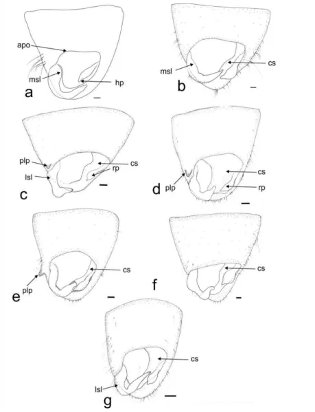
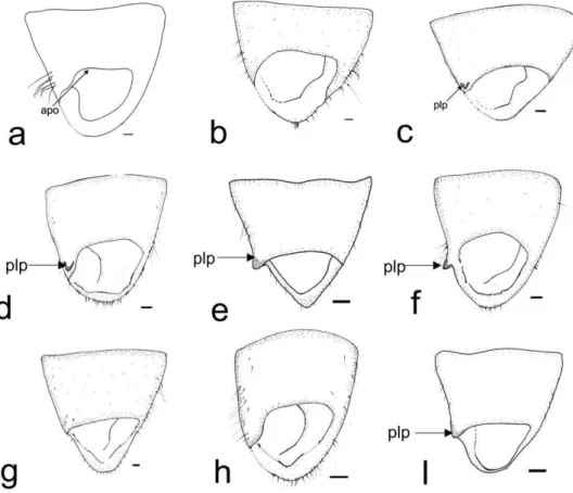
Pygophore
22 prolongation (plp), which can be large (Fig. 1 d, e) or small (Fig. 1 c). The cuplike sclerite (cs) has its apex and lateral margins strongly sclerotized (Fig. 1).
Figure 1. Phygophore in dorsal view. Collaria spp. a. C. boliviana. b. C. capixaba. c. C. improvisa. d. C. meilleurii. e. C.oculata. f. C.oleosa. g. C.scenica. Scale 0.1 mm. apo Anterior opening of pygophore, cs Cuplike sclerite, hp hypophysis, lsl lateral margin of basal sensory lobe of paramere, lp left paramere, msl medial margin of basal sensory lobe of paramere, plp prolongation of lateroposterior margin of pygophore, rp right paramere.
Parameres
23 basal sensory lobe (msl) of the left paramere varies from almost straight (Fig. 1 b) to clearly convex (Fig. 1 a), and the lateral margin of the basal sensory lobe (lsl) can be almost straight (Fig. 1g) or clearly convex. The right paramere (rp) is smaller than the left one, and can be: clubbed (Fig. 1c) or straight (Fig. 1a) with the apex of hypophysis small (Fig. 1 b-g) or large (Fig. 1 a).
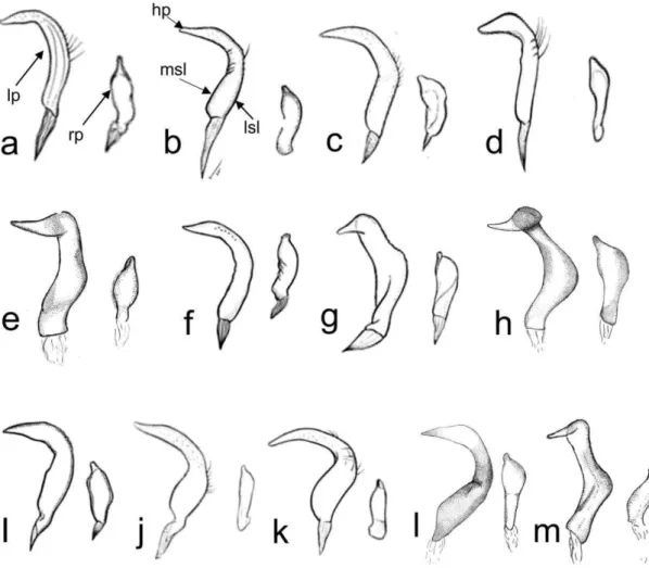
Aedeagus Phalloteca
The proximal phallotheca is little sclerotized and wraps the endosoma. The shape in all species is tube-like and the apical part is projected to the right left and does not show modifications (pt, Fig. 2 g).
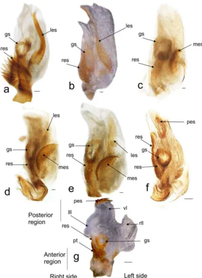
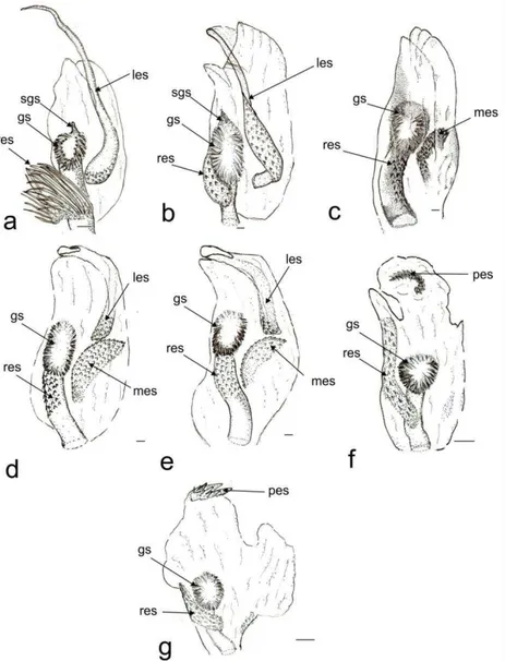
24 Endosoma
The endosoma is a tube-like membranous sac, with three lobes (lll, vll, rll, Fig. 2). When the endosoma was inflated it showed: two lateral lobes (right lateral lobe rll, left lateral lobe lll), and a ventral one (vl) (Fig. 2 f, g). The ventral lobe is larger than the lateral lobes (Fig. 2 f, g) or small (Fig. 2 a-e). The right lateral lobe is elongated (Fig. 2 b, f), or shorter than the ventral (Fig. 2 a, c, d, e, g). The left lateral lobe is short (Fig 2 a, b, c, f, g) but in some species it is elongate, narrow, and with a strongly curved apex (lll, Fig. 2 d, e).
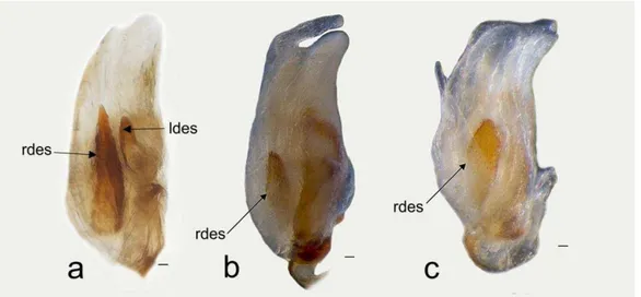
25 There are six endosomal sclerites: in ventral view we found the posterior endosomal sclerite (pes), the ribbonlike endosomal sclerite (res), the medial endosomal sclerite and the lobal endosomal sclerite (les). The posterior sclerite is found in the posterior region of th endosoma and can be c-shaped with several dorsal spines (Fig. 2, 3f) or comb-shape with about six thick spines joined at the base of different sizes (Figs. 2, 3 g). The lobal endosomal sclerite is near the periphery of the membranous lobe and with a left position in the endosoma; this sclerite has: smooth margins with microtrichia on the basal region and can be elongated (les, Figs. 2, 3 a), elongated with microtrichia in basal region (Fig. 2, 3 a, d, e), or on the basal and medial regions (Figs. 2, 3 b). The medial endosomal sclerite (mes) can be oval with serrate margins (Fig. 2, 3 c) or semicircular (= crescent) broad (Fig. 2, 3 d), or slender (Fig. 2, 3 e). The ribbonlike endosomal sclerite (res) is basal to the secondary gonopore enclosing the ductus seminis and antero-posteriorly oriented (Fig. 2, 3); can be brush-shaped with long spines (Fig. 2, 3, a), a short lobe with microtrichia (Fig. 2 b, c, d, e, g), or an expanded lobes that goes beyond the secondary gonopore (Fig. 2, 3 f). The right dorsal endosomal sclerite (rdes) can be: elongated without trichia on surface (Fig. 4a) fusiform with trichia on surface (Fig. 4b) or elliptical with trichia on surface (Fig. 4c). When is present the left dorsal endosomal sclerite (ldes) can be: fusiform with apex broad (Fig. 4a). Some species have a sclerite on its ventral surface (sgs) associated with the secondary gonopore, which can be relatively small (e.g., C. boliviana, Fig. 3a) or large (e.g., C. capixaba Fig 3b).
26 Female genitalia
The abdominal segments 8 and 9 contain the external female genitalia. The ovipositor consists of two pairs of sheets formed by the first and second gonapophyses; the sternal structures associated with abdominal segments eight and nine compose the entire Bursa copulatrix.
First Gonapophysis
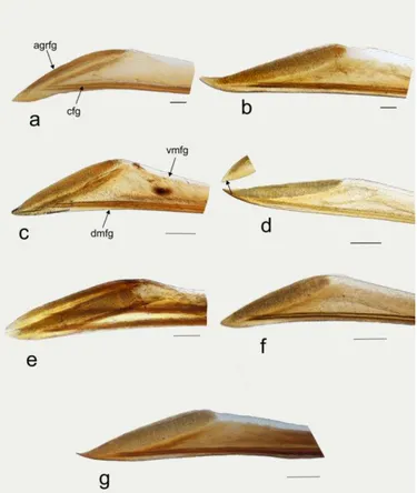
The first gonapophysis are enlarged, weakly sclerotized structures. The ventral margin (vmfg, Fig. 5 c) is membranous, and the dorsal one is sclerotized (dmfg, Fig. 5 c). The carina which is found sublateral to the dorsal margin of the first gonapophysis, (cfg, Fig. 5 a) reaches the apical grooved (or striated) region of the first gonapophysis (agrfg), and is strongly (Fig 5a, b, c, f, g) or weakly sclerotized (Fig. 5 d, e). The apex is sharpened with apical region grooved (agrfg) broad (Fig. 5 a, b, c, e, f, g) or acute with the grooved region narrower (Fig. 5 d).
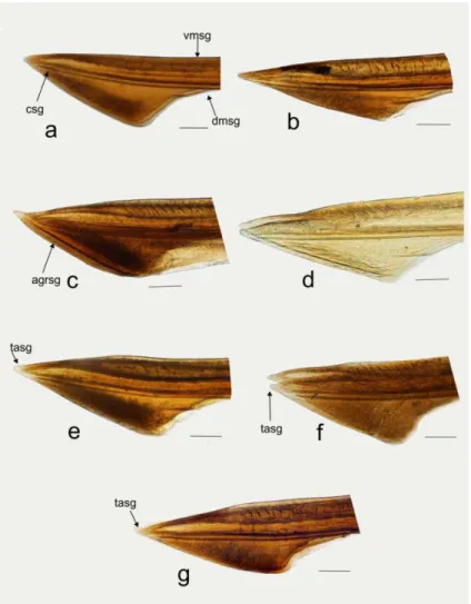
27 Second Gonapophysis
The second gonapophysis is smaller than first gonapophysis. It is spear shaped in lateral view, with a narrow region at base, and can be triangular (Fig. 6 a, c, e, f, g) or elongated apically (Fig. 6 b, d). The ventral margin is heavily sclerotized and smooth (vmsg, Fig. 6 a), and the dorsal margin is membranous (dmsg), being smooth (Fig. 6 a, b, d, e, g) or rugose (Fig. 6 f). The apex have teeth (tasg), varying in number, one dorsal (Fig. 6 b, c), one ventral (Fig 6 a), one dorsal and one ventral (Fig 6 e, g), or three in the apex (Fig 6 f). Thet also varying in size, being small (Fig. 6 b) or large (Fig. 6 c). The carina of t hesecond gonapophysis (csg) can be single (Fig 6 c) or double (Fig 6 a, b, d, e, f, g), reaching the apex of second gonapophysis. An apical grooved region (agrsg), present in at least in one species (Fig 6 c).
28 Bursa copulatrix
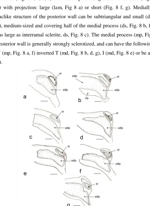
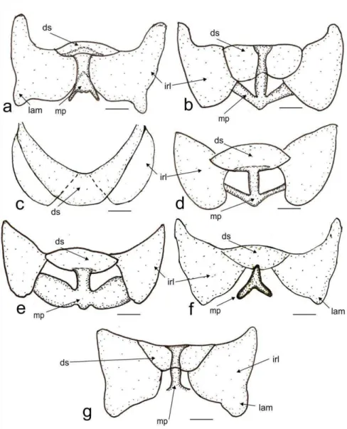
The dorsal labiate plate (dlp, Fig. 7 d), has a sclerite caudal to the sclerotized rings (sdlp) (Fig. 7 b), being small (Fig. 7 a) or large (Fig 7 b-g). The sclerotized rings of the dorsal labiate plate can be oblong (rs, Fig.7a -d), or ovoid (Fig. 7 f, g). The posterior wall has interramal lobes that can be triangular (Fig. 8 a, f, g) or rounded (Fig. 8 b, c, d, e). The lateroapical margin can be rounded (Fig. 8 b, c, d, e) or with projection: large (lam, Fig 8 a) or short (Fig. 8 f, g). Medially, the dorsal saclike structure of the posterior wall can be subtriangular and small (ds, Fig.8 a, d, e), medium-sized and covering half of the medial process (ds, Fig. 8 b, f, g), or large (as large as interramal sclerite, ds, Fig. 8 c). The medial process (mp, Fig. 8 d) of the posterior wall is generally strongly sclerotized, and can have the following shapes: an Y (mp, Fig. 8 a, f) inverted T (md, Fig. 8 b, d, g), I (md, Fig. 8 e) or be absent (Fig. 8 c).
29
Figure 8. Posterior wall with Dorsal Structure and Medial Process. Collaria spp. a. C. boliviana. b. C. capixaba. c. C. improvisa. d. C. meilleurii. e. C.oculata. f. C.oleosa. g. C.scenica. Scale 0.1 mm. ds Dorsal structure of posterior wall, irl interramal lobe of posterior wall, lam lateroapical margin of interramal lobe, mp medial process of posterior wall.
Discussion
A detailed study on the genitalia of Collaria was showed by the illustrations, photographs and comparisons of the male and female structures of representative species of Neotropical, Neartic and Afrotropical regions. Variations and documentation of new structures in both, males (e.g. endosomal sclerites, prolongation of margin of the pygophore), and females (e.g. apex of gonapohyses) different from those recorded by Carvalho and Fontes (1981) and Schwartz (2008)
30 constitute a starting point for the development of primary homologies and thus to elucidate the phylogenetic relationships of the species.
According to Carvalho and Fontes, (1981), Schwartz (2008), and the newly documented structures, the characteristic features for Collaria genitalia are summarized below: margin of the pygophore ranging from elongate to triangular, sometimes with lateroposterior margin projected in species belonging to Afrotropical and Neartic regions. Endosoma with six sclerites differening in form and position. Female with sclerotized rings of the dorsal labiate plate on the Bursa copulatrix oblong or ovoid, well developed, and dorsal structure of the posterior wall and medial process with different forms.
Terminology and structure of male and female genitalia
Interpretations of the morphological structures of the genitalia of Collaria, have varied between the decades of 1980's and 1990 's. Carvalho and Fontes (1981) used structures of the genitalia of males with names and illustrations that differ from those outlined by Carvalho and Carpintero (1989) and Carvalho (1990). These differences are presented by the manner in which each author has oriented and illustrated the genitalia of males and females. This has also been identified in other taxa, for example, Forero (2009) found that illustrations of the endosoma within the subfamily Orthotylinae were highly schematic in some of the species descriptions and had to provide more accurate drawings of the parameres, vesica, phallotheca, and genital capsule to facilitate comparisons with closely related new taxa.
Recently, the methodology of using photos has been applied to try to overcome this problem and display more clearly, precisely, and with a standardized orientation all structures of taxonomic significance in the genitalia of a given taxon (e.g., Chamorro and Lopes, 2014; Forero and Weirauch 2012). Here, we expose a representation of the morphological diversity of the genitalia of Collaria, and perceive differences in structures that would be almost impossible without extractions and observations in different orientations (e.g. dorsal, ventral positions). Therefore, the use of techniques such as photographs, can facilitate the display of components of genitalia, however it is also essential to make drawings in order to
31
Carvalho and Fontes (1981) used the name how “field of thorns” for sclerites
in the endosoma but they did not comment the variability and on which side (right or
left) of gonopore secondary they are were located. For female, they documented only
a “sclerotized area of the rings” (which is part of the dorsal wall of the Bursa
copulatrix), did not mention the structure of the dorsal wall proposed by other authors like Davis (1955).
Schwartz (2008) established that the male of Collaria has a basal sclerotized process on the endosoma with spiny and filamentous patches. We supported these observations, the endosoma has sclerites in all examined species in ventral view with a posterior, right and left position, and we found the sclerite in dorsal view; which had not been documented for the species of genus although being found in other Mirinae (e.g Gapon, 2014). The right dorsal endosomal sclerite (rdes) and left dorsal endosomal sclerite (ldes); were observed when looking at dorsal view of the structure in Afrotropical species like C. impovisa while in Nearctic species the only the right dorsal endosomal sclerite (rdes) is found, and in Neotropical species it is absent.
According to Schwartz (2008), the ribbonlike endosomal sclerite (res) is attached to the posterobasal portion of the ductus seminis; this sclerite is present in all the studied species of Collaria. Although the orientation is distinct between them, it is most easily observable in lateral view, where the sclerite lies in vertical position lengthened to apex in C. oleosa and C. scenica; whereas in other species is almost
horizontal (like in C. capixaba, C. improvisa, C. meulleurii). To explain the variety of orientations found, we can invoke the torsion hypothesis of the endosomal
spicules proposed by Cassis (2008), where the spicules or sclerites can migrate basally proximal to the endosomal membranous border with the apex twisting along the longitudinal axis of the endosoma, which probably occurred to species of Collaria.
32 we include additional characters such as the lateroposterior margin of the interramal lobes of posterior wall (irl), the medial process (mp), and the first and second
gonapophysis.
We herein add structures of the Collaria female genitalia not previously discussed by other authors, such as the apical grooved region and the teeth in the apex of gonapophysis. Davis (1955) mentioned that the cutting teeth are developed on the first and second gonapophyses and probably reflect the ovoposition habits of the each species, the data obtained in this study are congruent with those Davis (1955) and Fontes (1981) where the second gonapophysis has small teeth on their ventral margins near the apices.
Primary homology
We found evident primary homologies like the endosomal sclerites, parameres, and pygophore in the male, and the margin of interramal lobes,
gonapophyses, and dorsal structure of posterior wall, sclerotized rings and medial process of posterior wall in females. However in other cases, these homologies were difficult to determine. We initially found it difficult to propose primary homologies for the lobes of endosoma (lll, vl, rll) of the male; because they are hard to observe and in some cases we were unable to inflate the endosoma. However, when comparing the left lobe (lll) of C. meilleuri and C.oculata with other species, we can propose primary homology between the endosomal lobes. Nonetheless, more detailed studies with different techniques to inflate the endosoma are needed in order to establish variations of form and size. According to exposed, some proposed primary homologies are tentative and need to be tested on a phylogenetic analyses.
Species groups and genitalia
Schwartz (2008) recognized Collaria as the sister group of Nabidomiris based on characters of the head, pronotum and genitalia of both sexes. Although the
33 genitalias we propose that C. meulleurii and C. oculata form a natural group since they share the particular structure of the lobal endosomal sclerite, which is elongated
with a strongly curved apex, medial endosomal sclerite semicircular, right dorsal endosomal sclerite fusiform and elliptical, and the shape of the medial process of posterior wall in female.
Characters that might support a Neotropical clade composed by C. boliviana, C. capixaba, C. oleosa, and C. scenica are the presence and particular structure of the endosomal sclerites, and the shape of the interramal lobes and the medial process of the posterior wall in females. Collaria oleosa is probably more related to C. scenica because of the posterior position of endosomal sclerite, unlike the hypothesis proposed by Carvalho and Fontes (1981) in which C. oleosa would be more related to C. capixaba. The Afrotropical species examined exhibits unique features such as the strongly curved left paramere and two dorsal endosomal sclerites: the left dorsal endosomal sclerite is shorter than the right dorsal endosomal sclerite. In the female, the rounded interramal lobes and medial process are absent. It is likely that other Afrotropical species might exhibit some of these characters, however additional studies on more species from this region, will provide further contribution to the taxonomy of the group.
Finally, the detailed description of the genitalia of the genus Collaria increase new characters for the description, redescription and diagnosis of the genus. Future studies should emphasize on testing the primary homologies proposed here establish
the phylogenetic relationships of within the genus Collaria, and biogeographic relationships in the global range.
Acknowledgements
We thank the curators of the entomological collections that allowed us to study specimens of Collaria. We are also greateful to “Laboratorio de Orthoptera” and “Laboratorio de Sistemática Molecular-Beagle” (Departament of Animal Biology-UFV), where we took images, and to Yeisson Gutiérrez who helped us taking and editing the photographs. Financial support was provided by CAPES/CNPq—IEL Nacional—Brasil doctoral fellowship for the senior author.
References
34 Carvalho JC (1990) Mirideos neotropicais, CCCVII: novas espécies da Argentina e Bolivia (Hemiptera). Rev Bras Entomol (34): 445–452
Carvalho JC, (1945) Mirídeos neotropicais: Gêneros Dioniza Distant, Neella Reuter, Collaria Provancher, Falconia Distant e Ophthalmomiris Berg, com descrições de espécies novas. Rev Bras Entomol 16(1-2): 158–187
Chamorro J, Lopes C (2014) The phallus in Tettigoniidae (Insecta: Orthoptera: Ensifera): revision of STRUCTURE and terminology, and discussion on its taxonomic importance and evolution. Zootaxa 3815 (2): 151–199
Carvalho JC, Carpintero D (1989) Mirideos neotropicais, CCCIX: quatro espécies novas da tribo Stenodemini colecionadas na Republica Argentina (Hemiptera). Rev Bras Biol (49): 1101–1108
Carvalho JC, Fontes AV (1981) Mirídeos neotropicais CCXXV: Revisão do gênero Collaria Provancher no continente americano (Hemiptera). Experientae 27(2): 11–46 Carvalho JC, Jurberg J (1974) Neotropical Míridae, CLXXX: On the Horcias
complex (Hemiptera). Rev Bras Entomol 34(1): 49–65
Carvalho JC, Jurberg J (1976) Mirídeos Neotropicais, CCVI: Revisão do gênero Horciasinus Carvalho & Jurberg (Hemiptera). Rev Bras Biol 36(4): 811–834 Cassis G (2008) The Lattinova Complex of Austromirine Plant Bugs (Hemiptera : Heteroptera : Miridae : Orthotylinae). Proc Entomol Soc Wash 110(4): 845–939 Clayton RA (1982) A phylogenetic analysis of Lygocoris Reuter (Heteroptera: Miridae) with notes on life histories and zoogeography. Dissertation, University of Connecticut
Clayton R (1989) Preparation of phalluses of Miridae (Heteroptera) for scanning electron microscopy and a redescription of the vesica of Mirinae. Trans Am Microsc Soc 108(4): 419–423
Davis NT (1955) STRUCTURE of the female organs of reproduction in the Miridae (Hemiptera). Ann Entomol Soc Am 48: 132–150
Eyles A, Carvalho J (1975) Revision of the genus Dolichomiris, with a revised key to the genera of Stenodemini (Heteroptera: Miridae). J Nat Hist 9(3): 257–269
35 Fontes A (1981) Estudos comparativos da genitália da fêmea do gênero Notholopus Bergroth, 1922 (Hemiptera, Miridae). Arq Mus Nac 56: 137–183
Fontes A (1989) Contribuição ao estudo da genitália da fêmea de algumas espécies de Prepops Reuter, 1905 (Hemiptera, Miridae). Bol Mus Nac NS Zool 330: 1–31 Fontes A (1993a) Contribução ao estudo da genitália de fêmeas de Henicocnemis Stal (Hemiptera, Miridae). Rev Bras Zool 10(4): 605–612
Fontes A (1993b) Contribução ao estudo da genitália de fêmeas de Phytocoris Fallén (Hemiptera, Miridae). Rev Bras Zool 10(4): 595–604
Fontes A (1993c) Contribução ao estudo da genitália das fêmeas de doze espécies de Prepops Reuter, 1905 (Hemiptera, Miridae). Rev Bras Biol 53(3): 385–395
Forero D (2008) Revision and phylogenetic analysis of the Handronema group (Miridae: Orthotylinae: Orthotylini), with descriptions of new genera and new species, and comments on the Neotropical genus Tupimiris. Bull Am Mus Nat Hist 312:1-172
Forero D (2009) Description of one new species of Chileria and three new species of Orthotylus, with nomenclatural and distributional notes on Neotropical Orthotylinae (Heteroptera: Miridae: Orthotylini). Am Mus Novit 3642: 1–50
Forero D, Weirauch C (2012) Comparative genitalic STRUCTURE in the New World resing bugs Apiomerini (Hemiptera, Heteroptera, Redeviidae, Harpactorinae). Deutsch Entomol Z 59(1): 5–41
Gapon DA (2014) Revsion of the genus Polymerus (Heteroptera: Miridae) in the Eastern Hemisphere Part 1: Subgenera Polymerus, Pachycentrum sbgen. nov and new genus Dichelocentrum gen nov. Zootaxa 3787(1): 1–87
Henry T, Kim K (1984) Genus Neurocolpus Reuter (Heteroptera: Miridae): Taxonomy, economic implications, hosts and phylogenenetic review. Trans Am Entomol Soc 110: 1–75
Hernández LM, Henry TJ (2010) The plant bugs, or Miridae (Hemiptera: Heteroptera), of Cuba. Bulgaria
Kelton L (1955) Genera and subgenera of the Lygus complex (Hiemiptera: Miridae). Can Entomol 87: 277–301
36 Kelton L (1966) Review of the Species of Teratocoris Fieber, With Description of a new species from the Nearctic Region (Hemiptera: Miridae). Can Entomol 98(12): 1265–1271
Kelton L (1980) The Plant Bugs of the Prairie Provinces of Canada Heteroptera : Miridae. In In the Insects and Arachnids of Canada Agriculture Research Branch Publication, Quebec
Kerzhner I, Konstantinov FV (1999) Structure of the aedeagus in Miridae
(Heteroptera) and its bearing to suprageneric classification. Acta Soc Zool Bohem 63: 117–137
Konstantinov FV (2003) Male genitalia in Miridae ( Heteroptera ) and their significance for suprageneric classification of the family . Part I : general review , Isometopinae and Psallopinae. Belg J Entomol 5: 3–36
Konstantinov FV (2007) Male genitalia in Miridae : structure , terminology and application to phylogenetic inference . Critical comments on Cheng-Shing Lin & Chung-Tu Yang ’s ideas (Heteroptera). Zoosyst Rossica 16(22): 235–238
Kullenberg B (1947) Uber morphologie und Funktion des Kopulationsapparats der Capsiden und Nabiden. Zool Bidr Uppsala 24: 1–418
Lattin JD, Schwartz MD (1986) A review of Acetropis americana Knight in North America (Hemiptera: Miridae: Stenodemini). J NY Entomol Soc 94(1): 32–38 Lin C,Yang C (2005) External Male Genitalia of the Miridae (Hemiptera: Heteroptera). Nat Mus Natur Sci Spec Publ 9:1–174
Remane R, Günther H (2008) Acetropis stysi, a new species from Spain (Hemiptera : Heteroptera : Miridae). Acta Entomol Mus Nat Pragae 48(2): 389–394
Schuh RT (2014) On-line Systematic Catalog of Plant Bugs (Insecta: Heteroptera: Miridae). http://research.amnh.org/pbi/catalog/
Schwartz MD (1984) A revision of the black grass bug genus Irbisia Reuter (Heteroptera: Miridae). J NY Entomol Soc 92: 193–306
Schwartz MD (2008) Revision of the Stenodemini with a review of the included genera (Hemiptera: Heteroptera: Miridae: Mirinae). Proc Entomol Soc Wash 110(4): 1111–1201
37 and a new genus from Mexico, and a review of the genus Scalponotatus Kelton (Heteroptera: Miridae: Orthotylinae). Bull. Amer. Mus. Nat. Hist. 354:1- 290 Schwartz MD (2012) A New Species of Trigonotylus (Heteroptera : Miridae : Mirinae : Stenodemini ) from Central Oregon , United States. Entomologica Americana 118(1): 152–156
Scudder G, Schwartz MD (2001) The genus Leptopterna Fieber ( Heteroptera : Miridae : Stenodemini ) in North America. Proc Entomol Soc Wash 103:797–806 Scudder G, Schwartz MD (2012) Two new species of Trigonotylus ( Hemiptera : Heteroptera : Miridae : Stenodemini) from western Canada and northwestern United States. Zootaxa 3174: 51–58
Scudder G (1959) The Female of the Heteroptera: Structure and bearing on classification. Trans Entomol Soc Lond 111(14): 405–467
Singh-Pruthi H (1925) The structure of the male genitalia in Rhynchota. Trans Entomol Soc Lond 73(1-2): 127–267
Slater JA (1950) An investigation of the Female as taxonomic characters in the Miridae (Hemiptera). Iowa State Coll J Sci 25:1–81
Song H, Bucheli R (2010) Compararison of phylogenetic signal between male genitalia and non-genital characters in insect systematics. Cladistics 26: 23-35 Weirauch C, Schuh R (2011) Systematics and evolution of Heteroptera: 25 years of progress. Annu Rev Entomol 56: 487–510 doi:10.1146/annurev-ento-120709-144833 Wheeler AG (2001) Biology of the plant bugs (Hemiptera: Miridae). Pests,
Predators, Opportunists. Comstock, New York
Yasunaga T, Schwartz MD (2007) Revision of the mirine plant bug genus
Philostephanus Distant and allies (Heteroptera: Miridae: Mirinae: Mirini). Tijdschr v Ent 150: 101–180
38
CAPÍTULO II
Revision of Collaria Provancher, 1872 (Hemiptera: Miridae) with the description of a new species from the Afrotropical region
Manuscrito original, apresentado em formato de submissão para publicação na revista ZOOTAXA
IRINA T. MORALES-C 1, PAULO S. F. FERREIRA2, DIMITRI FORERO3
1 Programa de Pós-Graduação em Entomologia. Universidade Federal de Viçosa 36570-900 Viçosa, MG, Brasil. E-mail: [email protected]
2 Departamento de Entomologia, Universidade Federal de Viçosa. 36570-900 Viçosa, MG, Brasil. E-mail: [email protected]
3 Departamento de Biología, Laboratorio de Entomología, Unidad de Ecología y Sistemática, Pontificia Universidad Javeriana Bogotá, Colombia. E-mail: [email protected]
Abstract
Collaria (Mirinae: Stenodemini) is a genus of grass-feeding bugs with 14 recognized species. The present work includes redescriptions of species and the description of C. schwartzi sp. nov. The female genitalia of C. boliviana and C. villiersi, and male and female genitalias of C. improvisa and C. obscuricornis are described. New distribution records for Neotropical and Afrotropical regions are included. Finally, a key and ilustrations of male and female genitalias of all species are included.
Keys words: Heteroptera, Stenodemini, grass-feeding plant bugs, new record, male and female genitalia, taxonomy.
Introduction
39 descriptions were based in general color, with no illustrations and descriptions of male and female genitalia. Carvalho & Fontes (1981) partially revised the genus, including descriptions of three new species, diagnosis and illustrations of male and female genitalias, and a key to the Nearctic and Neotropical species.
Collaria is predominantly Neotropical, with seven species (C. boliviana Carvalho 1990, C. capixaba Carvalho & Fontes 1981, C. guaraniana Carvalho & Fontes 1981, C. husseyi Carvalho 1955, C. malonoi Carvalho & Carpintero 1989, C. oleosa (Distant 1883), and C. scenica (Stål 1859) from this region. The Neartic Region is represented by two species (C. meilleurii Provancher 1872 and C. oculata (Reuter 1876)), and five species from the Afrotropical Region, are currently recognized: C. danae Linnavuori 1974, C. improvisa Reuter 1893, C. nigra Linnavuori 1975, C. obscuricornis Poppius 1910, C. villiersi Carvalho 1953. Most of the taxonomy of Collaria is based on the Neotropical and Neartic species, and there are no keys for all species of the genus. This study provides a synopsis of the genus Collaria with description of a new species from the Afrotropical region, a key to species, and information concerning geographic distribution and plant associations. Material and Methods
Examined Specimens. A total of 1080 specimens were borrowed from the following
entomological collections and examined:
AMNH American Museum of Natural History, New York, USA. BPBM Bernice P. Bishop Museum, Hawaii, USA.
CAS California Academy of Sciences, San Francisco, USA. CTNI Colección Taxonómica de Insectos “Luis María Murillo”-
CORPOICA, Tibaitatá, Colombia.
DZRS Departamento de Zoologia, Universidade Federal do Rio Grande do Sul, Porto Alegre, Brazil.
DZUP Coleção Entomologica Pe. Jesus Santiago Moure do Departmento de Zoologia, Universidade Federal de Paraná, Curitiba, Brazil.
ICN Colección de Zoología – Instituto de Ciencias Naturales – Universidad Nacional de Colombia Bogotá, Colombia.
40 KU University of Kansas Natural History Museum, Lawrence, Kansas,
USA.
MCZN Museu Ciências Naturais Fundação Zoobotanica Rio Grande do Sul, Porto Alegre, Brazil.
MCN Coleção Zoológica Científica do Museu de Ciências Naturais do Centro Univates, Lajeado, Brazil.
MNRJ Museu Nacional, Universidade Federal do Rio de Janeiro, Rio de Janeiro, Brazil.
MPUJ-ENT Museo de Pontificia Universidad Javeriana, Laboratorio Entomología, Bogotá, Colombia.
MRAC Royal Museum for Central Africa, Tervurem, Belgium. NMSA Natal Museum, Pietermaritzburg, South Africa.
RBINS Royal Belgian Institute of Natural Sciences, Brussells, Belgium. UFES Universidade Federal do Espirito Santo, Vitoria, Brazil.
UFVB Museu Regional de Entomologia, Universidade Federal de Viçosa, Viçosa, Brazil.
UPTC Laboratorio de Entomología, Museo de Historia Natural “Luis
Gonzalo Andrade” Universidad Pedagógica y Tecnológica de
Colombia, Tunja, Colombia.
USU Insect Collection Utah State University, USA.
USNM National Museum of Natural History, Smithsonian Institution, Washington DC, USA.
ZMUC Zoological Museum, University of Copenhagen, Copenhagen, Denmark.
Besides of information gathered from labels, data from the online catalogue The Plant Bug Planetary Biodiversity Inventory Project (http://research.amnh.org/pbi/) were obtained for species C. meilleurii, C. oculata and C. obscuricornis.It concerns specimens held in the following collections:
CNC Canadian National Collection of Insects. Ontario, Ottawa, Canadá.
41 NCSU North Carolina State University Insect Collection, North Dakota,
Fargo, USA.
UDCC University of Delaware, Delaware, Newark,USA. UCR University of California, Riverside, USA.
MEMU Mississippi Entomological Museum, Mississippi State University, Mississippi, Mississippi, USA.
MHNCM Colección Nacional de insectos Dr. Alfredo Barrera Marín del Museo de Historia Natural de la Ciudad de México.
SANBI South African National Biodiversity Institute IZIKO.
Genitalic dissections. The genitalia was prepared following the methodology of Scudder and Schwartz (2012): the whole abdomen (male and female) was removed and immersed in a warm solution of 85% lactic acid, rinsed in distilled water and transferred to 70% ETOH. The dissections were carried out in glycerin following Forero & Weirauch (2012), under a Leica MZ8 stereomicroscope.
Imaging and measurements. A Zeiss Discovery V20 stereomicroscopy adapted with a Zeiss AxioCam MRc digital camera, and Olympus BX53 microscopy with an Olympus DP73 camera were used for obtaining photographs. Final images were stacked using Zerene Stacker. Measurements were made on Zeiss Discovery V20 stereomicroscopy (Table 1).
Terminology. We follow Schuh and Slater (1995) for the general morphology. The terminology for male and female genitalia follows Schwartz (2008) and Morales-C et al. (In prep.).
Georeferences and maps. The software ArcGis (10.2.1) was employed to generate the maps; the localities from label data were georeferenced using gazetteers (http://www.fallingrain.com) and Google Earth. In some cases these data are given in brackets to indicate that they were not directly taken from PBI database (http://research.amnh.org/pbi/maps/) and label data of examined species.
Genus Collaria Provancher, 1872
TYPE SPECIES: Collaria meilleurii Provancher, 1872 (Lectotype designed by