SYSTEMATICS, MORPHOLOGY AND PHYSIOLOGY
Description of the Third-Instar of
Anastrepha leptozona
Hendel
(Diptera: Tephritidae)
D
ANIELF
RÍASL
ASSERRE1, V
ICENTEH
ERNÁNDEZO
RTIZ2, L
ILIANAL
ÓPEZM
UÑOZ31Instituto de Entomología, Univ. Metropolitana de Ciencias de la Educación, Av. JoséPedro Alessandri 774, Código postal 7760197, Santiago, Chile;2Instituto de Ecología AC. Departamento de Entomología,
Apartado postal 63, km 2.5, carretera antigua a Coatepec nº351, Congregación El Haya, 91070 Xalapa, Veracruz,
México;3Programa Moscamed, Central Poniente nº14, Altos, Tapachula, Chiapas, México Edited by Roberto A Zucchi–ESALQ/USP
Neotropical Entomology 38(4):491-496 (2009)
Descrição do TerceiroÍnstar deAnastrepha leptozonaHendel (Diptera: Tephritidae)
RESUMO - A morfologia da larva do terceiroínstar deAnastrepha leptozonaHendelédescrita. São analisados, em microscopia de luz e de varredura, o complexo antenomaxilar, as margens da abertura oral, o bordo, oórgão oral, o esqueleto cefalofaríngeo, os espiráculos anterior e posterior e o segmento caudal. As larvas do terceiroínstar deA.leptozona têm um“esclerito ventral”abaixo do esclerito faríngeo, o qualécaracterizado pela primeira vez em espécies deAnastrepha.
PALAVRAS-CHAVE: Morfologia, esclerito ventral, mosca-das-frutas
ABSTRACT - The morphology of the third-instar larva ofAnastrepha leptozonaHendel is characterized using optical and scanning electron microscopy. The antennomaxillary complex, oral ridges, labium, stomal sensory organ, cephalopharyngeal skeleton, anterior and posterior spiracles and caudal segment are described and illustrated. Mature larvae ofA.leptozona present a “ventral sclerite”below the pharyngeal sclerite which is characterized for thefirst time inAnastrephaspecies.
KEY WORDS: Morphology, ventral sclerite, fruitfly
Anastrepha leptozona Hendel is a species widely distributed throughout the Neotropical region including records from Mexico, Guatemala, Honduras, Costa Rica, Panamá, Guyana, Venezuela, Trinidad, Bolivia and Brazil (Hernández-Ortiz & Aluja 1993). It has been recorded breeding in several host plants species of the families Anacardiaceae (1), Icacinaceae (1), Myrtaceae (1), Quinaceae (1), Rosaceae (1) and Sapotaceae (6) (Norrbom et al 1999).
In Mexico,A.leptozonahas been reported in the state of Chiapas, Morelos, Oaxaca, and Veracruz (Herná ndez-Ortiz 1992, 2007), usually breeding in the hostMicropholis mexicana(Sapotaceae) and less frequently inCrataegussp. (Rosaceae) ( Alujaet al1987)
This species belongs to the leptozona species group, along with four other species A. barnesi Aldrich, A. costalimaiAutuori,A.elongataFernández, andA.steyskali Korytkowski (Norrbomet al1999). Adults of this group may be recognized by the lateral surstylus short and somewhat boot-shaped, with laterally projecting apical lobe; and the vein M strongly curved apically.
Brief description of some larval characters ofA.leptozona was presented in studies by Carrollet al(2004). Also Steck
et al(1990) presented a key for the recognition of the third-instar larvae for 13Anastrephaspecies which includedA. leptozona. However, there is no information about the larval morphology of the other species belonging to theleptozona species group.
In this paper we made a detailed description of the morphology of the third-instar larva ofA.leptozonafor the
first time. Also we discuss the relevance of some distinctive characters, such as the“ventral sclerite”which has not been previously described in any otherAnastrephaspecies.
Material and Methods
The collected fruits were inspected, and the found larvae were kept alive for seven days in a maturation cage. At the end of this period the third-instar larvae were removed from the fruits and killed by dipping in hot water. In total, we obtained 350 larvae specimens, which were preserved in 70% ethanol. We selected 25 individuals which were prepared for analysis by optical and scanning electron microscopy (SEM). For optical study, the larvae were prepared following techniques described by Steck & Wharton (1988). Antennomaxillary complex, oral ridge, labium, stomal sensory organ, cephalopharyngeal skeleton, the anterior and posterior spiracles and the caudal segment were dissected and slide mounted for observations.
For scanning electron microscopy (SEM), samples were
fixed in 2.5% glutaraldehyde in 0.1 M cacodylate Buffer pH 7.4 for 2h at 4ºC and postfixed in 1% OsO
4 at room temperature and darkness. Tissues dehydrated in graded acetone were critical-point dried with CO2,mounted, and then coated with a layer of palladium-gold approximately 20 nm thick. Samples were examined using SEM JEOL JSM-5600-SV at Department of Entomology, Instituto de Ecologia (Xalapa, Veracruz), Mexico, and with JEOL JSM-25- SII at the Pontificia Universidad Católica de Chile (Santiago, Chile). The terminology of larval description follows White & Elson-Harris (1992), Whiteet al(1999) and Fríaset al(2006).Voucher specimens are deposited in the Instituto de Entomología, Universidad Metropolitana de Ciencias de la Educación (Santiago, Chile), in the Instituto de Entomología A.C. Department of Entomology (Veracruz, México) and in the Moscamed (Tapachula, Chiapas) collections.
Anastrepha leptozonaHendel
Description
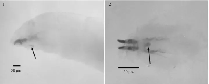
Third-instar larva. Body pale yellow, elongate, range of length 7.0-11.0 mm, mean length 8.6 ± 1.42 mm. There is a brown spot below the pharyngeal skeleton in ventral
view, between the intersection of segments T1 and T2. This character, that we coined as ventral sclerite, is exclusive forA.leptozonaand has not been previously described in Anastrepha(Figs 1-2).
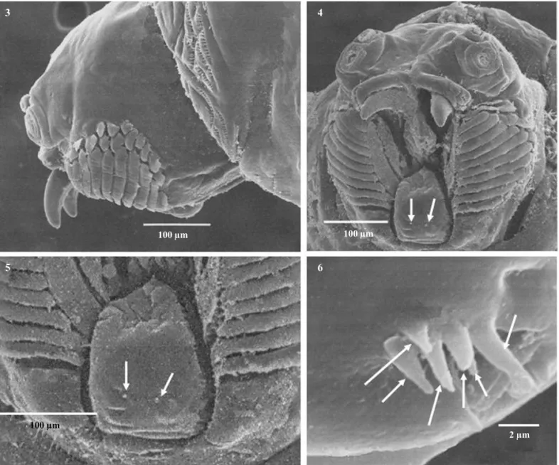
Head dorsally smooth, without spinules; mouth opening bordered with 10-11 rows of non-serrated oral ridges, with 10-15 small accessory plates (Figs 3-4). Labium broad triangular-shaped, with two papilla sensilla and a pair of medial pits (Figs 4-5). Stomal sensory organ elongated, apically rounded, with two secondary lobes and six peg sensilla (Figs 4, 6).
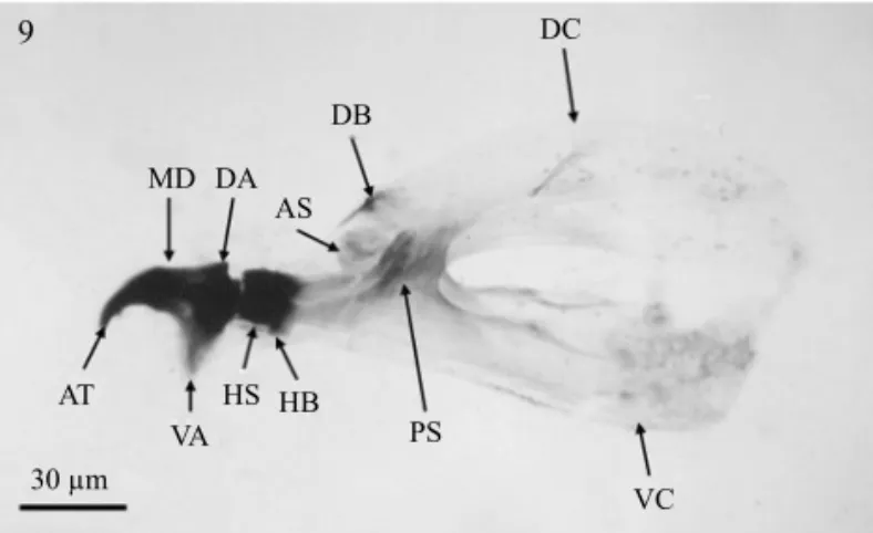
Antenna three-segmented; maxillary palpus with three papilla sensilla and two knob sensilla, dorsolateral group bearing two papilla sensilla close to maxillary palpus (Figs 7-8). Cephalopharyngeal skeleton with strongly sclerotized mandibles, apical tooth curved and black; ventral apodeme brown and broad, dorsal apodeme short; preapical teeth absent. Dental sclerite absent; labial sclerite short and brown. Hypopharyngeal sclerite anteriorly black and posteriorly yellow. Pharyngeal sclerite brown and moderately sclerotized; anterior sclerite, dorsal and ventral cornua yellow and weakly sclerotized; dorsal bridge of dorsal cornu brown, moderately sclerotized; ventral bridge of ventral cornu not evident (Figs 9-10). Below the pharyngeal sclerite, attached to the cuticle and bind with muscles to the ventral apodeme of mandible, there is a ventral sclerite that is a glove-shaped structure provided with four“fingers”-like protuberances; this character was
find by Liliana López (Figs 11-13). The ventral sclerite is not directly linked to the cephalopharyngeal skeleton therefore not shown in Fig 9.
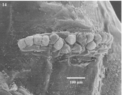
Anterior spiracles with 16-20 tubules arranged in a single row, mean 17.5±1.40, sometimes in the central area with a double row (Fig 14).
Caudal segment with two large lateral tubercles in intermediate area, intermediate sensilla I1 and I2 evident, sensilla I3 present. Lateral area with two tubercles and one lateral sensillum. Ventral area with two small tubercles over broad, triangular-shaped fold of cuticle (Fig15). Dorsal area with D1 and D2 sensilla, laterally two extraordinary
Figs 1-2 Third-instar larva ofA.leptozona: Cephalopharyngeal skeleton in lateral view (1). ventral view (2). Arrows showing the“ventral sclerite”.
1
30μm 30μm
Figs 3-6 Third-instar larva ofA.leptozona. Head in lateral view (3). Head in frontal view (4). Labium (5), arrows showing the papilla sensilla. Stomal sensory organ (6), arrows showing the peg sensilla.
Figs 7-8 Antennomaxillary complex of larva ofA.leptozona. Antenna and maxillary palpus (7). Maxillary palpus (8).
3
5
7 8
4
6
100μm
100μm
10μm
10μm
P2
K2 mxp
ant
P1
K2 P3
P4
P5
100μm
D3 and D4 sensilla. Anal lobes unilobate with anal opening surrounded by wrinkled surface with minute spinules (Figs Fig 9 Cephalopharyngeal skeleton of larva ofA.leptozona, lateral view. AS = anterior sclerite. AT = apical tooth. DA = dorsal apodeme. DB = dorsal bridge. DC = dorsal cornu. HB = hypopharyngeal bridge. HS = hypopharyngeal sclerite. MD = mandible. PS = pharyngeal sclerite. VA = ventral apodeme. VC = ventral cornu.
Fig 10 Cephalopharyngeal skeleton of larva ofA.leptozona, dorsal view. AT = apical tooth. DC = dorsal cornu. HB = hypopharyngeal bridge. HS = hypopharyngeal sclerite. LS = labial sclerite. MD = mandible. PS = pharyngeal sclerite. VC = ventral cornu.
Fig 11 Cephalopharyngeal skeleton of larva ofA.leptozona in lateral view. Ventral apodeme (VA); ventral sclerite (VS).
Figs 12-13 Ventral sclerite of larva ofA.leptozona. Ventral view (12). Lateral view (13).
15, 18). Posterior spiracles with long spiracular slits, nearly two times as long as wide, dorsal and ventral spiracular bundles with short spatulate hairs (Fig 17).
Remarks
Based on the morphology of the third-instar larva of A.leptozonasome traits could be used for the recognition of this species as follows: the presence of the ventral sclerite below the pharyngeal sclerite observed under optical microscopy appears glove-shaped and it is absent in all other larvae ofAnastrephapreviously described. The
9 11
12
13 10
30μm 30μm
10μm
10μm
30μm AT
MD DA AS
DB
DC
VA VA
VS HS HB
AT
LS MD
HS
VC
PS PB
DC HB PS
1988), in most otherAnastrephaspecies these tubules are arranged in a single row, e.g.A.fraterculus(Wiedmann), A.interruptaStone,A.limaeStone,A.ludens(Loew),A. obliqua(Macquart),A.serpentina(Wiedmann),A. striata Schiner andA.suspensa (Berg 1979, Steck & Wharton 1988, Carroll & Wharton 1989, White & Elson-Harris 1992, Frías et al 2006). In the posterior spiracles the bundle of hairs of the spiracular slits are shorter than width of spiracular slits, in other species such asA.fraterculus, A.interrupta,A.grandis,A.limae,A.ludens,A.obliqua, A.serpentina,A.striata A.suspensathese hairs are as long as or longer than the width of spiracular slits (Berg 1979, Steck & Wharton 1988, Steck & Malavasi 1988, Carroll & Wharton 1989, White & Elson-Harris 1992, Fríaset al 2006). The labium has two papilla sensilla and a small pair medial pits which have been previously described only in mature larva ofA.ludens(Carroll & Wharton 1989). The intermediate area of caudal segment presented two big papilla sensilla, which were never described before (Berg 1979, Steck & Malavasi 1988, Steck & Wharton 1988, Carrol & Wharton 1989, Stecket al1990, White & Elson-Harris 1992, Whiteet al1999, Carrollet al2004, Fríaset al2006). These traits may be diagnostic for the identification of third-instar larva ofA.leptozona. Fig 14 Anterior spiracle of larva ofA.leptozona.
Figs 15-18 Caudal segment of larva ofA.leptozona. Posterior view (15), D1, D2, D3, D4 = dorsal sensilla; I1, I2, I3 = intermediate sensilla, L = lateral sensillum. Lateral view (16). Posterior spiracles (17). Anal lobes (18).
anterior spiracles comprise 17-19 tubules in a single row laterally and double row centrally in all the individuals studied, similar to A. grandis (Macquart) (Steck & Wharton 1988) andA.bistrigataBezzi (Steck & Malavasi
14
15
17
16
18
100μm
100μm D4
D2 D1
D3
L
l3
l2 l1
100μm
100μm
Acknowledgments
We thank Pablo Montoya for improvements in earlier version of the manuscript. This paper was partially supported by the project FIBAS (06-08- DIUMCE).
References
Aluja M, Guillen J, Rosa G de la, Cabrera M, Celedonio H, Liedo P, Hendrichs J (1987) Natural host plant survey of the economically important fruitflies (Diptera: Tephritidae) of Chiapas, Mexico. Fla Entomol 70: 329-338.
Berg G H (1979) Clave ilustrada de larvas de moscas de la fruta de la familia Tephritidae. Organismo Internacional Regional de Sanidad Agropecuaria (OIRSA) San Salvador, República de El
Salvador, 36p.
Carroll L E, Norrbom A L, Dallwitz M J, Thompson F C (2004)
Anastrepha leptozonaHendel: 1-1. In Pest fruitflies of the world - larvae. Version: 13 April (2005. http://delta-intkey.com.
Carroll L E, Wharton R A (1989) Morphology of the immature stages ofAnastrepha ludens(Diptera: Tephritidae). Ann Entomol Soc Am 82: 201-214.
Frías L D, Hernández-Ortiz V, Vaccaro N, Bartolucci A, Salles L A (2006) Comparative morphology of immature stages of some frugiverous species of fruitflies (Diptera: Tephritidae). Isr J Entomol : 423-457.
García G H (1973) Modificaciones al sistema de clasificación climático Kopen UNAM México. Limusa, p. 215.
Hernández-Ortiz V (1992) El géneroAnastrepha Schiner en México (Diptera: Tephritidae). Taxonomía, distribución y sus plantas huéspedes. Instituto de Ecología, Sociedad Mexicana de Entomología, Xalapa, Veracruz, México, 162p.
Hernández-Ortiz V (2007) Diversidad y biogeografía del género
Anastrephaen México p.53-76. In Hernández-Ortiz V (ed) Moscas de la fruta en Latinoamérica (Diptera: Tephritidae): diversidad, biología y manejo. S y G editores, Distrito Federal, México, 167p.
Hernández-Ortiz V, Aluja M (1993) Listado de especies del género neotropicalAnastrepha(Diptera: Tephritidae) con notas sobre su distribución y plantas hospederas. Folia Entomol Mexicana 88: 89-105.
Norrbom A L, Zucchi R A, Hernández-Ortiz V (1999) Phylogeny of the genera Anastrepha and Toxotrypana (Trypetinae: Toxotrypanini) based on morphology p.299-342. In Aluja M, Norrbom A L (eds) Fruitflies (Tephritidae): phylogeny and evolution of behavior. CRC Press. Boca Ratón, 944p.
Steck G L, Carroll L E, Celedonio-Hurtado H, Guillen-Aguilar J (1990) Methods for identification ofAnastrephalarvae (Diptera: Tephritidae), and key to 13 species. Proc Entomol Soc Wash 92: 333-346.
Steck G J, Malavasi A (1988) Description of immature stages of
Anastrepha bistrigata(Diptera: Tephritidae). Ann Entomol Soc Am 81: 1004-1009.
Steck G J, Wharton R A (1988) Description of immature stages ofAnastrepha interrupta, A.limae andA. grandis(Diptera: Tephritidae). Ann Entomol Soc Am 81: 994-1003.
White I M, Elson-Harris M M (1992) Fruitflies of economic significance: their identification and bionomics. C A B International, United Kingdom, 601p.
White I M, Headrick D H , Norrbom A L, Carroll L E (1999) Glossary, p.881-920. In Aluja M, Norrbom A L (eds) Fruitflies (Tephritidae): phylogeny and evolution of behavior. CRS Press, City, 944p.