J. Evid. Based Med. Healthc., pISSN- 2349-2562, eISSN- 2349-2570/ Vol. 3/Issue 32/Apr. 21, 2016 Page 1522
FNAC AS A DIAGNOSTIC TOOL IN SALIVARY GLAND TUMOURS
Kalivarapu Paparatnam1
1Assistant Professor, Department of Pathology, RIMS Srikakulam, Andhra Pradesh.
ABSTRACT
BACKGROUND
FNAC of salivary gland tumours is an accurate, simple, rapid, inexpensive, well tolerated and harmless procedure. The success of FNAC depends on the adequacy of sample and high-quality preparation. FNAC of salivary gland neoplasms provides essential information in decision making and management.
AIM OF THE STUDY
Know the role of fine needle aspiration cytology in the diagnosis of benign and malignant lesions of salivary gland.
MATERIAL AND METHODS
This was a prospective study done at the tertiary care centre for a period of three years. A total number of 67 cases of clinically suspected salivary gland tumours were subjected to fine needle aspiration cytology and correlated with histopathology.
RESULTS
A total number of 67 cases, clinically suspected as salivary gland tumours were subjected to FNAC and compared with histopathology. The observations of the study were as follows:
Most of the tumours were observed between the age group of 31-40 years. The commonest gland involved was the parotid gland, 56 cases of benign, 10 cases of malignant and one case of inconclusive diagnosis was made on FNAC. In the present study, FNAC showed Sensitivity of 66.6%, Specificity of 98%, Positive predictive value; 90.9%, Negative predictive value; 91%, Percentage of false negative cases 33.3%, Percentage of false positive cases 1.9% and Overall Diagnostic Accuracy of 91%.
CONCLUSION
FNAC is a very useful, simple, cheap, accurate and repeatable technique in the preoperative diagnosis of various salivary gland neoplasms. Overall, diagnostic accuracy was 91%, in cystic lesions of salivary glands, combined FNAC and histopathology is essential for diagnosis.
KEYWORDS
FNAC, Salivary gland tumours, Role.
HOW TO CITE THIS ARTICLE: Paparatnam K. FNAC as a diagnostic tool in salivary gland tumours. J. Evid. Based Med.
Healthc. 2016; 3(32), 1522-1526. DOI: 10.18410/jebmh/2016/343
INTRODUCTION: A nodular or diffuse enlargement of the
salivary gland is quite frequent, which could be due to a cystic lesion, an inflammation, a degenerative process or a neoplasm of benign or malignant nature. Though salivary gland tumours account for only 2 to 6.5% of all head and neck tumours, their superficial location and easy accessibility makes FNAC a popular method with high diagnostic accuracy for evaluation of salivary gland tumours.
In adults, the majority of salivary neoplasms are epithelial in origin, pleomorphic adenoma being the most common. The parotid gland is the commonest site for occurrence of salivary gland neoplasms.
FNAC is a useful technique for evaluation of mass lesions of salivary glands. FNAC of salivary gland tumours is an
accurate, simple, rapid, inexpensive, well tolerated and harmless procedure. The success of FNAC depends on the fundamental requirements of representativeness, adequacy of sample and high-quality preparation. It produces a speedy result and can be easily repeated if necessary. FNAC possess a negligible risk of complications and in medical centres where the technique is routinely used, approximately a third of all patients with salivary gland lesions are spared of surgical intervention. FNAC of salivary gland neoplasms provides essential information in decision making and management.
High accuracy is obtained in cytological diagnosis of common lesions as pleomorphic adenoma. However, diagnostic errors occur particularly in rare neoplasms and cystic lesions. In such cases, cytological and histological examinations together give a more accurate diagnosis than either of the methods done individually.
MATERIAL AND METHODS: This was a prospective study
done at the tertiary care centre for a period of three years. A total number of 67 cases of clinically suspected salivary gland tumours were subjected to fine needle aspiration Financial or Other, Competing Interest: None.
Submission 23-03-2016, Peer Review 07-04-2016, Acceptance 17-04-2016, Published 21-04-2016. Corresponding Author:
Dr. Kalivarapu Paparatnam,
Plot No. 195/b, Govind Nagar Colony,
Near Abhyudaya Degree College, Srikakulam-532001. E-mail: [email protected]
J. Evid. Based Med. Healthc., pISSN- 2349-2562, eISSN- 2349-2570/ Vol. 3/Issue 32/Apr. 21, 2016 Page 1523 cytology. Patients were subjected to brief clinical
examination. A prior informed consent was taken from the patient after explaining the procedure. The procedure was repeated in cases where the aspiration was acellular or inconclusive. Excision biopsies were received in all the cases and the histopathology findings correlated with the FNAC findings and diagnoses.
RESULTS: A total number of 67 cases, clinically suspected as salivary gland tumours were subjected to FNAC and compared with histopathology. The observations of the study were as follows:
Most of the tumours were observed between the age group of 31-40 years. None of the tumours was seen after 80 years and before 10 years. Out of 67 cases, there were 32 male patients and 35 female patients with M:F of 1:1.1. (Table 1).
Age Male Female Total Percentage
10-20 1 4 5 7.5%
21-30 10 2 12 17.9%
31-40 8 8 16 23.8%
41-50 5 8 13 19.4%
51-60 5 6 11 16.4%
61-70 3 5 8 11.9%
71-80 0 2 2 2.98 %
Total 32 35 67 100%
Table 1: Age and Sex distribution
Out of 67 cases, 82%, 9%, 3%, 6% cases occurred in Parotid gland, Submandibular, Sublingual and Minor Salivary glands respectively. The commonest gland involved is the parotid gland. (Table 2). Out of 67 cases, 56 cases of benign, 10 cases of malignant and one case of inconclusive diagnosis were made by FNAC. (Table 3).
Site Male Female Total %
Parotid 23 32 55 82%
Submandibular 4 2 6 9%
Sublingual 1 1 2 3%
Minor salivary (Palate- 3, Floor of the mouth-1)
4 - 4 6%
Table 2: Site-Sex distribution
No. of Cases %
Benign 56 82%
Malignant 10 16%
Inconclusive 1 2%
Table 3: Analysis of tumours on FNAC
Out of 67 cases, there were 52(77.6%) cases of benign tumours and 15(22.38%) cases of malignant tumours on histopathology. Benign tumours were common in the age group of 21-40 years and malignant tumours were common in the age group of 41-60 years.
Age Benign Malignant Total %
10-20 5 0 5 7.5%
21-30 12 0 12 18%
31-40 12 3 15 22.9%
41-50 8 6 14 21%
51-60 7 4 11 16.4%
61-70 7 1 8 12%
71-80 1 1 2 3%
Total 52 15 67 100%
Table 4: Age-tumour distribution
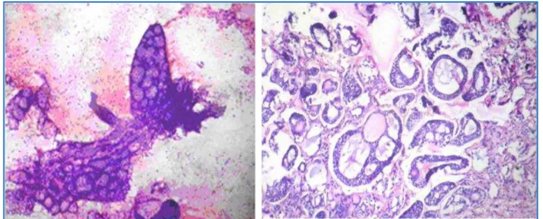
In aspirates with a diagnosis of pleomorphic adenoma, there was correlation seen in 86% of cases and 4 cases turned out to be malignant lesions on histopathology. Warthin’s tumour showed a correlation in 66.66% of cases. Eight cases with cystic aspirates on histopathology showed pleomorphic adenoma in 4 cases, basal cell adenoma in 1 case, Warthin’s tumour in 2 cases and one case with mucoepidermoid carcinoma. (Table 5).
Cytological diagnosis -Benign lesions Number of cases Histopathological diagnosis Pleomorphic
adenoma 43
37-Pleomorphic adenoma 2-Basal cell adenoma
2-Mucoepidermoid carcinoma 1-Adenoid cystic carcinoma 1-Polymorphous low grade Adenocarcinoma
Warthin’s tumour 3
2-Warthin’s tumour
1-Lymphoepithelial lesion
Basal cell
adenoma 2
1-Basal cell adenoma 1-Pleomorphic
adenoma
Cystic lesions 8
4-Pleomorphic adenoma 2-Warthin’s tumour 1-Basal cell adenoma
1-Mucoepidermoid carcinoma
Table 5: Cyto-histopathological correlation in Benign lesions
J. Evid. Based Med. Healthc., pISSN- 2349-2562, eISSN- 2349-2570/ Vol. 3/Issue 32/Apr. 21, 2016 Page 1524
Cytological diagnosis –Malignant lesions Number of cases Histopathological diagnosis
Mucoepidermoid carcinoma 4 All were Mucoepidermoid carcinoma
Adenoid cystic carcinoma 3 All were Adenoid cystic carcinoma
Acinic cell carcinoma 1 All were Acinic cell carcinoma
Carcinoma.Ex Pleomorphic adenoma 1 Pleomorphic adenoma
Adenocarcinoma. 1 Basal cell adenocarcinoma
Table 6: Cyto-histopathological correlation in Malignant lesions
Fig. 1: Mucoepidermoid carcinoma-both cytology and histopathology correlated
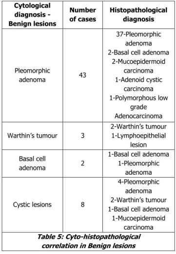
Fig. 2: Acinic cell carcinoma: both cytology and histopathology correlated
J. Evid. Based Med. Healthc., pISSN- 2349-2562, eISSN- 2349-2570/ Vol. 3/Issue 32/Apr. 21, 2016 Page 1525
Fig. 4: Polymorphous low grade adenocarcinoma: cytology reported as pleomorphic adenoma and Histopathology showed features of polymorphous low grade adenocarcinoma
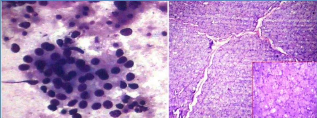
Fig. 5: Adenoid cystic carcinoma: both cytology and histopathology correlated
Fig. 6: Cytology reported as carcinoma. ex pleomorphic adenoma, histopathology showed pleomorphic adenoma
Out of 56 cases diagnosed as benign tumours on FNAC, 51 and 5 cases came out as benign and malignant tumours respectively on histopathological examination. Out of 11 cases diagnosed as malignant tumours on FNAC, 1 and 10 cases came out as benign and malignant tumours respectively on histopathological examination. (Table 7)
Benign on FNAC Malignant on FNAC
56 11
(Including inconclusive diagnosis) Benign on
HPE
Malignant on HPE
Benign on HPE
Malignant on HPE
51 5 1 10
Table 7: Correlation of cytology with histopathological diagnosis
In the present study FNAC showed Sensitivity of 66.6%, Specificity of 98%, Positive predictive value; 90.9%, Negative predictive value; 91%, Percentage of false negative cases 33.3%, Percentage of false positive cases 1.9% and Overall Diagnostic Accuracy of 91%.
DISCUSSION: Salivary gland tumours elicit considerable medical interest because of their multifacial clinical presentation, varied histological appearance, and difficulties in predicting prognosis.
J. Evid. Based Med. Healthc., pISSN- 2349-2562, eISSN- 2349-2570/ Vol. 3/Issue 32/Apr. 21, 2016 Page 1526 carcinomas of the salivary glands are uncommon accounting
for less than 0.33% of all cancers. Tumours have the highest chance of being malignant if they arise in retromolar area, the floor of the mouth, tongue and sublingual gland while only 20% of all parotid tumours are malignant.
FNAC of salivary gland tumour is advantageous to both the patient and the clinician because of its immediate results, accuracy, minimal complications and economy. Appropriate therapeutic management may be planned earlier, whether it is a local excision for a benign tumour, radical surgery for a malignant neoplasm or any other alternative treatment.
In the study by Shafkat Ahmad[1] et al, benign tumours
were common in the 3rd and 4th decade and malignant tumours in 4th and 5th decade. Pablo Agustin[2] et al and
the present showed similar features. The male:female ratio of salivary tumours in various studies were: Ma aita JK[3] et
al 1.6:1.2, Shafkat Ahmad[1] et al 1.17:1.3, Dilip K Das[4] et
al 1.28:1.4, Pablo Agustin[2] et al 1:2.5. In the present study
the ratio was 1: 1.1 with female preponderance.
The percentage of benign and malignant tumours in various studies: Patrick O’ Dwyer[5] et al; 80%, 20%, Pablo
Agustin[2] et al; 80%, 20%, Elagoz S[6] et al; 77.5%, 22.5%,
Nicholas Stow[7] et al; 67.3%, 32.7% respectively. In the
present study, benign tumours were: 77.6% and malignant tumours 22.4%, similar to other comparative studies.
In the present study, parotid gland was most common gland involved similar to other studies. M. Naderpour[8] et al,
Fernandes C[9] et al, Pablo[2] et al, Shafkat[1] et al and in the
present study pleomorphic adenoma and mucoepidermoid carcinoma were the most common benign and malignant tumours.
The majority of the cysts of major salivary glands are cystic neoplasms either benign or malignant. Warthin’s tumour and low-grade mucoepidermoid carcinoma are the commonest (Fig. 1), but pleomorphic adenoma and acinic cell carcinoma may also predominantly or partly cystic. Aspirated fluid from cystic neoplasm is often poor in cellularity and difficult to interpret. Sampling from solid area under ultrasound may solve the problem in selected cases. In a study done by M. Naderprour[8] et al, 10/124 cases were
diagnosed as cystic lesions on FNAC.
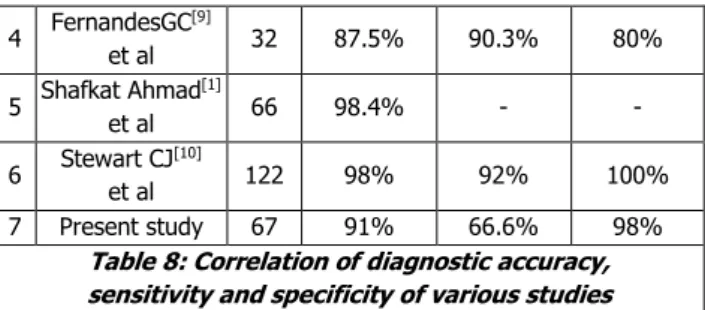
In the present study, we observed that Diagnostic accuracy-91 %, Sensitivity-66.6 % and Specificity–98 %. Our study showed less Sensitivity, more Specificity when compared to other studies. (Table 8).
Sl.
No Study
No. of cases
Diagnostic
Accuracy Sensitivity Specificity
1
Patrick O` Dwyer[5]
et al
249 90% 73% 94%
2 Dilip K Das
[4]
et al 45 91.1% 94.6% 75%
3 Nicholas Stow
[7]
et al 104 92.3% 88.6% 88.6%
4 FernandesGC
[9]
et al 32 87.5% 90.3% 80%
5 Shafkat Ahmad
[1]
et al 66 98.4% - -
6 Stewart CJ
[10]
et al 122 98% 92% 100% 7 Present study 67 91% 66.6% 98%
Table 8: Correlation of diagnostic accuracy, sensitivity and specificity of various studies
CONCLUSION: FNAC is a very useful, simple, cheap,
accurate and repeatable technique in the preoperative diagnosis of various salivary gland neoplasms. Overall diagnostic accuracy was 91%, Sensitivity was 66.6 % and Specificity was 98%, The results of present study were comparable to most other studies. We found that in cystic lesions of salivary glands, FNAC alone is not diagnostic. As cystic neoplasms may be benign or malignant, guided FNAC combined with biopsy is essential for specific diagnosis.
REFERENCES:
1. Shafkat Ahmad, Mohainmad Lateef, Rouf Ahmad.
Clinicopathological study of primary salivary gland tumours in Kashmir. JK-Practitioner 2002;9(4):231-233.
2. Pablo Agustin Vargas, Rene Gerhard, Vergílius JF Araújo Filho, et al. Salivary gland tumours in a Brazilian population: a retrospective study of 124 cases. Rev Hosp Clin Sao Paulo 2002;57(6):271-276.
3. Ma’ata JK, Al-Kaisi, A–tamimi S. Salivary gland tumours
in Jordan: a retrospective study of 221 patients, Croat Med J 1999;40(4):539-542.
4. Das DK, Petkar MA, Al-Mane NM, et al. Role of FNAC in the diagnosis of swellings in the salivary gland regions: a study of 712 cases. Med Princ Pract 2004;13(2):95-106.
5. O'Dwyer P, Farrar WB, James AG, et al. Needle
aspiration biopsy of major salivary gland tumours, its value. Cancer 1986;57(3):554-557.
6. Elagoz S, Gulluoglu M, Ozer H, et al. The value of fine-needle aspiration cytology in salivary gland lesions. ORL J Otorhinolaryngol 2007;69(1):51-56.
7. Nicholas Stow, David Veivers, Alan poole. FNAC in the management of salivary gland tumours: an Australian
experience. Ear, Nose and Throat Journal
2004;83(2):128-131.
8. Masoud Naderpour, Sahidi N, Daryani Amir. Evaluation
of diagnostic fine needle aspiration cytology in parotid masses. The Internet Journal of Head and Neck Surgery 2008;2(2):9.
9. Fernandes GC, Pandit AA. Diagnosis of Salivary gland
tumours by FNAC. Bombay Hospital Journal 2000;42(1):108-111.
10.Stewart CJ, Mackenzie K, McGarry GW. FNAC of