© Applied Science Innovations Pvt. Ltd., India Carbon – Sci. Tech. 5/1 (2013) 203 $ 210
! "# # $ "# # % & ' ( % ' ) ' *#
(A) Interactive Research School for Health Affairs (IRSHA), Bharati Vidyapeeth University, Katraj Dhankawadi, Pune Satara Road, Pune 411043, Maharashtra, India.
(B) Nanoscience group, Department of Chemistry, Post graduate & Research Center, MES Abasaheb Garware College, Karve Road, Pune 411004, Maharashtra, India.
#
Contributed equally; * + Ruchika Kaul Ghanekar, Tel: +91 20 24366929/+91 20 24366931; Fax: +91 20 24366929/+91 20 24366931; E mail: [email protected], [email protected]
We report a cost effective and eco friendly biosynthesis of gold nanoparticles (F AuNPs) using aqueous extract of Ficus religiosa as the reducing and stabilizing agent. These nanoparticles were characterized by various techniques such as UV Vis, XRD, TEM and FTIR. The characteristic surface plasmon peak was observed at 540 nm while XRD analysis suggested it to be a face centered cubic (fcc) structure with peaks at 38.06, 44.46, 64.75 and 77.56. FTIR studies indicated the capping of the nanoparticles with polyphenols, amines and carboxylates present in the extract of Ficus religiosa whereas TEM analysis showed spherical morphology with other shapes such as triangles and hexagons. The F AuNPs were found to be non toxic to HEK 293 cells, thereby suggesting their potential application in the field of nanobiotechnology.
, + Biosynthesis, gold nanoparticles, Ficus religiosa, cytotoxicity.
- + The field of nanotechnology has
recently witnessed significant advances in the physical and chemical methods of synthesis of nanomaterials ./# 01. Due to the use of toxic and aggressive chemicals as reducing and/or capping agents in such synthetic processes, large amounts of hazardous by products are released in the environment, thereby raising an alarming concern .2# 31. This has invited attention towards a clean, non toxic and environment friendly method of nanomaterial synthesis known as ‘Green Nanotechnology’ .2# 41. Biological methods of synthesis of nanomaterials are considered to be environmentally safe and sound compared to the conventional synthetic methods .5# 61.
A number of studies have used biomolecules such as proteins, amino acids, carbohydrates, and sugars; whole cells of bacteria, fungi, and algae; or different plant parts such as roots, leaves, flowers, bark powders, seeds, roots and fruits for the synthesis of metal nanoparticles .71. Since plants are rich in polyphenols, they serve as important materials for the production of metallic nanoparticles .81. Such type of synthesis using phytochemicals minimizes or eliminates chemical interventions, thereby resulting into truly green and non polluting eco friendly industrial process. This type of synthesis of nanoparticles reduces the cytotoxicity as well as increases the bioavailability of the nanoparticles.
- 9863 : 9435
© Applied Science Innovations Pvt. Ltd., India Carbon – Sci. Tech. 5/1 (2013) 203 $ 210
Currently, there has been a growing demand for the biogenic synthesis of gold nanoparticles to avoid toxicity problems, if such metal nanoparticles are intended for application to human beings ./1. Biosynthesis of gold nanoparticles has been reported with microbes such as bacteria ./91, fungi .//1 and actinomycetes ./01; as well as with phytochemicals present in hibiscus ./21, geranium ./31, lemon grass ./41, cinnamon ./51, neem ./61, Aloe vera ./71, tamarind ./81, oat, wheat .091, alfalfa .0/1, bengal gram .001, tea .81, cumin .021 and onion .031. Honey has also been reported to help in the synthesis of gold nanoparticles .041.
Ficus religiosa Linn. (Moraceae), has been known for medicinal properties due to presence of phenols, tannins, steroids, alkaloids and flavonoids, vitamin K, methyl oleanolate, n octacosanol, β sitosteryl d glucoside, lanosterol, stigmasterol, lupen 3 one .051. Most of these phytochemicals are water soluble and, hence are easily extracted in water without losing their properties. Recently, we have reported the antioxidant activity of Ficus religiosa that may be due to the presence of the phytochemicals .051. Such phytochemicals act as metal reducing and capping agents, thereby resulting into one step biosynthesis of metal nanoparticles.
In this study, we have reported an environmental friendly biosynthetic approach for the synthesis of gold nanoparticles (F AuNPs) with Ficus religiosa extract. The F AuNPs were synthesized by mixing an aqueous solution of Ficus religiosa extract with chloroauric acid solution. The as obtained nanoparticles were characterized by UV/Vis spectroscopy, X ray diffraction (XRD), transmission electron microscopy (TEM) and Fourier transform infrared spectroscopy (FTIR). Their stability was monitored and cytotoxicity was evaluated on human HEK 293 cells.
+
+ All the chemicals including HAuCl4, Analytical Reagent (AR) grade were procured from Sigma Aldrich, USA. Bark of Ficus religiosa L. was collected from Pune District, Maharashtra, India. A voucher specimen (MPCC
2417) of authentic plant species has been deposited at the herbarium of Medicinal Plants Conservation Center (MPCC), Pune, Maharashtra, India. Triple distilled water was used throughout the experimental work. For cell culture, Dulbecco’s Modified Eagle’s Medium (DMEM) and Fetal Bovine Serum (FBS) were purchased from Sigma Aldrich, USA. Penicillin / streptomycin and Trypsin were obtained from Gibco BRL, CA, USA, and L glutamine was bought from Himedia. All other common reagents including 3 (4,5 dimethylthiazol 2 yl) 2,5 diphenylthiazolium bromide (MTT) were bought from Sigma Aldrich, USA. HEK 293 cell line was obtained from NCCS, India.
+
/< = >
/</ > ?
+ The aqueous extract was prepared as described previously .051. Briefly, the bark of Ficus religiosa was chopped into small pieces, shade dried at ambient temperature and ground into coarse powder in a grinder. Aqueous extracts was prepared as per standard Indian Pharmacopoeia .061. The extract so obtained, was centrifuged at 13000 rpm for 15 min and the supernatant was used as reducing agent for synthesis of gold nanoparticles.
/<0 + Aqueous
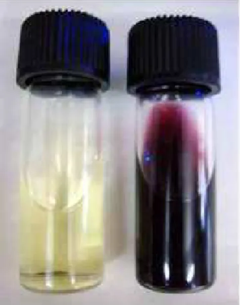
extract of F. religiosa was used for the biosynthesis of gold nanoparticles (F AuNPs). Around 10 ml of 1mM auric chloride salt was heated to 80°C in a rotamantle and stirred continuously for 20 min to which was added 600 Ml of 10 mg/ml concentration of F. religiosa aqueous extract. The reaction was carried out till the solution changed colour from yellow to wine red, indicating the formation of gold nanoparticles. The solution was further filtered through a 0.2 micron filter. The as obtained gold nanopaticles (F AuNPs) were characterized by UV Vis absorption spectroscopy, XRD, TEM and FTIR.
0< >
0</ @A A + UV Vis
© Applied Science Innovations Pvt. Ltd., India Carbon – Sci. Tech. 5/1 (2013) 203 $ 210
spectrophotometer. The spectra was recorded in the wavelength range of 450 1000 nm.
0<0 B % C B%C $ +
Determination of crystallinity, phase purity, lattice properties and identification of air dried F AuNPs was done by XRD studies using powder diffractometer with Cu Kα radiation, operating at 40 kV and a current of 40 mA (X Ray Diffractometer, Schimadzu, at Pune University, Pune). The crystallite size was calculated from the width of the XRD peaks by using Scherrer’s formula :
θ
β
λ
cos 9 . 0 =
D (1)
where, D is the average crystalline size, λ is the X ray wavelength used, β is full width at half maximum intensity and θ is the Bragg’s angle in degrees.
0<2 D $ = D=
$ + TEM was performed to elucidate the morphology as well as size of the biosynthesised F AuNPs on a JEOL model 1200EX instrument operated at an accelerating voltage at 80 kV. Colloidal solution F AuNPs, in triple distilled water, was ultrasonicated for 15 min and then coated onto ultraclean carbon coated copper grid for analysis.
2< $
D-% +FTIR studies of as synthesized F AuNPs were performed on Schimadzu IR Infinity (at SP College, Pune) to analyze their capping/bonding with polyphenols present in the extract of F. religiosa. The spectra have been recorded between 400 to 4000 wavelength range by mixing the dried F AuNPs with KBR powder and allowing the spectra to be run under controlled atmospheric conditions.
3< - + In vitro stability
studies of as obtained F AuNPs were performed by monitoring the UV Vis absorbance over a period of 0 h, 24 h and 7 days .71.
4< $ DD
+ The viability study was performed by using
MTT dye in non cancerous transformed human embryonic kidney cell line, HEK 293. The cells were grown in DMEM supplemented with 2 mM L glutamine, 100 units/ml of penicillin / streptomycin, and 10% fetal bovine serum incubated in a humidified 5% CO2 atmosphere at 37°C. The cells were seeded at 1×10⁵cells/ml density in 96 well plates (BD Falcon, USA). After 24 h, the cells were incubated with fresh medium containing F AuNPs added at concentrations ranging from 0 200 MM (each dose in triplicates) and the plates were incubated overnight at 37°C in 5% CO2 incubator. The MTT solution (5 mg/ml) was added to each well and the cells were cultured for another 4 h at 37°C in 5% CO2 incubator. The intensity of colored formazan derivative was determined by measuring optical density (OD) with the ELISA microplate reader (Biorad, Hercules, CA) at 570 nm (OD570–630 nm). The mean OD value of three wells was used for assessing the cell viability expressed as percentage of control.
×100
cells Control
cells treated le
Nanopartic =
%Viability (2)
5< + All the experiments
were performed in triplicates. The data has been represented as mean ±SD. Statistical analysis was conducted with the Graph Pad Prism 4 program using one way ANOVA. The p value used for the comparison was <0.05.
% C +
/< > + The production of
© Applied Science Innovations Pvt. Ltd., India Carbon – Sci. Tech. 5/1 (2013) 203 $ 210
Figure (1) : F AuNPs formation: A picture showing change in colour of 1mM HAuCl4 gold precursor (yellow colour: left vial) after gold nanoparticles formation (wine red colour: right vial) due to the addition of Ficus religiosa extract.
0< >
0</ @A A + The UV
Visible spectra of F AuNPs (Figure 2) showed prominent absorbance maxima at 540 nm, thereby confirming the production of gold nanoparticles. This peak was seen due to the excitation of surface plasmon vibrations in the gold nanoparticles.
Figure (2) : UV Vis spectra of as obtained F AuNPs: Graph of absorbance of F AuNPs plotted against 450 1000nm wavelength range showing prominent absorbance maxima at 540 nm.
0<0 B % C B%C $ +
XRD spectra of F AuNPs (Figure 3) exhibited diffraction peaks corresponding to (111), (200), (220) and (311) phases in the 2θ range of 30° 90°. This was in agreement with JCPDS 04 0784 data for gold nanoparticles. In the XRD pattern, diffraction peaks at angles of 38.1°, 40.4°, 65.3° and 77.4° could be assigned to face centered cubic (fcc) metallic gold (111), (200), (220) and (311) facets of the gold crystals, respectively. The average crystallite size of the F AuNPs was estimated from Scherrer’s formula to be ~30 nm indicating that the dispersed nanoparticles have uniform growth along this plane. It also suggested a spherical morphology for F AuNPs.
Figure (3) : XRD Spectra of F AuNps: The XRD pattern showing characteristic peaks at specific angles that are the signature of gold nanoparticles.
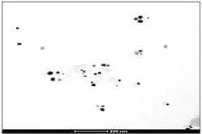
0<2 D= $ + The TEM image of
© Applied Science Innovations Pvt. Ltd., India Carbon – Sci. Tech. 5/1 (2013) 203 $ 210
Figure (4) : TEM Image of F AuNPs. The image shows mostly spherical F AuNPs as well as presence of other shapes such as triangles, pentagons and hexagons. Scale bar is 200 nm.
Figure (5) : FTIR spectra of F AuNPs: Graph of % Transmittance plotted against wavelength range of 400 4000nm. FTIR spectra of (a) as obtained F AuNPs and (b) Ficus religiosa extract.
0<3 $
D-% + FITR spectra of F AuNPs in the range 4000 400 cm1 (Figure 5) exhibited characteristic finger printing regions of various functional groups such as –OH, amide, amine and carboxylates with –C=O stretching vibrations. For example, in Figure 5 (a) of F AuNps, the peak at 3894 cm1 indicated the bonded –OH groups while the other splitted peak at 3637 cm1 corresponded to amine linkages from β sitosteryl d glucoside that has come from the F. religiosa extract. The shift in the peak at 3605 cm1 in F. religiosa extract (b) to 3497 cm1 in (a) indicated that F AuNPs were functionalized with amine groups. Other facet of the spectra indicated that the peak at 1745 cm1 (Figure 5b), due to
carboxylic acid functions present in the extract of Ficus religiosa, is shifted towards lower energy side i.e. 1658 cm1 (Figure 5a) for F AuNP’s. This shift clearly reflects the capping of Au naoparticles with carboxylic acid groups which are responsible for their aqueous dispersion.
FTIR data suggests that the polyphenols and aldehydes/ketones present in Ficus religiosa extract are not only responsible for the biosynthesis of F AuNPs but also govern their size due to their binding ability resulting into capping of the nanoparticles. Moreover, binding of the functional groups present in the aqueous extract to the nanoparticles is directing their morphology, thus, resulting into synthesis of different shapes.
2< - + The as synthesized
F AuNPs were kept on the shelf for a period of 0 h, 24 h and 7 days and UV absorbance was taken during these time periods (Figure 6). Besides slight changes in 600 700 nm region in 7 day period, there was no major change in the UV absorbance of F AuNPs at 540 nm over different time periods. Thus, the nanoparticles exhibited excellent stability over the time.
Figure (6) : UV Vis absorbance spectra: Graph of absorbance of F AuNPs in water at different time intervals (0h, 24h and 7 days) plotted against wavelength.
3< A DD + It was
© Applied Science Innovations Pvt. Ltd., India Carbon – Sci. Tech. 5/1 (2013) 203 $ 210
100% viability upto 80 MM concentration of F AuNPs and around 87 79% viability at concentrations varying between 100 200 MM. The cells showed more than 70% viability up to 200 MM concentration of the nanoparticles, thereby confirming that the as synthesized F AuNPs were non toxicand safe.
Figure (7) : Cell Viability Assay: Graph shows % viability of HEK 293 cells treated with different concentrations of F AuNPs.
+ The present report demonstrates a rapid green synthesis of gold nanoparticles using Ficus religiosa extract. The nanoparticles showed excellent stability and uniform capping due to the presence of polyphenols, amines and carboxylates present in the extract. The effective capping and subsequent dispersiblility of these nanoparticles in water enhanced their bioavailability, thereby making them available for various biomedical applications. Thus, biological methods of nanoparticle synthesis have been suggested as ecofriendly alternatives to chemical and physical synthetic methods since the latter involve toxic chemicals that could be detrimental to human health.
' , $ + We thank our Director, Professor P. K. Ranjekar as well as IRSHA, Bharati Vidyapeeth University, Pune, Maharashtra for funding our work. We also thank Physics Department, Pune University and Chemistry Department, S.P. College, Pune for helping us in characterization experiments.
% +
[1] V. Kumar, S. K. Yadav, Synthesis of variable shaped gold nanoparticles in one solution using leaf extract of Bauhinia variegata L., Digest Journal of Nanomaterials and Biostructures, 6(4) (2011) 1685 1693.
[2] J. A. Dahl, B. L. S. Maddux, J. E. Hutchison, Toward Greener Nanosynthesis, Chem. Rev., 107(6) (2007) 2228 2269. [3] J. E. Hutchison, Greener nanoscience: a
proactive approach to advancing applications and reducing implications of nanotechnology, ACS Nano., 2(3) (2008) 395 402.
[4] L. Hanying , J. D. Carter, T. H. LaBean, Nanofabrication by DNA self assembly, 12(5) (2009) 24 32.
[5] M. A. Albrecht, C. W. Evans, C. L. Raston, Green chemistry and the health implications of nanoparticles, Green Chem., 8 (2006) 417–432.
[6] D. Bhattacharya, R. K. Gupta, Nanotechnology and Potential of Microorganisms, 25(4) (2005) 199 204. [7] P. Mohanpuria, N. K. Rana and S. K.
Yadav, Biosynthesis of nanoparticles: technological concepts and future applications, Journal of Nanoparticle Research, 10(3) (2008) 507 517.
[8] G. Ghodake, C. Eom, S. W. Kim and E. Jin, Biogenic Nano Synthesis; towards the Efficient Production of the Biocompatible Gold Nanoparticles, Bull. Korean Chem. Soc., 31(10) (2010) 2771 2775.
[9] S. K. Nune , N. Chanda , R. Shukla , K. Katti , R. R. Kulkarni , S. Thilakavathi, S. Mekapothula , R. Kannan, K. V. Katti , Green Nanotechnology from Tea : Phytochemicals in Tea as Building Blocks for Production of Biocompatible Gold Nanoparticles, J Mater Chem., 19(19) (2009) 2912 2920.
© Applied Science Innovations Pvt. Ltd., India Carbon – Sci. Tech. 5/1 (2013) 203 $ 210
[11] P. Mukherjee, A. Ahmad, D. Mandal, S. Senapati, S. R. Sainkar, M. I. Khan, R. Parishcha, P. V. Ajaykumar, M. Alam, R. Kumar, M. Sastry, Fungus mediated synthesis of silver nanoparticles and their immobilization in the mycelial matrix: a novel biological approach to nanoparticle synthesis, Nano Lett, 1 (2001) 515–519. [12] A. Ahmad, S. Senapati, M. I. Khan, R.
Kumar, M. Sastry, Extracellular biosynthesis of monodisperse gold nanoparticles by a novel extremophilic actinomycete, Thermomonospora sp., Langmuir, 19 (2003) 3550–3553.
[13] P. Daizy, Green synthesis of gold and silver nanoparticles using Hibiscus rosa sinensis, Physica E: Low dimensional Systems and Nanostructures, 42(5) (2010) 1417–1424. [14] S. S. Shankar, A. Ahmad, R. Pasrichaa, M.
Sastry, Bioreduction of chloroaurate ions by geranium leaves and its endophytic fungus yields gold nanoparticles of different shapes, J Mater Chem, 13 (2003) 1822–1826. [15] S. S. Shankar, A. Rai, A. Ahmad, M. Sastry,
Controlling the optical properties of lemongrass extract synthesized gold nanotriangles and potential application in infrared absorbing optical coatings, Chem Mater 17 (2005) 566–572.
[16] J. Huang, Q. Li, D. Sun, Y. Lu, Y. Su, X. Yang, et al, Biosynthesis of silver and gold nanoparticles by novel sundried
Cinnamomum camphora leaf,
Nanotechnology 18:105 (2007) 105104– 105114.
[17] S. S. Shankar, A. Rai, A. Ahmad, M. Sastry, Rapid synthesis of Au, Ag, and bimetallic Au core–Ag shell nanoparticles using neem (Azadirachta indica) leaf broth, J Colloid Interf Sci, 275 (2004) 496–502.
[18] S. P. Chandran, M. Chaudhary, R. Pasricha, A. Ahmad, M. Sastry, Synthesis of gold nanotriangles and silver nanoparticles using Aloe vera plant extract, Biotechnol Prog 22 (2006) 577–583.
[19] B. Ankamwar, M. Chaudhary, M. Sastry, Gold nanotriangles biologically synthesized using tamarind leaf extract and potential application in vapor sensing, Synth React
Inorg Metal Org Nano Metal Chem, 35 (2005) 19–26.
[20] V. Armendariz, J. L. Gardea Torresdey, M. Jose Yacaman, J. Gonzalez, I. Herrera J. G. Parsons, Gold nanoparticles formation by oat and wheat biomasses, Waste Research Technology Conference at the Kansas City, Mariott Country Club Plaza July30–Aug1, (2002)
[21] J. L. Gardea Torresdey, E. Gomez, J. R. Peralta Videa, J. G. Parsons, H. Troiani, M. Jose Yacaman, Alfalfa sprouts: a natural source for the synthesis of silver nanoparticles, Langmuir 19 (2003) 1357– 1361.
[22] K. Ghule, A. V. Ghule, J. Y. Liu, Y. C. Ling, Microscale size triangular gold prisms synthesized using Bengal gram beans (Cicer arietinum L.) extract and HAuCl4 × 3H2O: a green biogenic approach, J Nanosci Nanotechnol, 6 (2006) 3746–3751.
[23] K. Kattia, N. Chandaa, R. Shuklaa, A. Zambrea, T. Suibramaniana, R. R. Kulkarni, R. Kannana, K. V. Katti, Green
Nanotechnology from Cumin
Phytochemicals: Generation of Biocompatible Gold Nanoparticles, Int J Green Nanotechnol Biomed., 1(1) (2009) B39–B52.
[24] U. K. Parida, B. K. Bindhani, P. Nayak, Green Synthesis and Characterization of Gold Nanoparticles Using Onion (Allium cepa) Extract, World Journal of Nano Science and Engineering, 1 (2011) 93 98. [25] P. Daizy, Honey mediated green synthesis of
goldnanoparticles, Spectrochimica Acta Part A: Molecular and Biomolecular Spectroscopy, 73(4) (2009) 650–653.
[26] A. S. Choudhari, S. Suryavanshi, H. Ingle, R. Kaul Ghanekar, Evaluating the antioxidant potential of aqueous and alcoholic extracts of Ficus religiosa using ORAC assay and assessing their cytotoxic activity in cervical cancer cell lines, Biotechnol. Bioinf. Bioeng., 1(4) (2011) 443 450.
© Applied Science Innovations Pvt. Ltd., India Carbon – Sci. Tech. 5/1 (2013) 203 $ 210
[28] C. Singh, R. K. Baboota, P. K. Naik, H. Singh, Biocompatible synthesis of silver and gold nanoparticles using leaf extract of Dalbergia sissoo, Adv. Mat. Lett., 3(4) (2012) 279 285.
[29] T. Elavazhagan, K. D. Arunachalam, Memecylon edule leaf extract mediated green synthesis of silver and gold nanoparticles, International Journal of Nanomedicine 6 (2011) 1265–1278.
[30] H. Bar, D. K. Bhui, G. P. Sahoo, P. Sarkar, S. P. De, A. Misra, Green synthesis of silver nanoparticles using lstex of Jatropha curcus, Colloids and Surfaces A : Physicochem. Eng. Aspects 339 (2009) 134–139.
[31] S. S. Shankar, A. Rai, B. Ankamwar, A. Singh, A. Ahmadand M. Sastry, Biological synthesis of triangular gold nanoprisms, Nature Materials 3 (2004) 482 – 488.