RUTHENIUM-106 BRACHYTHERAPY FOR UVEAL MELANOMAS –
PRELIMINARY RESULTS: A SINGLE INSTITUTIONAL EXPERIENCE*
Rodrigo Souza Dias1, Adelmo José Giordani1, Clélia Maria Erwenne2, Helena Regina Comodo Segreto1, Luiz Fernando Teixeira2, Roberto Araujo Segreto1
OBJECTIVE: To analyze the early response of uveal melanomas in patients treated with ruthenium-106 brachytherapy. MATERIALS AND METHODS: In the period between April 2002 and July 2003, 20 patients diagnosed with uveal melanoma were submitted to ruthenium-106 brachytherapy. The calculated dose de-livered at the apex of the tumor ranged between 55 Gy and 100 Gy. Patients with lesions greater than 5 mm were submitted to transpupillary thermotherapy concomitantly with ophthalmic plaque insertion. RESULTS: As regards the lesions site, 75% of the lesions were located in the choroid, 15% in the iris, and the remain-der 10% in the ciliary body. In a median 19-month-follow-up, the progression-free survival for brachytherapy was 69%, and 87% for associated brachytherapy and transpupillary thermotherapy. A significant tumor height reduction was observed after treatment. No patient was submitted to enucleation. CONCLUSION: Our pre-liminary results show that ruthenium-106 brachytherapy is an appropriate method for conservative treat-ment of patients with uveal melanomas in terms of local managetreat-ment, ocular and visual acuity preservation with an acceptable complications incidence rate.
Keywords:Uveal melanoma; Ruthenium plaque therapy; Conservative therapy; Brachytherapy.
Braquiterapia com rutênio-106 em melanomas uveais – resultados preliminares: experiência uni-institucio-nal.
OBJETIVO: Analisar os resultados preliminares da braquiterapia com rutênio-106 em pacientes portadores de melanomas uveais. MATERIAIS E MÉTODOS: No período de abril de 2002 a julho de 2003, 20 pacientes com diagnóstico de melanoma uveal foram tratados com braquiterapia com rutênio-106. A dose calculada no ápice tumoral variou de 55 Gy a 100 Gy. Pacientes com lesões com altura maior que 5 mm foram sub-metidos a termoterapia transpupilar concomitante à colocação da placa oftálmica. RESULTADOS: Quanto à localização da lesão, esta se encontrava na coróide em 75% dos casos, na íris em 15% e no corpo ciliar em 10% dos pacientes. Com seguimento mediano de 19 meses, a sobrevida livre de progressão para a braqui-terapia e para a associação com a termobraqui-terapia transpupilar foi de 69% e 87%, respectivamente. Observou-se redução significante da altura tumoral após o tratamento. Nenhum dos pacientes foi submetido a enuclea-ção. CONCLUSÃO: Nossos resultados preliminares mostram que a braquiterapia com rutênio-106 é uma opção adequada para o tratamento conservador de melanomas uveais em termos de controle local, manutenção do globo ocular e visão útil, com índice aceitável de complicações.
Unitermos: Melanoma uveal; Placa de rutênio; Tratamento conservador; Braquiterapia. Abstract
Resumo
* Study developed in the Radiotherapy Unit of Department of Medicine and Department of Ophthalmology at Universidade Federal de São Paulo/Escola Paulista de Medicina (Unifesp/EPM), São Paulo, SP, Brazil.
1. Radiotherapy Unit of Department of Medicine – Universi-dade Federal de São Paulo/Escola Paulista de Medicina (Unifesp/ EPM), São Paulo, SP, Brazil.
2. Department of Ophthalmology – Universidade Federal de São Paulo/Escola Paulista de Medicina (Unifesp/EPM), São Paulo, SP, Brazil.
Mailing address: Prof. Dr. Roberto Araujo Segreto. Universidade Federal de São Paulo – Setor de Radioterapia, Departamento de Medicina. Rua Botucatu, 740, Vila Clementino. São Paulo, SP, Brazil, 04023-0900. E-mail: segreto.dmed@epm.br
Received February 22, 2006. Accepted after revision August 21, 2006.
INTRODUCTION
Although rare, uveal melanoma is the most common primary intra-ocular malig-nancy in adults, with an approximate
fre-quency of six cases per million inhabitants per year in the United States(1,2), seven
cases per million inhabitants per year in the Western Europe(3). Estimates for Brazil are
not available.
Up to the eighties, the treatment for pa-tients with uveal melanoma was surgical, consisting in enucleation, still the mainstay of therapeutical intervention for extensive lesions. However, other therapies aiming at preserving the vision and the ocular globe have been proposed since 1930(4).
In 1930, Moore utilized radon seeds di-rectly implanted into the ocular tumor of a patient(4). In 1960, Stallard initiated the
treatment for these tumors with cobalt-60 episcleral plaques. Since then, other
iso-topes like iodine-125, iridium-192, gold-198, paladium-103 and ruthenium-106 started being utilized(6–8).
Randomized prospective studies like those developed by the Collaborative Ocu-lar Melanoma Study (COMS) since 1986, showed that the mortality rate of patients with melanomas between 2.5 mm and 10 mm in elevation was similar when com-pared with the mortality rate for patients submitted to iodine-125 brachytherapy and to enucleation(6). Yet, in 1986, Lommatzsch
reported the utilization of ruthenium-106 beta-radiation for treatment of uveal mela-nomas with results comparable to those from other radioisotopes(7). So, ocular
pa-tients with uveal melanomas with up to 10 mm in elevation, allowing results similar to those from more radical procedures like enucleation, while preserving the visual acuity and the ocular globe.
Amongst the radioisotopes most fre-quently utilized in ocular brachytherapy, cobalt-60 is a gamma-radiation emitter with 1.25 MeV energy and 5.2-year half-life, whose main disadvantages are the higher radiation exposure of the medical team and higher radiation dose in adjacent organs, and iodine-125, a gamma-radiation emitter with 0.028 MeV energy and a rela-tively short 59.6-day half-life. As regards ruthenium-106, this isotope presents as main characteristic the beta-radiation emis-sion, allowing higher dose concentration in the tumor, and lower dose in adjacent ar-eas. Besides, the medical team involved in the procedure is submitted to lower expo-sure to radiation, and organs in risk receive lower radiation doses, consequently opti-mizing the radiological protection(3,8).
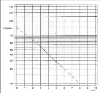
Considering that ruthenium-106 is a beta-radiation emitter, it is best indicated in the treatment of small melanomas, with up to 6 mm in elevation and a long half-life, allowing longer periods of use and there-fore lower cost compared with other iso-topes. Ruthenium radiation dose calcula-tion is performed with basis on tables pro-vided by the manufacturer, and otherwise expensive computer planning systems are not necessary(9) (Figure 1).
Main complications associated with brachytherapy are: retinopathy, cataract, neovascular glaucoma and maculopathy which may occur in 9% to 27% of cases in a three-year period. Main influencing fac-tors which seem to be related with such complications are tumor elevation, TMN stadium, and the proximity of the tumor with the fovea and optic disc(10).
Most recently, transpupillary thermo-therapy has started being utilized. It is a form of therapy utilizing infrared laser di-ode for inducing hyperthermia with a varia-tion between 45°C to 60°C. This therapeu-tic modality is exclusively indicated in cases of small melanomas in conjunction with brachytherapy allowing the treatment of lesions > 5 mm, or even as an adjuvant therapy in cases where regression is not achieved or where there is a regression of the disease(11). Transpupillary
thermo-therapy is characterized by a deep penetra-tion with an immediate cytotoxic effect, re-sulting in necrosis of the tumor up to 6 mm in depth in experiments with animals, and 4.7 mm in melanomas of the choroid in humans(12).
The ideal therapy for ocular melanomas still remains controversial, and ocular brachytherapy and external radiotherapy with proton beam are adequate therapeutic options aiming at preserving the ocular globe and the vision(13,14).
So, the present study is aimed at analyz-ing the preliminary results from
ruthenium-106 brachytherapy in patients with uveal melanoma.
MATERIALS AND METHODS
This is a retrospective study performed on 20 cases of patients with unilateral uveal melanoma, referred to the Radiotherapy Unit by the Department of Ophthalmology of Universidade Federal de São Paulo, for being submitted to ocular brachytherapy, in the period between April 2002 and July 2003.
The diagnosis was made by clinical and ophthalmological examinations. Fluores-cein angiography and ocular ultrasound A-and B-scans were requested. Both modes were employed for evaluating the tumors elevation. For evaluation of the tumors lo-calized in the iris and ciliary body ultra-sonic biomicroscopy was performed.
The staging of patients aiming at char-acterizing the absence of metastatic lesions included chest x-ray, abdominal ultra-sound, hemogram and biochemical analy-sis. The patients were staged according to TNM(15) and COMS(16–18) criteria.
For the present study, the following in-clusion criteria were taken into consider-ation: diagnosis of unilateral melanoma of the choroid, iris or ciliary body; tumor ≤ 6 mm in elevation; base diameter ≤ 16 mm; patients with age ≥ 21 years. Patient’s in-formed consent. Exclusion criteria were the following: diagnosis of another malig-nancy; patients undergoing immunosup-pressive therapy; patients incapable of re-turning for follow-up; patients with severe co-morbidities; multifocal or diffuse le-sions with scleral infiltration; previously treated patients; patients with metastasis; lesions that cannot be delimited by ocular ultrasound.
Bebig GmbH (Berlin, Germany) ruthe-nium-106 ophthalmic plaques were uti-lized. The main feature of these plaques is beta-radiation emission, with maximum 3.54 MeV energy, and half-life of 365 days. Two plaque models were employed: COB (round notched) and CCB (round), both with 20 mm in diameter (Figure 2). Pa-tients with lesions proximal to the optic nerve were submitted to brachytherapy with the COB plaque, while in the other the CCB plaque was utilized. Patients with
Figure 1. Calculation of
were constructed. The t-test was employed for evaluating variables related to local management and visual acuity, and the paired t-test for comparing pre- and post-therapy tumor elevation. Contingency tables were elaborated, and the Fisher’s exact test was employed for evaluating.
Kaplan-Meier curves were constructed to estimate survival curves of interest in re-lation to the progression-free survival.
In all of the tests, the null hypothesis rejection level was fixed in 0.05 or 5% (p < 0.05), significant values being marked with an asterisk, and the non-significant values, with (NS).
RESULTS
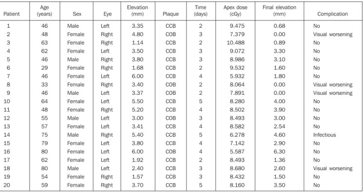
The patients’ ages ranged between 29 and 80 years (median, 56 years). The ma-jority of patients were Caucasian (95%), and women (70%) (Table 1).
As regards tumor characteristics, the right eye was involved in 45%, and the left eye in 55% of cases. Choroid and/or cili-ary body melanomas were diagnosed in 85% of patients, and iris melanoma in 15%. The lesion elevation ranged between 1.14 mm and 6 mm (median, 3.45 mm) (Table 1). TMN staging demonstrated 20% of
pa-Figure 2. RU-106 plaque models: CCB (round) and
COB (round notched).
lesions with more than 5 mm in elevation (five patients) were submitted to transpu-pillary thermotherapy delivered to the apex of the tumor, associated with placement of the ophthalmic plaque. The prescribed ra-diation dose was calculated on the apex of the lesion, from the inner face of the sclera. The present study covered the follow-ing aspects: demographic data (age and sex of patients), tumor characteristics (lesion site, affected eye, TNM and COMS stag-ing, tumor elevation), and treatment char-acteristics (type of plaque, radiation dose to the apex, base and sclera).
After the treatment, the patients were clinically evaluated every three months
during the first two years of follow-up, with the purpose of verifying the local manage-ment, progression-free survival, the lesion elevation, rate of preservation of the ocu-lar globe, visual acuity and presence of metastases and complications. Ocular ul-trasound and fluorescein angiography were performed every three months, and chest x-ray and abdominal ultrasound, every six months. The local management was de-fined as stabilization or decrease in the le-sion elevation at imaging study in compari-son with the pre-treatment study. The pres-ence of lesions in other sites was defined as metastasis. Progression-free survival corresponds to absence of local or distance failure. Treatment-related adverse effects were considered as complications. The fol-low-up period started in the date of the plaque placement.
Parameters regarding demographic, tu-mor and therapy characteristics, rate of preservation of the ocular globe, presence of metastasis and complications were sub-mitted for a descriptive analysis.
With the purpose of comparing numeri-cal variables in relation to the lonumeri-cal man-agement, lesion elevation before and after the therapy, and visual acuity, summary measures were calculated and box-plots
Table 1 Demographic and evolutive characteristics of patients submitted to ruthenium brachytherapy.
Patient 1 2 3 4 5 6 7 8 9 10 11 12 13 14 15 16 17 18 19 20 Age (years) 46 48 63 62 46 29 46 33 46 64 48 55 57 75 79 80 62 80 54 59 Sex Male Female Female Female Male Female Female Female Male Female Female Male Female Male Female Female Female Male Female Female Eye Left Right Right Left Right Right Left Right Left Left Right Left Left Right Left Left Left Left Right Right Elevation (mm) 3.35 4.80 1.14 3.50 3.80 1.68 6.00 3.40 3.37 5.50 5.20 3.00 3.41 5.40 3.80 6.00 1.92 2.40 1.57 3.70 Plaque CCB COB CCB CCB CCB CCB CCB COB COB CCB CCB COB CCB CCB CCB COB CCB CCB CCB CCB Time (days) 2 3 2 3 3 2 4 2 2 5 4 3 4 5 4 4 2 3 3 5 Apex dose (cGy) 9.475 7.379 10.488 9.072 8.986 9.532 5.932 8.064 7.891 8.280 8.502 8.493 8.582 6.278 7.142 5.587 8.493 8.680 8.432 8.160 Final elevation (mm) 0.68 0.00 0.89 3.30 3.10 1.60 1.80 0.00 0.00 4.00 3.90 3.00 2.54 4.60 2.90 6.30 1.36 2.60 1.50 3.50 Complication No Visual worsening No No No No No Visual worsening Visual worsening No No No No Infectious No No No Visual worsening No No
tients staged as T1, and 80% as T2. Accord-ing to COMS criteria, 10% of lesions were small-sized, and the remaining 90%, inter-mediate. As regards the therapy, the radia-tion dose to the apex ranged between 55.8 Gy and 104.8 Gy (median, 84.6 Gy). The median of the radiation-dose to the tumor base and sclera was respectively 248.3 Gy and 319.4 Gy. The CCB plaque was uti-lized in 75% of cases. The follow-up pe-riod ranged between 9 and 23 months (me-dian, 19 months).
During the clinical follow-up, an in-crease in the tumor dimensions was ob-served in five cases (25%). Of these pa-tients, three were submitted to adjuvant transpupillary thermotherapy, and currently are under follow-up, with no evidence of disease progression. Two patients could not undergo transpupillary thermotherapy be-cause of the lesion localization. One of them is under observation, and enucleation is schedule for the other. Table 2 shows the local management with brachytherapy and associated with transpupillary thermo-therapy. The initial elevation of the lesion
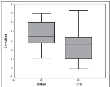
(p = 0.883), the prescribed radiation-dose to the tumor apex (p = 0.879) and the le-sion localization (p = 1.000) did not con-stitute significant factors for the local man-agement of the disease. The progression-free survival with brachytherapy and with adjuvant transpupillary thermotherapy may be observed in Kaplan-Meier survival curves (Figure 3). One patient progressed to death nine months after therapy due to unrelated causes. Comparing the initial el-evation of the lesion with the elel-evation af-ter the therapy, a significant reduction (p = 0.001) was observed (Figure 4). No patient was submitted to enucleation. As regards visual acuity at the moment of the latest evaluation, 70% of patients presented a vi-sual acuity higher than 20/200, and 30% presented visual acuity equivalent or lower than 20/200. The prescribed radiation dose to the tumor base and sclera did not con-stitute a prognosis factor of significance for the visual acuity.
During the follow-up period distant metastases were not detected. Complica-tions were observed in 25% of cases. In
four patients, visual acuity worsening was observed in one patient, and another pre-sented infectious complication.
DISCUSSION
Results regarding demographic com-pare favorably with previous studies pub-lished in the literature, demonstrating a predominance of uveal melanoma in Cau-casian patients with median age of 60 years. Singh and Tophan, in an epidemio-logical analysis of melanomas in the United States reported a 97.8% incidence in Cau-casian individuals. In this same study, the ages of the population studied ranged be-tween 6 and 100 years, 59.4 year for men, and 61.5 for women (median, 59.4 years). As regards sex, uveal melanoma seems to present a slightly high incidence in men. Notwithstanding this fact has not been ob-served in the present study, this may be jus-tified by the low number of patients in com-parison with studies performed for inci-dence evaluation(1).
In the analysis of the tumor character-istics, melanomas most frequently were located in the choroid. Also, a higher num-ber of cases of iris melanomas were ob-served in the present study in relation to the literature where the reported incidence achieves only 2%–3% for lesions localized in this topography(19)
. The mild predomi-nance in the left eye observed in the present
Table 2 Local management after brachytherapy and brachytherapy with transpupillary thermotherapy.
Treatament
Brachytherapy
Brachytherapy + TTT
N
15
18
Rate
75%
90%
N
5
2
Rate
25%
10%
N
20
20 Rate
100%
100%
Local management Failure Total
TTT, transpupillary thermotherapy.
Figure 4. Initial tumor elevation in relation to the final elevation.
E
le
va
ti
o
n
Initial Final
Figure 3. Kaplan-Meier estimation of progression-free survival in patients
submitted to ruthenium-106 brachytherapy and adjuvant transpupillary ther-motherapy.
Kaplan-Meier curve
P
ro
b
a
b
ili
ty
o
f
fr
e
e
-p
ro
g
re
ss
io
n
s
u
rv
iv
a
l
Time (months)
study also is reported by some studies in the literature. Hermann et al. observed that in 56% of cases, the tumors were localized in the left eye(20). However, this difference
was not observed in studies involving a higher number of cases(18)
.
All of the lesions were staged as T1 or T2N0M0 by the TNM (6th edition)(15), and
as intermediate by the COMS. In other publications about the employment of ru-thenium-106 in melanomas therapy, a pre-dominance of tumors staged as T1 and T2 also is observed. This is due the fact that ruthenium-106 is a Beta-radiation emitter with low penetrability, besides not being indicated for more extensive lesions. Also, it may be added that the majority of stud-ies was based on the staging method rec-ommended by the International Union Against Cancer (UICC) in 1997(21),
classi-fying choroid melanomas with > 5 mm in elevation and/or >15 mm in diameter as T3. According to the TMN 6th edition, only those lesions with > 16 mm in its larger diameter and/or > 10 mm in elevation would be classified as T3. So, considering the new staging, one can say that the inci-dence of T1 and T2 tumors described in the literature probably is higher. This also can be confirmed by the comparison between the median elevation of the lesions in the present study (3.45 mm) and that reported in the literature, ranging from 3 mm to 5.2 mm(22,23)
.
The most utilized plaque was the round-shaped CCB. The greatest majority of stud-ies performed with ruthenium-106 plaques do not describe the plaque models. In the present study, the CCB model was utilized in the treatment of patients with central lesions, proximal to the optic nerve, while the COB plaque was utilized in the remain-ing. As regards the lesions localization, the literature reports a lower incidence of cen-tral lesions in comparison with paracencen-tral, peripheral or yet ciliary body and iris le-sions. In the study performed by the COMS, lesions at 0–2 mm from the optic disc corresponded to only 14.9% of cases(24)
. On the other hand, in other pub-lications, central lesions were observed in 35% of cases, these results being similar to the ones from the present analysis(17)
. The ruthenium-106 plaque time of per-manence ranged between two and five days
(median, three days), and was directly pro-portional to the prescribed radiation-dose delivered to the tumor apex, and inversely proportional to the plaque activity. The prescribed dose delivered to the tumor apex ranged between 55 Gy and 105 Gy (me-dian, 85 Gy), and to the base, the prescribed dose ranged between 148 Gy and 450 Gy. The dose delivered to the sclera ranged between 191 Gy and 578 Gy. The radiation dose to be prescribed to the tumor apex with ruthenium-106 is still to be defined in the literature. In an analysis of several stud-ies in the literature about the use of ruthe-nium plaque for treatment of melanomas, one can observe that most of times the dose ranges between 80 Gy and 100 Gy, but studies with doses from 60 Gy to 160 Gy may be found. Rouberol et al. have treated 213 patients affected by choroid and cili-ary body melanoma with a 60 Gy dose(25)
. Shields et al., in a study about visual acu-ity in 1,106 patients with uveal melanoma submitted to brachytherapy with iodine-125, ruthenium-106, cobalt-60 or iridium -192, have described a median dose of 90.9 Gy to the apex, and 330 Gy to the tumor base(26). Seregard, in a meta-analysis with
1,066 patients treated with ruthenium-106 has observed a dose to the apex ranging between 80 Gy and 100 Gy(27)
. A study describing the Dutch experiment with ru-thenium for uveal melanoma utilized doses up to 160 Gy delivered to the tumor apex, and doses to the sclera from 220 Gy to 950 Gy(3). Hermann et al., analyzing the effect
of the dose-assignment in relation to results from ruthenium-106 plaque brachytherapy, utilized doses of 120 Gy to the apex of the tumor(20). Shields et al., in a study with
con-comitant transpupillary thermotherapy and brachytherapy, describe a dose to the apex ranging between 55 Gy and 124 Gy (me-dian, 90 Gy), and 254 Gy to the base(11). As
regards radiation dose to be utilized, the American Brachytherapy Society recom-mends the dose of 85 Gy to the apex of the lesion when iodine-125 is utilized as radio-isotope(14). However, these same guidelines
recommend a dose from 120 Gy to 160 Gy aiming at maximizing the healing effect when rutheium-106 is utilized. Meanwhile, the American Brachytherapy Society sug-gests that this large dose gradient may re-sult in high doses to the sclera, which may
cause a higher number of complications(14) . In a randomized prospective study devel-oped by the COMS utilizing iodine-125 in the treatment of choroid melanomas, in 49.9% of cases, the dose to the apex was 85.1 Gy to 120 Gy, and in 41.3% of patients the dose to the sclera ranged between 293 Gy and 409.9 Gy(27)
. So, studies evaluating dose-assignment excepted, the doses uti-lized in the present study are compatible with data in the literature regarding pre-scribed radiation dose to the apex of the lesion, to the tumor base and sclera.
Our results in relation to local manage-ment and progression-free survival com-pare to previous studies, despite the diffi-culty to evaluate retrospective data from different centers, considering variations in follow-up periods, tumors dimensions and doses delivered. Summanen et al., evaluat-ing 100 patients treated with ruthenium-106, have observed local failure in 19% of cases with 0.1 to 2.7 years of follow-up (median, 0.7 year)(28). This leads to the
con-clusion that local failures occur preco-ciously, especially in the first years of fol-low-up, and are very significant for the interpretation of our results. Studies with similar follow-up periods demonstrate a complete tumor regression in 57.1% of cases and local recidivation in 31% of pa-tients(29)
. Foerster et al., evaluating the ru-thenium therapy in100 patients affected by uveal melanoma, have observed local re-cidivation of 12%, with a median two-year follow-up period(30). Seregard, in a review
of the Swedish experiment with transpu-pillary thermotherapy as an adjunct to ru-thenium brachytherapy, has observed in a 20-month follow-up period that in 70.3% of cases there was a partial or complete regression of the lesion, a result very simi-lar to the 75% local management observed in the present study(31)
.
Lommatzsch et al., analyzing 141 pa-tients submitted to ruthenium brachyther-apy at a dose of 100 Gy, have observed a disease-free survival rate corresponding to 89.6% in five years(32). Seregard, in a
meta-analysis involving 1,066 patients treated with ruthenium at doses between 80 Gy and 100 Gy, has observed a disease-free sur-vival of 86% in five years(27). In the present
transpupillary thermotherapy is analyzed, one may observe that our results are simi-lar to those in the literature, with a longer follow-up period and higher number of cases.
Studies evaluating the dose-assignment effect on the local management show con-troversial results. Tjho-Heslinga et al., uti-lizing a dose to apex of 160 Gy in 101 pa-tients affected by uveal melanoma, have observed complete and partial remission in respectively 62.6% and 31.3% of cases, these results being slightly superior to those described in the literature with the usual doses. In the present study, transpupillary thermotherapy also was utilized in 25 pa-tients, allowing a reduction in the radiation dose and, consequently, in the risk of ret-inopathy(3)
. However, in another study with a 120 Gy dose to the apex of the lesion in patients with choroid melanomas, the ben-efit from the dose-assignment to the sur-vival has not been observed(20).
In a literature review, elevation and larger basal diameter of the tumor, dose livered to the tumor apex and retinal de-tachment play a significant role in relation to the local management and disease-free survival(27)
. In the present study, factors like lesion elevation, radiation dose, and tumor localization did not affect the local man-agement of the disease. This may have oc-curred because of the number o patients and number of failures observed, since this is a rare malignant neoplasm. Also, it may be added that the concomitant employment of transpupillary thermotherapy and brachytherapy in patients with lesions > 5 mm may have contributed to the favorable and similar results of patients with larger lesions who received lower radiation dose to the apex of their tumors.
The post-therapy tumor elevation was significantly lower than that before the treatment, evidencing that the therapy was effective, allowing a decrease in the tumor thickness of about 65% of the initial eleva-tion. In a literature review in terms of re-duction of tumor elevation after ruthenium brachytherapy, a reduction around 3%/ month is observed, and the majority of le-sions do not present a complete regression, stabilizing at a constant value around 61% of the initial elevation after approximately 24 months(23). In our series, results
concern-ing enucleation and visual acuity are slightly better than those in the literature. . Quivey et al. have observed that 58% of 239 patients treated with iodine plaques maintained a vision ≥ 20/200(33). The COMS Report no. 16 has evaluated the visual acuity of patients three years after iodine brachytherapy and observed an enucleation rate of 6.2% and visual acuity ≤ 20/200 in 43% of cases. In this same study, after a 24-month follow-up, com-pared with our series, 4% of the patients had been enucleated, and 33% presented visual acuity ≤ 20/200(24). In another
analy-sis of visual acuity after brachytherapy, Shields et al. have observed that in 51% of cases the visual acuity was ≤ 20/200(26).
Notwithstanding our results have shown that the prescribed radiation dose does not affect the visual acuity, probably because of the number of cases, the factors that seem to be related to the visual acuity after the therapy are: the isotope utilized, the presence of diabetes, the tumor elevation, the lesion localization, the type of plaque utilized, and the presence of retinal detach-ment(24).
The presence of metastasis is observed in about 10%-20% of patients with uveal melanoma, and is related mainly to the tu-mor size(34). In our series, the presence of
metastasis was not observed. This may be explained by the fact that the present study includes only patients at the initial phase of the disease, with tumors ≤ 6 mm in eleva-tion.
Analyzing this series as a whole, despite the short follow-up period and the small number of patients, our results show that ruthenium brachytherapy allowed a good local management of the disease and a rea-sonable progression-free survival in the studied population. Notwithstanding enu-cleation would be the therapy indicated in our institution, the use of this radioisotope allowed, in all of the cases, the preserva-tion of the ocular globe, and the mainte-nance of the visual acuity at a certain ex-tent.
This is the first study about the use of ruthenium in our country, and we consider that this radioisotope may be exclusively utilized in the treatment of uveal melano-mas with up to 5 mm in elevation, while patients with lesion with more than 6 mm
in elevation are candidates to brachyther-apy with other radioisotopes. In cases of melanomas with 5 mm to 6 mm in eleva-tion, ruthenium brachytherapy must, if possible, be associated with transpupillary thermotherapy.
REFERENCES
1. Singh AD, Topham A. Incidence of uveal mela-noma in the United States: 1973–1997. Ophthal-mology 2003;110:956–961.
2. Shields JA, Shields CL. Introduction to melano-cytic tumors of the uvea. In: Shields JA, Shields CL, editors. Intraocular tumors: a text and atlas. Philadelphia: Saunders, 1992;45–60. 3. Tjho-Heslinga RE, Davellar J, Kemme HM, et al.
Results of ruthenium irradiation of uveal mela-nomas: the Dutch experience. Radiother Oncol 1999;53:133–137.
4. Moore RF. Choroidal sarcoma treated by the in-traocular insertion of radon seeds. Br J Ophthal-mol 1930;14:145–146.
5. Stallard HB. Radiotherapy for malignant mela-noma of the choroid. Br J Ophthalmol 1966;50: 147–155.
6. Collaborative Ocular Melanoma Study Group. The COMS randomized trial of iodine 125 brachytherapy for choroidal melanoma, III: ini-tial mortality findings. COMS Report No.18. Arch Ophthalmol 2001;119:969–982. 7. Lommatzsch PK. Results after β-irradiation
(106Ru/106Rh) of choroidal melanomas: 20 years’
experience. Br J Ophthalmol 1986;70:844–851. 8. Lommatzsch PK, Werschnik C, Schuster E. Long-term follow-up of Ru-106/Rh-106 brachytherapy for posterior uveal melanoma. Graefes Arch Clin Exp Ophthalmol 2000;238:129–137. 9. Bebig Isotopen-und Medizintechnick GmbH.
Aplicadores oftalmológicos rutênio-106: instru-ções de emprego. Berlim: Bebig, 2001;04:01–08. 10. Summanen P, Immonen I, Kivela T, Tommila P, Heikkonen J, Tarkkanen A. Radiation related complications after ruthenium plaque radio-therapy of uveal melanoma. Br J Ophthalmol 1996;80:732–739.
11. Shields CL, Cater J, Shields JA, et al. Combined plaque radiotherapy and transpupillary thermo-therapy for choroidal melanoma: tumor control and treatment complications in 270 consecutive patients. Arch Ophthalmol 2002;120:933–940. 12. Journée-de Korver JG, Oosterhuis JA, de
Wolff-Rouendaal D, Kemme H. Histopathological find-ings in human choroidal melanomas after trans-pupillary thermotherapy. Br J Ophthalmol 1997; 81:234–239.
13. Gragoudas ES, Goitein M, Verhey L, Munzen-reider J, Suit HD, Koehler A. Proton beam irra-diation. An alternative to enucleation for intraocu-lar melanomas. Ophthalmology 1980;87:571– 581.
14. Nag S, Quivey JM, Earle JD, et al. The American Brachytherapy Society recommendations for brachytherapy of uveal melanomas. Int J Radiat Oncol Biol Phys 2003;56:544–555.
15. Greene FL, Page DL, Fleming ID, editors. In: AJCC Cancer Staging Manual. 6th ed. New York: Springer-Verlag, 2002;377–382.
of small choroidal melanoma: COMS Report No. 5. Arch Ophthalmol 1997;115:1537–1544. 17. Diener-West M, Earle JD, Fine SL, et al. The
COMS randomized trial of iodine 125 brachy-therapy for choroidal melanoma, II: characteris-tics of patients enrolled and not enrolled. COMS Report No. 17. Arch Ophthalmol 2001;119:951– 965.
18. The Collaborative Ocular Melanoma Study (COMS) randomized trial of pre-enucleation ra-diation of large choroidal melanoma I: character-istics of patients enrolled and not enrolled. COMS Report No. 9. Am J Ophthalmol 1998;125:767– 778.
19. Shields CL, Shields JA, Materim M, Gershen-baum E, Singh AD, Smith A. Iris melanoma: risk factors for metastasis in 169 consecutive patients. Ophthalmology 2001;108:172–178.
20. Hermann RM, Pradier O, Lauritzen K, Ott M, Schmidberger H, Hess CF. Does escalation of the apical dose change treatment outcome in β -radia-tion of posterior choroidal melanomas with 106Ru
plaques? Int J Radiat Oncol Biol Phys 2002;52: 1360–1366.
21. Sobin LH, Wittkind C. International Union Against Cancer. TNM classification of malignant tumors. 5th ed. New York: Wiley-Liss, 1997. 22. Stoffelns BM, Kutzner J, Jochem T.
Retrospec-tive analysis of ruthenium-106 brachytherapy for small and medium-sized malignant melanoma of
the posterior choroid. Klin Monatsbl Augenheilkd 2002;219:216–220.
23. Kaiserman I, Anteby Y, Chowers I, Blumenthal EZ, Kliers E, Pe’er J. Changes in ultrasound find-ings in posterior uveal melanoma after ruthenium 106 brachytherapy. Ophthalmology 2002;109: 1137–1141.
24. Melia BM, Abramson DH, Albert DM, et al. Col-laborative Ocular Melanoma Study (COMS) ran-domized trial of I-125 brachytherapy for medium choroidal melanoma. I. Visual acuity after 3 years. COMS Report No. 16. Ophthalmology 2001;108: 348–366.
25. Rouberol F, Roy P, Kodjikian L, Gerard JP, Jean-Louis B, Grange JD. Survival, anatomic, and functional long-term results in choroidal and cili-ary body melanoma after ruthenium brachyther-apy (15 years’ experience with beta-rays). Am J Ophthalmol 2004;137:893–900.
26. Shields CL, Shields JA, Cater J, et al. Plaque ra-diotherapy for uveal melanoma: long-term visual outcome in 1106 consecutive patients. Arch Ophthalmol 2000;118:1219–1228.
27. Seregard S. Long-term survival after ruthenium plaque radiotherapy for uveal melanoma. A meta-analysis of studies including 1,066 patients. Acta Ophthalmol Scand 1999;77:414–417. 28. Summanen P, Immonen I, Heikkonen J, Tommila
P, Laatikainen L, Tarkkanen A. Survival of pa-tients and metastatic and local recurrent tumor
growth in malignant melanoma of the uvea after ruthenium plaque radiotherapy. Ophthalmic Surg 1993;24:82–90.
29. Busse H, Muller RP. Techniques and results of
106Ru/106Rh radiation of choroidal tumours. Trans
Ophthalmol Soc UK 1983;103(Pt 1):72–77. 30. Foerster MH, Bornfeld N, Wessing A, Schulz U,
Schmitt G, Meyer-Schwickerath G. Treatment of malignant melanomas of the uvea with 106-ruthe-nium applicators. Report on the first 100 Essen cases. Klin Monatsbl Augenheilkd 1984;185: 490–494.
31. Seregard S, Landau I. Transpupillary thermo-therapy as an adjunct to ruthenium plaque radio-therapy for choroidal melanoma. Acta Ophthal-mol Scand 2001;79:19–22.
32. Lommatzsch PK, Werschnik C, Schuster E. Long-term follow-up of Ru-106/Rh-106 brachytherapy for posterior uveal melanoma. Graefes Arch Clin Exp Ophthalmol 2000;238:129–137. 33. Quivey JM, Char DH, Phillips TL, Weaver VA,
Castro JR, Kroll SM. High intensity 125-iodine (125I) plaque treatment of uveal melanoma. Int J
Radiat Oncol Biol Phys 1993;26:613–618. 34. Kleineidam M, Guthoff R, Bentzen SM. Rates of