Department of Orthopedics and Traumatology Faculty of Medicine, University of São Paulo /SP, Brazil.
Email: [email protected]
Received for publication on January 16, 2006 Accepted for publication on April 19, 2006
ORIGINAL RESEARCH
PROGNOSTIC FACTORS IN PATHOLOGIC
FRACTURES SECONDARY TO METASTATIC TUMORS
Douglas Kenji Narazaki, Carlos Coelho de Alverga Neto, André Mathias Baptista, Marcelo Tadeu Caiero Olavo Pires de Camargo
Narazaki DK, Alverga Neto CC, Baptista AM, Caiero MT, Camargo OP. Prognostic factors in pathologic fractures secondary to metastatic tumors. Clinics. 2006;61(4):313-20.
OBJECTIVE: Pathological fractures caused by metastases sharply decrease the quality of life and increase mortality rates for patients with malignant neoplasias. Orthopedic advances in osteosynthesis and endoprosthesis have been beneficial in the prevention and treatment of such fractures. The objective of our study was to determine which prognostic factors for pathologic fractures treated in our Service were significant.
METHOD: This was a retrospective study enrolling 112 patients treated for pathologic fractures secondary to metastatic tumors between April 1994 and December 2004 in our Service. Patients were analyzed according to sex, age, bone metastasis site, visceral metastases, origin of primary tumor, treatment type, serum hemoglobin, and survival.
RESULTS: The most affected site was the femur (44%), the most frequent primary tumor was breast cancer (25%); the most frequently employed surgical treatment was unconventional endoprosthesis (66%). Sex, age, primary tumor, site affected, non-bone metastasis, and clinical versus surgical treatment variables were not good predictors for survival. The only significant predictor was the type of surgery employed. Patients who received an endoprosthesis presented a worse prognosis (21.6 months) than patients undergoing osteosynthesis (47.8 months).
CONCLUSION: Patients undergoing osteosynthesis, with a less morbid surgical technique and earlier rehabilitation, had longer survival times than patients who received endoprostheses. Our case series is similar to international ones, where the most frequent primary tumor is breast tumor, followed by tumors of undetermined origin, prostate, and lung tumors.
KEYWORDS: Pathological fractures. Neoplasia. Metastasis. Prognosis. Surgery.
INTRODUCTION
According to data issued by the Brazilian Ministry of Health in 2002, neoplasias are the second most frequent, well-defined cause of mortality in Brazil, the first being cardiocirculatory system diseases.
Metastatic disease is the main cause of death among cancer patients, the third most common site of metastases being the bone, after liver and lungs. Breast, lung, thyroid, prostate, and kidney tumors are most likely to metastasize
to bone. Approximately 50% of all patients dying from can-cer have bone metastases.1
New oncologic treatments available allow these patients’ survival times to increase, and the oncologic orthopedist must be able to evaluate and treat the different complica-tions secondary to bone metastases. These problems include pain, pathologic fractures, and medullary compression.2
Pathologic fractures cause a sharp decrease in the qual-ity of life of these patients and increase their mortalqual-ity. Or-thopedic advances in osteosynthesis and endoprosthesis have benefited prevention and treatment of such fractures. Other beneficial adjuvant treatments include, locally, the use of cement and radiotherapy and, systemically, chemo-therapy, radiochemo-therapy, and hormone therapy.2-6
in-clude the vertebral column, hip, ribs, femur, and skull. Pathologic fractures occur in 9% to 29% of patients with bone metastases.7,8 The average survival of patients with metastatic disease secondary to breast, prostate, and lung adenocarcinoma is 34 months, 24 months, and 4 months, respectively.8,9
In bone metastases caused by renal cell carcinoma, pa-tients with limb, not axial skeleton, injuries have longer survival times (i) when there is a disease-free interval longer than 2 years between nephrectomy and the onset of metastases, (ii) when there is a single, versus multiple, bone injury, and (iii) when there is an absence of metastasis at early disease diagnosis.10
In bone metastases secondary to breast adenocarcinoma, the only significant prognostic factors are the presence of associated visceral metastases and duration of symptoms less than 3 months, determining shorter survival.6,9
With the increase in the global survival of patients with bone metastasis, it is important to establish defined protocols for clinical and surgical approaches aiming to improve the quality of local control of the bone injury, even before the occurrence of a pathologic fracture, and to main-tain such stabilization for many years.
The objective of our study was to determine the prog-nostic factors of pathologic fractures caused by metastatic tumors of various origins treated in our Service.
Determining which patients have a better prognosis is important, since this will determine the choice of surgical treatment type. Patients with a better expectation of sur-vival require a more aggressive treatment with wide and marginal resection of the bone tumor associated with endoprosthesis + cement or osteosynthesis + graft/cement and postoperative radiotherapy. This approach is necessary because patients with longer survival, typically greater than 2 years, present higher failure indexes of the synthetic ma-terial when used alone, without tumor resection.1,4,5,7-12,14 However, patients with low expectations for survival may benefit from a less aggressive, less morbid treatment with internal fixation using interlocking nails associated with adjuvant radiotherapy.
METHODS
This is a retrospective study enrolling 112 patients treated for pathologic fractures secondary to metastatic tumors between April 1994 and December 2004 in our Service. It is important to emphasize that the best inclu-sion criterion is histological confirmation of metastatic tumor, not merely the presence of the clinical tumor. This means that all patients undergoing conservative treatment were biopsied.
Data were obtained from medical dossiers and from a review of slices of histopathological samples from the meta-static bone tumor.
The treatment strategy for each patient depended upon factors such as the prognosis of the primary disease and the injury size and site. Therefore, metastatic tumors with poor prognosis and difficult surgical access were treated conservatively with radiotherapy and chemotherapy, while metastatic tumors with good prognosis, large injury caus-ing instability of the segment, and easy surgical access were treated surgically.
Patients were analyzed according to sex, age, site of bone metastasis, visceral metastases, origin of primary tumor, treatment type, serum hemoglobin, and sur-vival.3,4,6,10,14,15
Later, a statistical study was performed using the Log Rank test, Breslow test, Tarone-Ware test, and the Kaplan-Meier life-table of survival, a descriptive procedure to evaluate the distribution of time-dependent variables. Thus, it is possible to compare the distribution by levels, or pro-duce a separate analysis for each layer.4,10
RESULTS
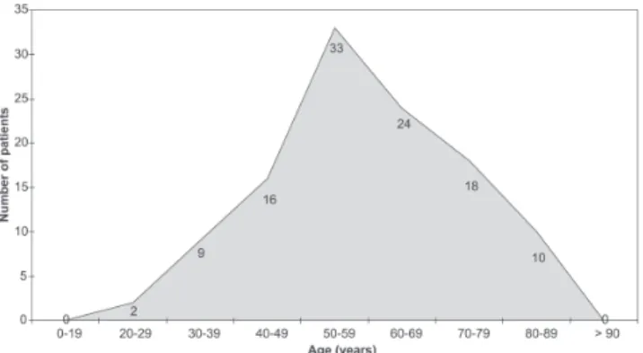
Of the 112 patients studied, 53 (47%) were men and 59 (53%) were women. Figure 1 displays the age distribu-tion of patients: 33 were aged between 50 and 59 years, while the range of the entire population was 20-88 years).
Primary tumors (Figure 2) were (in descending order) breast (29 patients), undetermined origin (18), prostate (16), lung (15), thyroid (7), kidney (6), collum uteri (5), esophagus (4), bowel (4), pharynx (3), myometrium (2), skin (2), stomach (1), liver (1), larynx (1), and ovary (1).
Figure 3 displays the sites of pathological fracture by segment, as follows: the lower limbs, 63 patients (47%); spine, 40 patients (30%); upper limbs, 16 patients (12%); and pelvic girdle, 14 patients (11%). In the extremities (Fig-ure 4), the most affected site was the femur (59 patients,
44%) followed by the humerus (15 patients, 11%). In the spine, the most affected portion was the lumbar area (22 patients, 17%); followed by the thorax (10 patients, 7.5%); sacrum, (10 patients, 4.5%) and cervical spine (2 patients, 1.5%). The pelvic girdle was affected in 11 patients (8.3%) and the scapula in 2 patients (1.5%).
We observed that 15% of patients presented metastases in other organs, and that the most affected site was the lung (59%), followed by liver (15%), as shown in Figure 5.
The treatment administered to all 112 patients ranged from clinical only to clinical and surgical. Forty-one percent were treated clinically only, with immobilizations and/or rest associated with radiotherapy, chemotherapy, hormone
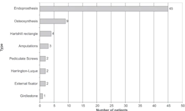
therapy, and/or biphosphonates. Fifty-nine percent underwent surgical treatment in addition to clinical treatment. Regard-ing surgical treatment of the extremities (Figure 6), 45 pa-tients had endoprostheses (66%), 9 had osteosyntheses (13%), 3 had amputations (4.4%), 2 had external fixators (3%), and 1 had a Girdlestone arthroplasty (1.5%). Concern-ing the spine, 4 patients underwent instrumentation with the Hartshill rectangle (5.8%), 2 had pediculate screws (3%), and 2 had Harrington-Luque instrumentation (3%).
The endoprostheses employed were as follows: 41 par-tial hip, 1 total hip, 1 total knee, 1 parpar-tial shoulder, and 1 humeral diaphyseal endoprosthesis.
Serum hemoglobin laboratory tests showed that 99% of the patients presented Hb >7 g/dL.
We were able to obtain survival data for 43 out of the 112 patients: 33 had died and 10 were still alive at the end of the study. These patients were analyzed using the Kaplan-Meier life-table, a descriptive procedure to exam-ine the distribution of time-dependant variables, which al-lows comparisons of the distribution by levels; or, a sepa-rate analysis for each stratum was produced.
Therefore, initially, we evaluated the distribution of sur-vival time according to the levels of each study variable. Vari-ables analyzed included sex, age at diagnosis, treatment type, fracture site, primary tumor, and metastases in other organs:
Sex
Of these 43 patients, 20 were men and 23 were women. Eighteen of the 20 men had died and 2 were alive. Fifteen
Figure 2- Distribution by primary tumor
Figure 3- Distribution according to fracture site per segment
Figure 4- Distribution according to site of pathological fractures per bone.
Figure 5- Distribution according to the site of non-bone metastases
of the 23 women had died and 8 were alive. The mean sur-vival time among men was 24.2 months and among women 31.7 months; however, this was not a significant difference (P > 0.05). (Table 1)
Age at diagnosis
The distribution by age at diagnosis shows a larger number of patients within the fifth life decade, totaling 15 patients. The age range (Table 2) with longest survival time was between 20 and 39 years (mean 90.6 months); followed by the 6th decade (35.3 months), 4th decade (30.3 months), 5th decade (26.1 months), 7th decade (9.5 months), and 8th decade (5.7 months); however, this distribution did not ex-hibit significant differences (P > 0.05).
Initial bone injury
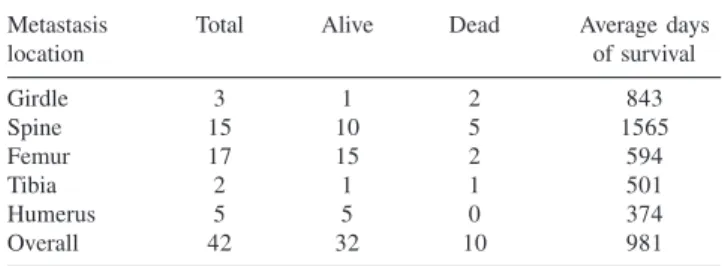
The most common site of pathologic fracture (Table 3) was the femur (17 patients, 15 died and 2 are alive). The next most common were the vertebral column (15 patients, 10 died and 5 are alive); humerus (5 patients, all died); pelvic girdle (3 patients, 1 died and 2 are alive); and tibia (2 patients, 1 died and 1 is alive). The longest survival time was observed for patients with pathologic vertebral column fractures (mean 52.1 months), and the shortest survival times were observed for patients with pathologic humeral fractures (12.4 months). Again, no significant differences were found (P > 0.05).
Primary tumor
Regarding primary tumors (Table 4), we observed a higher incidence of breast tumors (9 patients, 5 dead and
4 alive), followed by lung tumors (7 cases, 6 dead and 1 alive), prostate tumors (6 cases, all dead) and thyroid (5 cases, 2 dead and 3 alive). Other tumors comprised 16 cases (14 dead and 2 alive). Patients with breast tumors had the longest survival time (mean 43.3 months), followed by thy-roid (25.1 months), prostate (21.5 months), and lung (17.4 months, the shortest survival times); however, no signifi-cant differences were found (P > 0.05).
Non-bone metastasis
The 11 cases with non-bone metastases had survival times of 32 months. The 32 cases without non-bone metastases had a mean survival time of 28.4 months. There was no significant difference between these mean survival times (P > 0.05) (Table 5).
Treatment type
Of the 43 patients, 16 underwent exclusively clinical treatments. Eleven of them died and 5 are alive. Twenty-seven patients underwent clinical and surgical treatment; 22 died and 5 are alive. Longer survival times (Table 6) Table 4- Days of survival by primary tumor site
Primary Total Alive Dead Average of
tumor site Survival
Breast 9 5 4 1300
Prostate 6 6 0 646
Lung 7 6 1 525
Thyroid 5 2 3 753
Other 16 14 2 755
Overall 43 33 10 970
Table 3- Days of survival by location of the bone metastasis
Metastasis Total Alive Dead Average days
location of survival
Girdle 3 1 2 843
Spine 15 10 5 1565
Femur 17 15 2 594
Tibia 2 1 1 501
Humerus 5 5 0 374
Overall 42 32 10 981
Table 2- Days of survival by age
Age Total Alive Dead Average days
of survival
20/39 3 1 2 2718
40/49 7 5 2 911
50/59 15 11 4 784
60/69 7 6 1 1061
70/79 8 8 0 286
80/89 3 2 1 172
Overall 43 33 10 970
Table 1- Days of survival by sex
Sex Total Alive Dead Average days
of survival
Female 23 15 8 951
Male 20 18 2 725
Overall 43 33 10 970
Table 5- Days of survival by presence of non-bone metastasis
Non-bone Total Alive Dead Average days
metatasis of survival
No 32 24 8 853
Yes 11 9 2 962
were observed among clinically treated patients (mean: 34.1 months). Those surgically treated had a survival time of 28.3 months. There were no significant differences be-tween these mean survival times (P > 0.05).
Surgical type
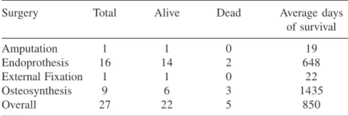
Regarding surgically treated patients (Tables 7 and 8; Figure 7), 16 had endoprostheses (14 dead, 2 alive); 9 had osteosynthesis (6 dead, 3 alive); 1 had an amputation (dead); and 1had an external fixator (dead). Patients treated with osteosynthesis had longer survival times (mean: 47.8 months), followed by endoprosthesis (21.6 months). These means were significantly different (P < 0.05).
DISCUSSION
Although we obtained survival data from only 43 of the 112 patients studied, it was important to analyze both groups, since the larger sample allows us to know whether the profile of patients treated at the Instutute of Orthpedics and Trauma of Hospital das Clínicas, São Paulo University Medical School is similar to that of the population studied in international lit-erature. This allowed us to reduce our sample bias.
Based on the 112 patients, we observed an agreement
with reported data in terms of incidence of primary tumors that metastasize mostly to the bone, ie, breast, prostate, lung, and thyroid primary tumors.12 The most affected age range was the 5th decade. The incidence of diagnosed non-bone metastases was 15%, the lung being the most common.
The most affected site in the patients treated in our Service were the lower limbs (47%), more specifically the femur, and this differs from most reported studies.2,3 A pos-sible explanation is that vertebral column fractures are less frequently referred to the Tumor Group of our Service and/ or they are underdiagnosed.
Surgical treatment was performed in most patients (59%), although it is more indicated for pathological femo-ral and humefemo-ral fractures. Exclusively clinical treatment was the most indicated for pathological fractures of the ver-tebral column and pelvic girdle (41%).
Among surgically treated patients, more endoprostheses were performed (66%) than any other type of procedure because there was higher incidence of proximal femoral pathological fractures (44%).
Currently, interlocking nails are preferred over endoprostheses in cases of proximal femoral pathological fractures, due to their lower morbidity in cases with less destruction of bone matrix. This procedure is invariably supplemented by postoperative radiotherapy.1,2,7
Hemoglobin was not a relevant datum in our study be-cause only 1 patient in our sample had Hb < 7g/dL.
By analyzing the subgroup formed by 43 patients for whom survival data was available, we found that sex, age, primary tumor, site affected, non-bone metastasis, and clini-cal versus surgiclini-cal treatment variables are not good pre-dictors for survival. We found that the evidence of visceral metastasis in patients with pathological fractures does not necessarily mean a poorer prognosis, nor does the fracture site or the primary tumor. A possible explanation for this Table 8. Statistical analysis for the different types of surgery,
as specified in Table 7.
chi-square df P
Log Rank (Mantel-Cox) 13.193 3 0.004
Breslow (Generalized Wilcoxon) 11.572 3 0.009
Tarone-Ware 12.284 3 0.006
Table 7- Days of survival by type of surgical treatment
Surgery Total Alive Dead Average days
of survival
Amputation 1 1 0 19
Endoprothesis 16 14 2 648
External Fixation 1 1 0 22
Osteosynthesis 9 6 3 1435
Overall 27 22 5 850
Table 6- Days of survival by type of treatment: surgical vs clinical
Treatment Total Alive Dead Average day
type of survival
Surgical 27 22 5 850
Clinical 16 11 5 1025
Overall 43 33 10 970
would be the fact that the fracture itself represents a strong determinant for survival decrease in this population.
However, the type of surgery performed was signifi-cantly associated with length of survival, which supported our hypothesis that surgery type would be the largest de-terminant of survival decrease in these cancer patients. Pa-tients who received an endoprosthesis (mean survival time, 21.6 months) had worse prognoses than those undergoing osteosynthesis (mean survival time, 47.8 months). This may be explained either by the higher severity of proximal femoral fractures that were more frequently treated with endoprostheses, or by the fact that the surgical technique employed in osteosynthesis is less aggressive, produces less morbidity, and is more prone to rehabilitation than the tech-nique involving an endoprosthesis.
CONCLUSION
We conclude that the treatment of choice for pathologi-cal diaphyseal femoral and proximal femoral fractures is internal fixation using interlocking medullary nailing, which is less aggressive to soft tissue and patients.
This is also shown by the current international litera-ture;1,2,7 the profile of the population in this study was simi-lar to those in other studies.1,4,5,7-14
Whenever it can be indicated, osteosynthesis with inter-locking nails for pathological fractures is more compatible with patients’ survival, since it reduces hospital stay, rehabili-tation time, surgical time and bleeding, and makes it possible to stabilize the entire femur, compared to endoprostheses.2,14
RESUMO
Narazaki DK, Alverga Neto CC, Baptista AM, Caiero MT, Camargo OP. Fatores prognósticos nas fraturas patológicas por tumores metastáticos.Clinics. 2006;61(4):313-20.
OBJETIVO: As fraturas patológicas por metástase óssea determinam uma queda abrupta na qualidade de vida dos pacientes com neoplasias malignas e também aumentam sua mortalidade. Os avanços ortopédicos de osteossíntese
e endopróteses têm beneficiado a prevenção e tratamento dessas fraturas. O objetivo de nosso estudo é determinar quais são os fatores prognósticos dessas fraturas patológicas tratadas no nosso serviço.
CASUÍSTICA E MÉTODOS: Foram estudados 112
quanto ao sexo, idade, local de metástase óssea, metástases viscerais, origem do tumor primário, tipo de tratamento, hemoglobina sérica e sobrevida.
RESULTADOS: O local mais acometido foi o fêmur (44%), o tumor primário mais freqüente foi o câncer de mama (25%), o tratamento cirúrgico mais realizado foi a endoprótese não convencional (66%). As variáveis sexo, idade, tumor primário, local acometido, mestástase não-óssea e tratamento clínico versus cirúrgico não são bons preditores para sobrevida. Os pacientes operados com endoprótese (21,6 meses) apresentaram pior prognóstico que os pacientes submetidos à osteossíntese (47,8 meses).
DISCUSSÃO E CONCLUSÃO: Os pacientes submetidos à osteossíntese, com uma técnica cirúrgica menos mórbida e de reabilitação mais precoce, apresentaram maior sobrevida em relação aos pacientes submetidos à endopróteses. Observamos que nossa casuística é seme-lhante à internacional, na qual aparece como tumor primário mais freqüente o de mama, os de origem indeterminada, próstata e pulmão.
UNITERMOS: Fraturas patológicas, Neoplasias, Metás-tases, Prognóstico, Cirurgia.
REFERENCES
1. Swanson KC, Pritchard DJ, Sim FH. Surgical treatment of metastatic disease of the femur. J Am Acad Orthop Surg. 2000;8:56-65. 2. Reich CD. Advances in the treatment of bone metastases. Clin J Oncol
Nursing. 2003;7:641-6.
3. Cheng EY. Prospective quality of life research in bony metastatic disease. Clin Orthop. 2003;415(Suppl):289-97.
4. Habermann ET, Lopez RA. Metastatic disease of bone and treatment of pathological fractures. Orthop Clin North Am. 1989;20:469-86. 5. Kelly CM, Wilkins RM, Eckardt JJ, Ward WG. Treatment of metastatic
disease of the tibia. Clin Orthop. 2003; 415(Suppl):219-29.
6. Wedin R, Bauer HC, Rutqvist LE. Surgical treatment for skeletal breast cancer metastases: a population-based study of 641 patients. Cancer. 2001;92:257-62.
7. Bocchi L, Lazzeroni L, Maggi M. The surgical treatment of metastases in long bones. Ital J Orthop Traumatol. 1988;14:167-73.
8. Buggay D, Jaffe K. Metastatic bone tumors of the pelvis and lower extremity. J Surg Orthop Adv. 2003;12:192-9.
10. Althausen P, Althausen A, Jennings LC, Mankin HJ. Prognostic factors and surgical treatment of osseous metastases secondary to renal cell carcinoma. Cancer 1997;80:1103-9.
11. Iwamoto Y, Sugioka Y, Chuman H, Shiba K, Yuge I. Surgical treatment of metastatic tumors of long bones and the spine. Adv Exp Med Biol. 1992;324:295-303.
12. [No authors listed]. British Association of Surgical Oncology Guidelines. The management of metastatic bone disease in the United Kingdom. The Breast Specialty Group of the British Association of Surgical Oncology. Eur J Surg Oncol. 1999;25:3-23.
13. Schmidt RG. Management of extremity metastatic bone cancer. Curr Probl Cancer. 1995;19:166-82.
14. Weber KL, Lewis VO, Randall RL, Lee AK, Springfield D. An approach to themanagement of the patient with metastatic bone disease. Instr Course Lect. 2004; 53:663-76.