197
Echocardiography has contributed to the diagnosis and thera-peutic management of patients suspected of having infective
endo-carditis, both through the transthoracic1 and transesophageal
techniques2.
Despite the progress observed, situations occur in which the diagnosis of infective endocarditis is difficult, such as in patients with acute disease, infection in cardiac valvular prostheses 3,4,
prosthetic infection in the immediate postoperative period 5,6,
pa-tients with an artificial cardiac pacemaker 7,8, patients receiving
previous antimicrobial therapy, or those whose blood cultures are negative 9,10.
In the different recent published studies 11-15 about the use of
transesophageal echocardiography in patients suspected of having infective endocarditis, we observe case series ranging from 23 to 180 patients studied using biplanar transducers, or part of the case series being studied with multiplanar probes. In addition, they did not include the diagnostic categorization of patients.
Therefore, this study aimed at assessing the echocardiographic findings, both through the transthoracic and multiplanar transe-sophageal techniques, in a greater case series of patients suspected of having infective endocarditis, classified according to diagnostic categories, which also include situations of greater diagnostic difficulty.
Methods
Two hundred sixty-two patients were prospectively studied in 266 episodes of the disease, in which the differential diagnosis included the diagnostic hypothesis of infective endocarditis. The patients’ age ranged from 10 to 85 years (mean, 47.6; standard deviation, 17.9), and 139 (52.2%) episodes occurred in men and 127 (47.8%) in women.
The echocardiographic examination was indicated by the clini-cian responsible for the patient. The patients studied initially under-went the transthoracic study, and, on the occasion of the transe-sophageal investigation, the examination was preceded by a new transthoracic study. All patients underwent transthoracic and transe-sophageal echocardiographic investigation.
The diagnosis of infective endocarditis was based on clinical findings (fever, predisposing heart disease, illicit use of intravenous drugs, cardiac murmur, vascular or immunologic phenomena), identification of the etiologic agent in at least 2 blood cultures, identification of endocardial impairment on transthoracic or tran-sesophageal echocardiography, and histological evidence of infective
Original Article
Echocardiographic Findings in Patients with
Suspected Infective Endocarditis
Marcelo Luiz Campos Vieira, Max Grinberg, Pablo M. A. Pomerantzeff, José L. de Andrade,
Alfredo J. Mansur
São Paulo, SP - Brazil
Instituto do Coração of the Hospital das Clínicas of the FMUSP Mailing address: Marcelo Luiz Campos Vieira – Rua Cardoso de Melo, 463/21 - 04548-002 - São Paulo, SP, Brazil
E-mail: [email protected] Received for publication: 10/9/03 Accepted for publication: 2/18/04 English version by Stela Maris Costalonga
Objective
To assess the echocardiographic findings in patients with suspected infective endocarditis.
Methods
Two hundred sixty-two patients with suspected infective endo-carditis underwent transthoracic and transesophageal echocar-diographic investigation. Images of vegetations, valvular absces-ses, and acute periprosthetic insufficiency were analyzed, and the correlation with clinical and laboratory data, diagnostic cate-gory, and hospital evolution was assessed.
Results
The diagnosis of endocarditis was categorized as defined in 127 (47.8%) episodes, possible in 81 (30.4%), and rejected in 58 (21.8%). In patients with the defined diagnosis, the following images were identified: 135 vegetations, 37 abscesses, and 6 periprosthetic insufficiencies. Vegetations were more frequent in patients with endocarditis due to streptococci of the viridans group and enterococci (P=0.02), and with symptom duration < 10 days (P=0.001). Abscesses were more frequent in patients with symptom duration < 10 days (P=0.001). Periprosthetic insufficiency was associated with a greater need for surgical treatment (P=0.001). In patients with the possible diagnosis of endocarditis, 8 echocardiographic images considered compatible with vegetations were identified. In patients whose diagnosis of endocarditis was rejected, no vegetations, valvular abscesses, or periprosthetic insufficiencies were demonstrated.
Conclusion
Our echocardiographic findings varied according to the diag-nostic category. The contribution to both the diagnosis and prog-nostic evaluation should consider the pretest probability of the diagnosis of infective endocarditis.
Keywords
198
endocarditis on surgical or autopsy findings. The diagnosis of infec-tive endocarditis followed the recommendations of the previously published criteria 1 and were categorized as defined, possible,
and rejected.
The examinations were performed according to the routine of our service and previously recommended techniques 16,17. The
tran-sesophageal echocardiography was performed with electrocar-diographic and oximetric monitoring, and no antibiotic prophylaxis
was administered 17. The mean duration of the transesophageal
examination was 28.2 minutes (standard deviation of 6.4 minutes). The examinations were performed on Advanced Technology Laboratory devices, Apogee CX 200, HDI 3000, and HDI 5000 models (Bothell, Washington, USA). The transesophageal exa-minations were performed with multiplanar transducers. The trans-thoracic examinations that precede the transesophageal exami-nations and the transesophageal examiexami-nations were recorded on VHS videotapes.
The examinations were analyzed by the physician who performed them, and then by a second researcher, who ignored the previous clinical diagnosis. Agreement between the different observers in the analysis of the transesophageal examinations was greater than 95% for the presence of defined images, such as vegetations, abscesses, and acute periprosthetic insufficiency (dehiscence of cardiac valvular prosthesis). When divergence in identification of the images occurred, a third observer also analyzed the examinations.
The echocardiographic findings were diagnosed according to previously published criteria 18-20.
The images obtained on transthoracic examinations were cate-gorized according to quality, using the identification of the endo-cardium and the valvular structures as a criterion, as follows: a) adequate; b) regular; c) inadequate 21.
The following characteristics of the vegetations were examined: a) location in the cardiac structure; b) dimension in their greater diameter (until 5 mm, from 6 to 10 mm, > 10 mm); c) refringence
≥ that of the endocardium (with the gain of the echocardiographic device in minimum values capable of identifying the image defined as a vegetation); d) mobility (vegetation firmly adhered to the underlying structure, fixed base with free mobile border, and prolapsing).
The following characteristics were studied: a) demographic and clinical aspects: age, sex, time between symptom onset and hospitalization, previous use of antimicrobial agents, diagnostic categorization of endocarditis, etiologic agents, heart disease, treatment, evolution; b) echocardiographic findings: vegetation, perivalvular abscess; periprosthetic insufficiency; fistula between the cardiac chambers; thrombus; and pericardial effusion.
The descriptive analysis of the continuous variables was perfor-med using the minimum and maximum values, mean, and standard deviation. The descriptive analysis of the categorical variables was performed using the absolute and relative frequencies. The comparative analysis was performed using the chi-square test or the Fisher exact test. The data were processed with the statistical analysis system of the SAS Institute, Cary, North Carolina, USA. The P values < 0.05 were considered significant.
The project was approved by the Committee on Ethics in Research of the Hospital das Clínicas of the Medical School of the Universidade de São Paulo.
Results
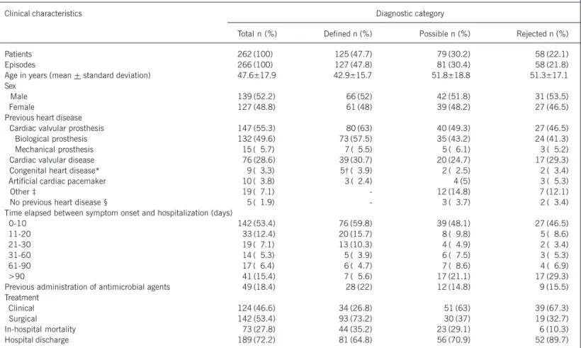
The demographic and clinical characteristics, including labo-ratory data, complications, and hospital evolution are shown in table I. The population studied was characterized by the predo-minance of patients with cardiac valvular prostheses, and time elapsed between symptom onset and hospitalization < 10 days. More than half of the patients required surgical treatment during in-hospital evolution.
The diagnosis was categorized as defined in 127 (47.8%) episodes as follows: 112 (88.2%) through clinical criteria, and 15 (11.8%) through anatomicopathological criteria. The diagnosis was categorized as possible in 81 episodes (30.4%), and as rejected in 58 (21.8%) episodes.
The microbiologic agents identified in the 127 episodes with the defined diagnosis of infective endocarditis were: coagulase–
negative staphylococcus in 26 (20.5%); staphylococcus aureus
in 21 (16.5%); streptococcus of the viridans group in 20 (15.7%); streptococcus not pertaining to the viridans group in 6 (4.7%); gram-negative bacteria in 10 (7.9%); enterococcus faecalis in 5 (3.9%); fungi in 3 (2.4%); and association of 2 infective agents in 3 (2.4%) episodes. No infective agents were identified in blood cultures in 33 (26%) episodes, and, in 22 patients with 33 episodes, no etiologic agent could be identified, because they had cardiac valvular prostheses.
The quality of the images of transthoracic examinations that preceded the transesophageal examinations were considered as follows: adequate in 256 (77.6%) of 330 examinations; regular in 52 (15.7%); inadequate in 22 (6.7%).
In patients with the defined diagnosis of endocarditis, 135 images of vegetations were identified as follows: through trans-thoracic analysis in 61 (48%) of the 127 cases of the diagnosis defined as infective endocarditis; and through transesophageal analysis in 112 (88.2%) cases. Therefore, vegetations were iden-tified through transthoracic analysis in 61 (54.4%) of the 112 cases identified through transesophageal examination.
The images of vegetations were identified on mitral valve pros-thesis in 46 (36.1%) of 127 episodes; on aortic valve prospros-thesis in 33 (26%) episodes; on pulmonary valve prosthesis in 1 (0.8%); on native mitral valve in 15 (11.8%); on native aortic valve in 9 (7.1%); on tricuspid valve in 2 (1.6%); on native pulmonary valve in 1 (0.8%); on mitral valve and on aortic valve in 15 (11.8%); on the extension of artificial cardiac pacemakers in 3 (2.4 %); and on ventricular endocardium in 2 (1.6%) episodes.
The dimensions of the images identified as vegetations were as follows: < 5 mm in 24 of 135 (17.8%); between 6 and 10 mm in 78 (57.8%); and > 10 mm in 33 (24.4%). The refrin-gence of the images of the vegetations was equal to that of the endocardium in 132 (97.8%) images, and greater than that of the endocardium in 3 (2.2%). The images of the vegetations had a fixed base with free mobile border in 45 (33.3%) characteriza-tions, and prolapsing structures in 90 (66.7%) images.
The distribution of the presence of vegetations differed with the different etiologies (more frequent for streptococci of the viridans group and enterococci) (P=0.02).
199
for the time between symptom onset and hospitalization up to 10 days) (P=0.001).
In patients with the possible diagnosis of endocarditis, images interpreted as vegetations were identified on transesophageal exa-mination in 8 (9.8%) of 81 episodes, and in 2 (2.5%) with the use of transthoracic echocardiography. Of those 8 cases, all had negative blood cultures, 5 received antibiotic therapy for a short period of time (under 14 days), with remission of the findings and maintenance of the images on later transesophageal examination. They were discharged from the hospital and clinically followed up for at least 6 months, with no other possible diagnosis of infective endocarditis. Of the 8 patients with an image interpreted as vege-tation, 3 underwent surgical treatment, and neither macroscopic, nor histological material compatible with vegetation was found. The image saw as vegetation on echocardiography was then attri-buted to surgical suture thread. Of the 8 cases, 7 had a biopros-thesis (5 in the mitral position, 2 in the aortic position). One patient had an image on the native aortic valve. All 8 patients had no previous history of an episode of infective endocarditis.
In the episodes in which the diagnosis of endocarditis was rejec-ted, no image of vegetation was identified on echocardiography.
In patients with the defined diagnosis of infective endocarditis, 357 complications due to infection occurred as follows: acute renal failure, 67 episodes; congestive heart failure, 52 episodes; cardiac valve abscess, 31 episodes; stroke, 29 episodes; extrace-rebral embolism, 21 episodes; complete atrioventricular block, 13 episodes; cerebral embolism, 7; mycotic aneurysm, 7; pulmo-nary embolism, 3; and other complications, 127. Stroke was
more frequent in patients whose vegetations were > 10 mm (P = 0.02).
No greater need for surgical treatment was observed in patients in whom vegetations were identified.
The mortality rate was not greater in patients with vegetations. No statistical significance was observed between refringence and the following parameters: sex, age, abscess, flow of acute periprosthetic insufficiency, time elapsed between symptom onset and hospitalization, previous use of antibiotic therapy, complica-tions, treatment, and condition at hospital discharge.
In the episodes with the defined diagnosis of infective endo-carditis, 5 cases of valvular ring abscesses were identified by using transthoracic echocardiography (4 in the aortic ring, and 1 in the mitral ring). On transesophageal echocardiography, 37 images of valvular ring abscess were identified as follows: 24 in the aortic ring, 7 in the mitral ring, and 6 in other locations. A perivalvular abscess was identified during surgery in 31 cases (22 in the aortic ring, 7 in the mitral ring, and 2 in the mitroaortic junction). In the 6 cases, in which a perivalvular abscess was identified on transesophageal echocardiography, but was not confirmed during surgery, previous surgical manipulation had occurred.
Valvular ring abscess was more frequent in patients with endo-carditis, who had vegetations with dimensions > 10 mm (P=0.01) and prolapsing mobility (P=0.02).
No statistical significance was found between the presence of a perivalvular abscess and the different etiological agents.
The distribution of the presence of abscesses differed according to the different times between symptom onset and hospitalization
Table I – Clinical characteristics of 262 patients with 266 episodes of suspected infective endocarditis
Clinical characteristics Diagnostic category
Total n (%) Defined n (%) Possible n (%) Rejected n (%)
Patients 262 (100) 125 (47.7) 79 (30.2) 58 (22.1)
Episodes 266 (100) 127 (47.8) 81 (30.4) 58 (21.8)
Age in years (mean + standard deviation) 47.6±17.9 42.9±15.7 51.8±18.8 51.3±17.1 Sex
Male 139 (52.2) 066 (52) 42 (51.8) 31 (53.5)
Female 127 (48.8) 061 (48) 39 (48.2) 27 (46.5)
Previous heart disease
Cardiac valvular prosthesis 147 (55.3) 080 (63) 40 (49.3) 27 (46.5)
Biological prosthesis 132 (49.6) 073 (57.5) 35 (43.2) 24 (41.3)
Mechanical prosthesis 015 (05.7) 007 (05.5) 5 (06.1) 3 (05.2)
Cardiac valvular disease 076 (28.6) 039 (30.7) 20 (24.7) 17 (29.3)
Congenital heart disease* 009 (03.3) 05† (03.9) 2 (02.5) 2 (03.4)
Artificial cardiac pacemaker 010 (03.8) 003 (02.4) 4 (5) 3 (05.3)
Other ‡ 019 (07.1) - 12 (14.8) 7 (12.1)
No previous heart disease § 005 (01.9) - 3 (03.7) 2 (03.4)
Time elapsed between symptom onset and hospitalization (days)
0-10 142 (53.4) 76 (59.8) 39 (48.1) 27 (46.5)
11-20 033 (12.4) 20 (15.7) 8 (09.8) 5 (08.6)
21-30 019 (07.1) 13 (10.3) 4 (04.9) 2 (03.4)
31-60 014 (05.3) 5 (03.9) 6 (07.5) 3 (05.3)
61-90 017 (06.4) 6 (04.7) 7 (08.6) 4 (06.9)
>90 041 (15.4) 7 (05.6) 17 (21.1) 17 (29.3)
Previous administration of antimicrobial agents 049 (18.4) 28 (22) 12 (14.8) 9 (15.5) Treatment
Clinical 124 (46.6) 34 (26.8) 51 (63) 39 (67.3)
Surgical 142 (53.4) 93 (73.2) 30 (37) 19 (32.7)
In-hospital mortality 073 (27.8) 44 (35.2) 23 (29.1) 6 (10.3)
Hospital discharge 189 (72.2) 81 (64.8) 56 (70.9) 52 (89.7)
200
(time between symptom onset and hospitalization up to 10 days) (P=0.001). No statistical significance was found between the presence of abscesses, sex, and age.
In patients with the possible diagnosis of endocarditis or in those whose diagnosis of endocarditis was rejected, no perivalvular abscess was identified on echocardiography.
Renal failure was more frequent in patients with the defined diagnosis of infective endocarditis who had an abscess of the valvular ring (P=0.04).
The need for surgical treatment was greater among patients in whom perivalvular abscesses were identified (P=0.004).
The mortality rate was greater in patients with perivalvular abscesses (P=0.003).
In patients with a defined diagnosis of infective endocarditis, periprosthetic insufficiency was evidenced in 2 cases on transtho-racic echocardiography, and in 6 on transesophageal echocardio-graphy (tab. II). Of the 6 episodes, a new cardiac murmur was reported in 3. The greater number of transthoracic and transeso-phageal examinations predominated in patients with the defined diagnosis of endocarditis, with a relative decrease in the number of those examinations in patients with the possible and rejected diagnoses of endocarditis.
A greater need for surgical treatment was observed among patients in whom acute periprosthetic insufficiency was identified (P=0.001).
No statistical significance was observed between the presence of acute periprosthetic insufficiency and the following parameters: sex, age, and the different etiologic agents. No statistical signifi-cance was observed between the different times between symptom onset and hospitalization. Neither the presence of complications, nor the mortality rate was greater. In patients with the possible or rejected diagnosis of endocarditis, no acute periprosthetic insuffi-ciency was identified on echocardiography.
In the cases with the defined diagnosis of infective endocardi-tis, in addition to vegetations, the following characteristics were observed on echocardiography: thrombi in 6 cases (all of them with surgical confirmation, 2 on transthoracic examination);
sponta-neous contrast in 10 cases (2 on transthoracic echocardiography); perforation of the mitral leaflet in 2 (with surgical confirmation and no evidence on transthoracic examination); early closure of the mitral valve in the case of acute aortic valve insufficiency in 1 (only transthoracic echocardiography was performed); destruction of the mitroaortic junction in 8 (all of them with surgical confirmation, and identification on transthoracic examination in 1 case); fistulas from the aorta to the right atrium in 2 (1 case identified on transtho-racic echocardiography and confirmed on surgery); and rupture of the chordae tendineae in 5 (all with surgical confirmation and identification on transthoracic echocardiography).
Discussion
The case series studied was characterized by predominance of patients with cardiac valve prostheses (63%), patients under-going surgical treatment (73.2%), and a high mortality rate (35.2%), which show the severity of the disease in those cases. The data obtained by use of transthoracic echocardiography contributed to the diagnosis in 61 (48%) of the 127 patients with the defined diagnosis of endocarditis. The information provided by transesophageal echocardiography enabled the addition of diag-nostic information in 112 (88.2%) of these 127 patients, obtaining the diagnostic of vegetationin 51 (40,1%) other cases, more than the 61 diagnosed on the transthoracio examination. This diagnostic increment takes into consideration the population involved in the study, in which patients with cardiac valvular prostheses predo-minate. In specific groups, such as that of patients with cardiac valvular prostheses, the transesophageal echocardiographic investigation may be regarded as a method of initial diagnostic investigation, due to the greater diagnostic accuracy of the transesophageal technique as compared with that of transthoracic echocardiography 2,14. Patients with greater diagnostic difficulty require
repetition of the echocardiographic examinations 2. In the present
study, the transthoracic echocardiographies were repeated until the sixth examination, and the transesophageal echocardiographies until the fifth examination. No addition of diagnostic information
Table II – Transthoracic and transesophageal echocardiographic findings in 262 patients with 266 episodes suspected of infective endocarditis
Clinical characteristics Diagnostic category
Category Total n (%) Defined n (%) Possible n (%) Rejected n (%)
Episodes 266 (100) 127 (47.8) 81 (30.4) 58 (21.8)
Transthoracic examinations (n/mean per group) 629 / 2.4 363 / 2.8 170 / 2.1 96 / 1.6
Vegetation 63 (23.7) 61 (48) 2 (2.5)
-Cardiac perivalvular abscess 5 (01.9) 5 (03.9) -
-Acute periprosthetic insufficiency 2 (0.75) 2 (01.6) -
-Thrombus 4 (01.5) 2 (01.6) 1 (1.2) 1(1.7)
Pericardial effusion 26 (09.8) 16 (12.6) 6 (7.4) 4 (6.9)
Intercavitary fistula 1 (00.4) 1 (00.8) -
-Rupture of the chordae tendineae 5 (01.9) 5 (03.9) -
-Transesophageal examinations (n/mean per group) 330 / 1.2 183 / 1.4 87 / 1.1 60 / 1
Vegetation 120 (45.1) 112 (88.2) 8 (9.9)
-Cardiac perivalvular abscess 37 (13.9) 37 (29.1) -
-Acute periprosthetic insufficiency 6 (02.2) 6 (04.7) -
-Thrombus 18 (06.8) 6 (04.7) 3 (3.7) 3 (5.1)
Pericardial effusion 27 (10.1) 16 (12.6) 6 (7.4) 5 (8.6)
Intercavitary fistula 2 (00.7) 2 (01.6) -
-Rupture of the chordae tendineae 5 (01.9) 5 (03.9) -
201
was observed after the third examination, either transthoracic or transesophageal 22.
A greater frequency of stroke was observed in patients with endocarditis in whom vegetations with dimensions > 10 mm were identified, although a greater frequency of peripheral embolic pheno-mena correlated with the dimensions of the vegetations was not observed. A greater frequency of stroke or peripheral embolism correlated with the mobility of the vegetations or location of the vegetation was not observed. Up to 65% of the embolic events may affect the central nervoussystem 2, with a lower frequency in
the different organs and places 23,24. Previous studies 24-30 about
the frequency of embolic phenomena in regard to the dimensions and mobility of vegetations are considered controversial. Some studies 24-27 report a greater frequency of embolic phenomena in
patients with vegetations with dimensions > 10 mm, with great mobility, and with involvement of the mitral valve 26. Other series
do not report a greater frequency of embolic phenomena in patients with vegetations > 10 mm 28-30 and with great mobility 28,30. The
different series diverge in regard to the results and the echocardio-graphic methods used for identifying the vegetations. Some series used only the transthoracic investigation 25,27,29, while others the
transesophageal 24,26,28,30.
Patients with time between symptom onset and hospitalization < 10 days had a greater incidence of the following: vegetations and cardiac valvular abscesses, defined diagnosis of endocarditis, 76 (59.8%) of 127 cases, heart failure, stroke, renal failure, and extracerebral embolic phenomena, 28 (53.8%) of 52 cases, 19 (65.5%) of 29 episodes, 37 (55.2%) of 67, and 12 (57.1%) of 21, respectively. These data show the importance of the initial clinical signs and symptoms and of the diagnostic investigation in patients in whom the diagnosis of infective endocarditis is possible.
The identification of 132 (97.8%) of 135 images of vegetation with refringence similar to that of the endocardium shows recent endocardial involvement, because the greater structural refringence may represent calcium accumulation due to the greater time of occurrence of the event.
The identification of cardiac perivalvular abscesses was also more frequent in patients with vegetations > 10 mm and with greater mobility. The vegetations with greater dimensions and mobility may represent greater endocardial involvement, with a greater potential to develop cardiac valvular abscesses.
The diagnosis of perivalvular abscesses and periprosthetic insuffi-ciency was very important because of therapeutic and prognostic
implications. Patients with perivalvular abscesses had a greater need for surgical treatment and a higher incidence of renal failure, with higher mortality, and, patients with periprosthetic insuffi-ciency had a higher need for surgical treatment.
In 8 patients with the possible diagnosis of endocarditis, struc-tures identified as vegetations due to infective endocarditis were visualized on transesophageal echocardiography. These structures were either not confirmed on the anatomicopathological exami-nation or had clinical improvement after treatment for a short period of time. In the series of patients in which echocardiographic criteria were introduced for the diagnostic categorization of endo-carditis 1, images defined as vegetations were identified in 11
(7%) cases with the possible diagnosis of endocarditis and in 4 (8%) cases with the rejected diagnosis of endocarditis. The diffe-rential diagnosis of vegetations resulting from infective endocarditis is made with natural structures, such as valvular thickening and degeneration, chordae tendineae, and nodular structures found mainly in the native aortic valve, such as the Lambl excrescences and the Arantius nodule. Structures with morphology similar to that of the vegetation of infective endocarditis may also be found in the following conditions: marantic (thrombotic) endocarditis; noninfective Libman-Sacks endocarditis; small tumors, such as papillary fibroelastomas; and in the presence of cardiac thrombi. The morphological differentiation of small vegetations from surgical suture thread or structures denominated fibrin filaments is more difficult 31,32. In our study, all structures identified as vegetations,
which underwent surgery and were not confirmed as such, were surgical suture thread with a postoperative period < 2 months.
In our case series, most patients had cardiac valvular prostheses with the suspected diagnosis of infective endocarditis and echo-cardiographic investigation performed in a tertiary hospital specia-lizing in cardiology, and this may not represent other clinical cir-cumstances with a possible diagnosis of endocarditis. The indica-tions for performing echocardiographic investigation were hetero-geneous and based on the need for diagnostic investigation and appreciation of the attending physician.
In conclusion, in patients with suspected infective endocarditis undergoing echocardiographic assessment, the echocardiographic findings varied according to the following diagnostic categories: defined, possible, and rejected. The contribution of the echocar-diographic information, both for the diagnosis and prognostic assessment, should take into consideration the pretest probability of the diagnosis of infective endocarditis.
1. Durack DT, Lukes AS, Bright DK. New criteria for diagnosis of infective endocardi-tis: utilization of specific echocardiographic findings. Am J Med 1994; 96: 200-9. 2. Bayer AS, Bolger AF, Taubert KA et al. Diagnosis and management of infective
endocarditis and its complications. Circulation 1998; 98: 2936-48.
3. Vered Z, Mossinson D, Peleg E, Kaplinsky E, Beker B. Echocardiographic assess-ment of prosthetic valve endocarditis. Eur Heart J 1995; 16 [suppl B]: 63B-67B. 4. San Roman JA, Vilacosta I, Sarriá I. Clinical course, microbiologic profile, and diagosis of periannular complications in prosthetic valve endocarditis. Am J Cardiol 1999; 83: 1075-9.
5. Aslamaci S, Dimitri WR, Williams BT. Operative considerations in active native valve infective endocarditis. J Cardiovasc Surg 1989; 30: 328-33.
6. Farina G, Vitale N, Piazza L, de Vivo F, de Luca L, Cotrufo M. Long term results of surgery for prosthetic valve endocarditis. J Heart V Dis 1994; 3: 165-71.
References
7. Victor F, de Place C, Camus C et al. Pacemaker lead infection: echocardiographic features, management, and outcome. Heart 1999; 81: 82-7.
8. Voet JG, Vandekerckhove YR, Muyldermans LL, Missault LH, Matthys LJ. Pace-maker lead infection: report of three cases and review of the literature. Heart 1999; 81: 88-91.
9. Tunkel AR, Kaye D. Endocarditis with negative blood cultures. N Engl J Med 1992; 326: 1215-7.
10. Hoen B, Selton-Suty C, Lacassin F, Etienne J, Briancon S, Leport C. Infective endo-carditis in patients with negative blood cultures: analysis of 88 cases from a one-year nationwide survey in France. Clin Infect Dis 1995; 20: 501-6.
202
12. Irani WN, Grayburn PA, Afridi I. A negative transtoracic echocardiogram obviates the need for transesophageal echocardiography in patients with suspected native valve active infective endocarditis. Am J Cardiol 1996; 78: 101-3.
13. Lindner JL, Case RA, Dent JM, Abbot RD, Scheld WM, Kaul S. Diagnostic value of echocardiography in suspected endocarditis. An evaluation based on the pretest probability of disease. Circulation; 1996; 93: 730-6.
14. Thalme A, Nygren AT, Julander I, Freyschuss U. Endocarditis: clinical outcome and benefit of transesophageal echocardiography. Scand J Infect Dis 2000; 32: 303-7.
15. Roe MT, Abranson MA, LI J et al. Clinical information determines the impact of transesophageal echocardiography on the diagnosis of infective endocarditis. Am J Cardiol 2000; 139: 945-51.
16. Henry WL, de Maria A, Gramiak R, King DL, Kisslo JA, Popp RL. Report of the American Society of Echocardiography Committee on Nomenclature and Stan-dards in Two-Dimensional Echocardiography. Circulation 1980; 62: 212-5. 17. Seward JB, Khanderia BK, Oh JK, Abel MD, Hughes Jr. RW, Edwarda WD.
Tran-sesophageal echocardiography: technique, anatomic correlations, implementa-tion, and clinical applications. Mayo Clin Proc 1988; 63: 649-80.
18. Dillon JC, Feigenbaum H, Konecke LL, Davis RH, Chang S. Echocardiographic ma-nifestations of valvular vegetations. Am Heart J 1973; 86: 698-704.
19. Ellis SG, Goldstein J, Popp RL. Detection of endocarditis-associated perivalvular abs-cesses by two-dimensional echocardiography. J Am Coll Cardiol 1985; 5: 647-53. 20. Wilson BH, Schillig S. Infective perivalvular abscess of the aortic ring:
echocardio-graphic features and clinical course. Am J Cardiol 1990; 66: 102-5.
21. Shively BK, Gurule FT, Roldan CA, Leggett JH, Schiller NB. Diagnostic value of transesophageal compared with transthoracic echocardiography in infective endocarditis. J Am Coll Cardiol 1991; 18: 391-7.
22. Vieira MLC, Grinberg M, Pomerantzeff PM, Andrade JL, Mansur AJ. Repeated
echocardiographic examinations in patients with suspected infective endocarditis. Heart 2004. No prelo.
23. Heinle SK, Kisslo J. The clinical utility of transesophageal echocardiography in pa-tients with left-sided infective endocarditis. Am J Card Imaging 1995; 9: 199-202. 24. Di Salvo G, Habbib G, Pergola V et al. Echocardiography predicts embolic events
in infective endocarditis. J Am Coll Cardiol 2001; 37: 1069-76.
25. Mügge A, Daniel W. Echocardiographic assesment of vegetations in patients with infec-tive endocarditis: prognostic implications. Echocardiography 1995; 12: 651-61. 26. Rohmann S, ErbeL R, George G. Clinical relevance of vegetation localization by
transe-sophageal echocardiography in infective endocarditis. Eur Heart J 1992; 13: 446-52. 27. Sanfilippo AJ, Picard MH, Newell JB. Echocardiographic assessment of patients with infectious endocarditis: prediction of risk for complications. Am J Coll Cardiol 1991; 18: 1191-202.
28. Heinle S, Wilderman N, Harrison K et al. Value of transthoracic echocardiography in predicting embolic events in active infective endocarditis. Am J Cardiol 1994; 74: 799-801.
29. Lutas EM, Roberts RB, Devereux RB, Prieto LM. Relation between the presence of echocardiographic vegetations and the complication rate in infective endocar-ditis. Am Heart J 1986; 112: 107-13.
30. De Castro S, Magni G, Beni S. Role of transthoracic and transesophageal echo-cardiography in predicting embolic events with active infective endocarditis invol-ving native cardiac valves. Am J Cardiol 1997; 80: 1030-4.
31. Stoddard MF, Dawkins PR, Longaker RA. Mobile strands are frequently attached to the St. Jude medical mitral valve prosthesis as assessed by two-dimensional transesophageal echocardiography. Am Heart J 1992; 124: 671-4.