The accuracy of multidetector Computed Tomography
for preoperative staging of renal cell carcinoma
Yinghua Liu, Turun Song, Zixing Huang, Suchuan Zhang, Yuan Li
Department of Pediatric Surgery (YL, YL), Department of Urology (TS), Department of Radiology (ZH) and Lab department of Zigong First Hospital (SZ), West China Hospital, Sichuan University, China
ABSTRACT ARTICLE INFO
_________________________________________________________ ___________________
Key words:
Carcinoma, Renal Cell; Multidetector Computed Tomography; Surgery
Int Braz J Urol. 2012; 38: 627-36
__________________
Submitted for publication: March 08, 2012
__________________
Accepted after revision: July 27, 2012
Purpose: The purpose of this study was to evaluate the accuracy of multidetector com-puted tomography (MDCT) in the preoperative staging of renal cell carcinoma (RCC). Materials and Methods: We retrospectively reviewed the clinical and pathological re-cords of 312 patients with RCC who underwent staging MDCT before surgery. Radio-graphic findings were compared to the findings at surgery and pathological examina-tion. All staging used 2009 updated TNM classificaexamina-tion.
Results: The difference in tumor size between radiographic and pathological findings was 0.21cm. In T1a group, the difference was 0.33cm. Agreement between MDCT and histopathological findings was moderate for T staging (Kappa = 0.469), fair for N sta-ging (Kappa = 0.322), and excellent for M stasta-ging (Kappa = 0.932). The sensitivity and specificity of MDCT in detecting perinephric fat invasion were 32.26% and 85.87%, in detecting tumor thrombosis were 84% and 100%, in detecting adrenal gland invasion were 60% and 95.79%, in detecting lymph node involvement were 50% and 96.36%, in detecting distant metastasis were 100% and 99.67%, respectively. In regard to stage grouping, 237 of 314 patients were correctly staged by MDCT, with an overall accuracy of 75.48%.
Conclusions: MDCT with a dynamic contrast protocol is able to delineate RCC with high accuracy. However, a great portion of tumors were overstaged by MDCT because of overestimation of tumor size and poor visualization of infiltration of the perinephric fat. In addition, nodal metastatic lesion evaluation relies on node size only and remains a difficult task.
INTRODUCTION
Renal cell carcinoma (RCC) is the most common primary renal malignant neoplasm in adults. It accounts for approximately 90% of re-nal tumors and 3.8% of all adult malignancies.1 Approximately 58,240 new cases of RCC and 13,040 deaths are expected to have occurred in the United States in 2010 (1). With the increas-ing use of cross-sectional imagincreas-ing modalities, incidence of serendipitously discovered RCC has
risen dramatically (2). Of the incidentally discov-ered tumors, a prominent proportion were con-sidered small (< 4 cm) (2).
tumor location, size, organ confinement, pres-ence and extent of tumor thrombus in vena cava, lymph node involvement and visceral metastasis are important for planning of surgical approach and providing accurate prognostic information for patient.
Staging of RCC is the most important fac-tor affecting the prognosis and survival of pa-tients. Currently, the most applied staging system for RCC is TNM classification including the most prominent histopathological features, such as tu-mor size, tutu-mor extension and tutu-mor thrombus. In preoperative staging of the RCC, imaging mo-dalities are expected to adequately evaluate these parameters. Although a variety of examinations (ultrasound [US], magnetic resonance imaging [MRI], angiography) can be used in the workup of patients with suspected RCC, the preferred meth-od of imaging these patients is dedicated renal computed tomography (CT) (4). Since the intro-duction of multidetector computed tomography (MDCT) in late 1990s, it has won popularity in preoperative imaging of RCC for its high spatial resolution, high speed of acquisition and imag-ing reformattimag-ing in any plans which can provide excellent anatomical details (5,6).
The aim of the present study was to eval-uate the accuracy of MDCT in preoperative stag-ing of RCC, by takstag-ing the postoperative histo-pathological staging as the reference method.
MATERIALS AND METHODS
We retrospectively reviewed the clinical and pathological records of patients with RCC who underwent radical nephrectomy or NSS be-tween January 2008 and June 2010. All patients who had triphasic enhanced MDCT scan done at our institution within two weeks prior to surgery were included. Patient with cystic lesion, pre-operative arterial embolization, positive surgi-cal margins or known hereditary disease such as Von Hippel-Lindau and tuberous sclerosis were excluded. Papillary RCC was defined as a tumor with largest diameter larger than 5 mm and those of less than 5 mm considered papillary adenoma were excluded. In patients with multiple uni-lateral tumors, the largest tumor was included.
When tumors were found in bilateral kidneys, both were taken into consideration. Both radio-graphic and histopathologic staging used 2009 updated TNM classification.
MDCT imaging
All MDCT scans were performed using a 64-slice MDCT scanner (Philips Brilliance, Ger-many) with a 0.5 second gantry rotation speed, a tube voltage of 120 KV, and a tube current of 250 mAs. In all patients, four phases image were obtained: an unenhanced scan from the thorax to the kidney to identify possible lung metasta-sis, renal calcification and intratumoural fat; a arterial phase from diaphragm to lower pole to evaluate the renal cortex, renal arteries, and tu-mor vascularization; a parenchymal phase from diaphragm to lower pole to detect small lesions and assess renal venous drainage; and a excre-tory delayed phase from lower pole to bladder to evaluate the relationship between the tumor and collecting system.
When performing unenhanced CT scan, a collimation of 5 mm, thickness of 5 mm, a table speed of 5 mm per revolution, and an image re-construction interval of 5 mm were used. Con-trast enhanced scanning was performed using collimation of 5 mm, thickness of 2 mm, recon-struction interval of 1 mm, and a table speed of 5 mm. For each study, 120-200 mL (2 mL/kg) of io-dinated contrast agent (Iopromide, Ultravist 320 mgl/mL, Bayer Schering Pharma AG, Guangzhou, China) was injected intravenously at 3 mL/sec through an antecubital vein. Arterial-phase and parenchymal venous phase images were obtained after a 30-second delay and a 60-second delay, respectively. The excretory phase was acquired 5 minutes after the beginning of the injection.
Image evaluation
by the presence of small hyperdense strands and nodules surrounding the lesion); involvement of the adrenal gland or satellite lesions within the Gerota’s fascia; presence and extent of tumor thrombus, lymph node involvement and visceral metastasis. Renal hilar, paraaortic, and paraca-val lymph nodes with short-axis diameter > 1 cm were considered to be positive.
Surgical Results
All histopathological specimens were re-viewed by urological pathologists and histologi-cal subtype was classified following the 2004 WHO classification of RCCs. All tumors were graded according to the 1982 Fuhrman grad-ing system. Pathological size was defined as the maximal transaxial diameter on specimen.
Statistical Analysis
All radiographic findings were compared with operative and pathological findings. Tumor size was analyzed as a continuously variable and analyzed by either the two-tailed Student t test or one-way analysis of variance, when appropri-ate. Agreement between the two staging systems was determined using the kappa statistic (0.00-0.20, poor; 0.20-0.40, fair; 0.40-0.60, moderate; 0.60-0.80, good; and 0.80-1.00, excellent). Sta-tistical analysis was performed using SPSS soft-ware package version 16.0 (Statistical Package for Social ScienceTM, Chicago, IL, USA) and p < 0.05 was considered to be statistical significant.
RESULTS
Surgical findings
312 patients with histopathologically con-firmed 314 RCC were included: in two men, bi-lateral tumors were found. The mean age of all included patients was 54.99 ± 1.41 years old (range, 10-83 year). Patients with both bilateral tumors and another 91 patients underwent NSS and 219 patients were submitted to unilateral radical nephrectomy. Histological characteristic was evaluated in all RCC, revealing the follow-ing results: clear cell (n = 285), papillary cell (n
= 12), chromophobe (n = 10), unclassified (n = 3), multiple cystic (n = 3), XP11.2 translocation (n = 1). Fuhrman grade I (n=11), grade II (n = 154), grade III (n = 121), grade IV (n = 27). The mean pathological tumor size was 4.92 ± 2.58 cm. The mean size of T1a tumors (n = 158) was 3.14 ± 0.77 cm, of T1b tumors (n = 87) 5.47 ± 0.68cm, of T2a tumors (n = 20) 8.47 ± 0.95 cm, of T2b tumors (n = 8) 11.38 ± 1.09 cm, of T3a tumors (n = 30) 7.56 ± 3.92 cm, of T3b tumors (n = 4) 7.75 ± 2.84cm, of T3c tumors (n = 2) 7.75 ± 1.06 cm, of T4 tumors (n = 5) 8.20 ± 3.09 cm. There were 31 tumors with perinephric fat invasion (9.87%), 25 tumors with renal vein or vena cava thrombo-sis (7.96%), 5 tumors with adrenal involvement (1.59%). 12 tumors were detected with lymph node invasion (3.82%) (4 N1, 8 N2). Metastatic lesions were found in 7 tumors with three in lung, two in vertebrae or ribs and two in liver.
MDCT findings
The mean radiographic size of all includ-ed tumors was 5.13 ± 2.52 cm, 0.21 cm larger than the mean pathological size (P = 0.001). Pri-mary T1 and T2 RCC are defined as tumors lim-ited to the kidney. As pointed out by Catalano (7), presence of a well-defined pseudocapsule is an important finding to predict confined renal tumor (Figure-1). In the T1a group, the increment of tumor size was 0.33 cm (P < 0.001). 124 of 314 tumors were staged as T1a (34.59%), 75 tumors as T1b (23.89%), 24 tumors as T2a (7.64%), 3 tu-mors as T2b (0.96%), 69 tutu-mors as T3a (21.97%), 1 tumors as T3b (0.32%), 1 tumor as T3c (0.32%), 17 tumors as T4 (5.41%). With respect to primary T staging, 196 tumors were correctly staged by MDCT and the overall accuracy was 62.42% (Ta-ble-1). In T1a tumors, 45/158 were overstaged; in T1b tumors, 34/87 were overstaged, 10/87 were understaged; in T2a tumors, 7/20 were over-staged, 2/20 were understaged; in T2b tumors, 4/8 were overstaged, 2/8 were understaged; in T3a tumors, 4/30 were overstaged, 5/30 were un-derstaged; in T3b tumors, 3/4 were overstaged; in T3c tumors, 1/2 were understaged; in T4 tumors, 1/5 were understaged.
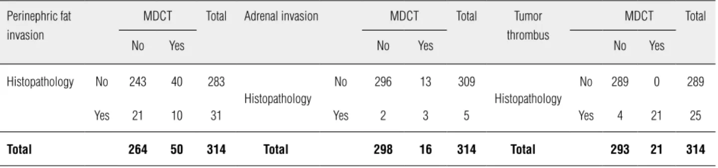
(Figure-2) (Table-2); only 10 were confirmed by specimen examination. In the patients with tumor thrombosis, MDCT was able to correctly identify and localize the presence and level of the thrombus in 21 patients. Of which, 13 were of thrombosis in renal vein (Figure-3), 7 in inferior vena cava (Figure-4) and 1 in inferior vena cava
and pulmonary vein. Though focal enhancement of venous wall or infiltration of adjacent soft tissue is suggestive of venous wall infiltration, especially in vena cava, MDCT failed to detect another 4 tumors with venous wall invasion (2 in renal vein, 2 in inferior vena cava). Direct inva-sion of ipsilateral adrenal gland was suspected in 16 patients on MDCT (Table-2) and only 3 had tumor involvement on specimen. Another two patients with ipsilateral adrenal gland invasion were not detected on imaging evaluation.
In the evaluation of lymph node involve-ment which included renal hilar, paraaortic, or paracaval lymph nodes, 297 tumors were staged
Figure 1 - Confined renal tumor with a well-defined pseu-docapsule (arrow).
Table 1 - Histopathological information of all included tumors.
No. tumors Tumor size(cm)
Included tumors 314 4.92 ± 2.58
Histopathological subgroup
Clear cell 285
Papillary cell 12
Chromophobe 10
Unclassified 3
Multiple cystic 3
XP11.2 translocation 1
Fuhrman grade
I 11
II 154
III 121
IV 27
Primary T stage
T1a 158 3.14 ±0.77
T1b 87 5.47 ±0.68
T2a 20 8.47±0.95
T2b 8 11.38 ± 1.09
T3a 30 7.56± 3.92
T3b 4 7.75 ±2.84
T3c 2 7.75 ±1.06
T4 5 8.20± 3.09
Table 2 - Histopathology and multidetector computed tomography (MDCT) staging of tumors (T).
MDCT Total
T1a T1b T2a T2b T3a T3b T3c T4
Histo-pathologic T1a 113 27 1 0 17 0 0 0 158
T1b 10 43 9 0 20 0 0 5 87
T2a 0 2 11 1 6 0 0 0 20
T2b 0 0 2 2 3 0 0 1 8
T3a 1 3 1 0 21 0 0 4 30
T3b 0 0 0 0 0 1 0 3 4
T3c 0 0 0 0 1 0 1 0 2
T4 0 0 0 0 1 0 0 4 5
Total 124 75 24 3 69 1 1 17 314
Figure 3 - A) Thrombus in right renal vein (Red arrow); B) Thrombus in right renal vein (Blue arrow).
N0, 8 tumors were staged N1 and 9 tumors were staged N2 by MDCT(Table-3). 295 tumors (93.95%) were correctly staged. 11 tumors (3.5%) were overstaged and 8 tumors (2.55%) were un-derstaged. In the 11 tumors with false-positive lymph nodes involvement by MDCT, the nodes were larger than 1 cm in short-axis diameter but were characterized as reactive hyperplasia on pathology. In the 6 tumors with false negative lymph nodes, microfocuses of cancer cell metas-tasis were identified. Similarly, another two tu-mors staged N2 were understaged as N1 by CT scan because malignant cell were also found in more than one paraaortic lymph node even with a diameter less than 1 cm. With respect to eval-uation of distant metastatic disease, 8 patients were suspected in arterial phase or parachymal phase and all were confirmed by pathological ex-amination (Table-4) (Figure-5).
Statistical findings
Agreement between MDCT and histo-pathologic findings was moderate for T
stag-ing (Kappa = 0.469), fair for N stagstag-ing (Kappa = 0.322), excellent for M staging (Kappa = 0.932), fair for stage grouping (Kappa = 0.502). 237 of 314 patients were correctly staged by MDCT, with an overall accuracy of 75.48%. The sensitivity and specificity of MDCT in detecting perinephric fat invasion were 32.26% and 85.87%, in detect-ing venous thrombosis were 84% and 100%, in detecting adrenal gland invasion were 60% and 95.79%, in detecting lymph node involvement were 50% and 96.36%, in detecting distant me-tastasis were 100% and 99.67%. In stage group-ing, 237 of 314 patients were correctly staged by MDCT, with an overall accuracy of 75.48%.
DISCUSSION
Since nephrectomy is still the only cura-tive method in the treatment of RCCs, preopera-tive evaluation of RCCs is of great importance. MDCT now serves as the most preferable imaging modality in determining tumor location, tumor size, tumor extension, thrombosis, lymph node
Table 3 - Histopathology and multidetector computed tomography (MDCT) staging of perinephric fat invasion, adrenal invasion and tumor thrombus.
Perinephric fat invasion
MDCT Total Adrenal invasion MDCT Total Tumor
thrombus
MDCT Total
No Yes No Yes No Yes
Histopathology No 243 40 283
Histopathology
No 296 13 309
Histopathology
No 289 0 289
Yes 21 10 31 Yes 2 3 5 Yes 4 21 25
Total 264 50 314 Total 298 16 314 Total 293 21 314
Table 4 - Histopathology and multidetector computed tomography (MDCT) staging of nodal (N) and distant metastasis (M).
Nodal metastasis MDCT Total Distant metastasis MDCT Total
N0 N1 N2 M0 M1
Histopathology
N0 291 6 5 302
Histopathology
M0 306 1 307
N1 4 0 0 4 M1 0 7 7
N2 2 2 4 8
involvement and distant metastasis. Previous studies have demonstrated that the accuracy of MDCT for detection and staging of renal mass is up to 90% (7), however, it is not of limitation.
Tumor size is known as the primary com-ponent of the 2009 updated TNM classification and an important prognostic variable for RCC. NSS or partial nephrectomy is now recommend-ed to patients with small localizrecommend-ed tumors (< 4 cm) (2), and that preoperative radiographical size estimation is an essential parameter to se-lect the appropriate treatment for RCC. Although CT measurement of the renal tumor size corre-lates well with the actual size of the tumor, CT scan tends to overestimate the tumor size (8,9). We have found an average overestimation of 0.21 cm on CT scan with significant difference (p < 0.001). In T1a group, the overestimation was even greater, of O.33 cm (p < 0.001). This may be the most reasonable explanation to the fact that 28 of 158 (17.72%) T1a tumors were overstaged as T1b and 9 of 87 (10.34%) T1b tumors overstaged
as T2a. Similarly, Nazim found 10 of 14 (71.43%) T1a tumors were overstaged as T1b and 14 of 44 (31.82%) T1b tumors were overstaged as T2a (10). Kanofsky also reported that the overestimation in tumor size was enough to upstage the tumor by TNM system in 16% of clear cell RCCs (11). Since a large portion of patients were overstaged preoperatively, these nephron sparing approaches should be considered in patients with tumor size slightly larger than 4 cm on CT scan without any other metastatic sign.
In prior studies, it has been shown that imaging using CT had low accuracy rates for the detection of perinephric tumor extension, as stranding in the perinephric fat is non-specific and can be due to edema, vascular engorgement or previous inflammation (12,13). The presence of enhancing nodules in the perinephric fat is now considered the most reliable finding of perinephric invasion (12). Comparing with the spiral CT used before, MDCT has proved to have higher spatial resolution and better anatomy detail delineation.
Catalano reported that MDCT had 95% accuracy for perinephric fat infiltration with sensitivity of 96% and specificity of 93% (7). However, even with MDCT and three dimensional technology, Hallscheidt and Türkvatan suggested the evalu-ation of renal tumor extension in to perineph-ric fat remains a difficult task (14,15). Türkvatan reported that 1 of 26 T1 tumors and 4 of 11 T2 tumors were overstaged as T3a. In our study, 17 of 158 T1a tumors, 20 of 80 T1b tumors, 6 of 20 T2a tumors, 3 of 8 T2b tumors were overstaged as T3a. As indicated by a recent retrospective analy-sis of 5339 patients, 5 years cancer specific sur-vival was 94.9% in pT1a, 92.6% in pT1b, 85.4% in pT2a, 70% in pT2b and 64.7% in pT3a (16), and patients with different staged tumors may require different treatments. Accurate stage of pT1a tu-mors is essential because infiltration to the peri-nephric fat is a contraindication to NSS. NSS is most appropriate for tumors located over the up-per or lower pole or in a up-peripheral location and with a clear demarcation to the renal vasculature and collecting system. In our study, 158 patients with pT1a tumors; 83 were submitted to radical nephrectomy, indicating that 52.54% patients were overtreated, and 43.37% of them were due to overstaging. However, although perinephric invasion characterized by perinephric stranding and enhancing nodule in perinephric fat have not a good sensitivity and accuracy, it still should be reserved in imaging evaluation, because under-staged tumors receiving more conservative thera-py may lead to disastrous clinical outcome.
Approximately 23% of RCC invade the re-nal veins and 7% invade the inferior vena cava (17). Accurate definition of the presence and level of tumor thrombus preoperatively is critical for surgical planning and patient counseling. Pa-tients with the level of tumor thrombus located inferior to the diaphragm only require laparot-omy, while the detection of supradiaphragmatic extension will require a thoracoabdominal surgi-cal approach. Although MRI has been proved su-perior to other modalities in tumor thrombus de-tecting and predicting the tumor thrombus level (18,19), it is not easily available and not proper for patients with pacemaker or altered cardio-pul-monary function. In a prospective study,
Halls-cheidt found no difference in tumor thrombus staging of 23 patients who underwent MRI plus MDCT preoperatively (20). More recently, Guzzo reported that accuracy rate of MDCT in predicting the superior level of tumor thrombus is 96% (21). A low attenuation filling defect within the vein seen after injection of contrast material is the most prominent feature for venous involvement on CT scan. In our series MDCT correctly identi-fied and localized the extent of the tumor throm-bus in all patients, and the agreement between MDCT and pathological finding was excellent. However, four patients with venous wall inva-sion were not detected by MDCT, probably due to the local extension. Invasion of the inferior vena cava will significantly complicate surgical proce-dure because prosthetic reconstruction is usually required. Though negative vascular margins were achieved in all four cases, it is important to note that no imaging modality is 100% accurate and the surgeon must be prepared if more advanced disease is noted than anticipated.
Because of the low incidence of ipsilat-eral adrenal gland involvement (16), the current surgical trend is to spare adrenal gland during surgery. Türkvatan reported that MDCT correctly identified all six cases of adrenal involvement (15). In our study, only 3 of 13 (18.75%) sus-pected adrenal involvement were confirmed by pathological findings. Because direct extension of large RCC into adrenal always compresses it into a thin tiny organ and causes local inflam-mation, it is difficult to distinguish it from the tumors. Our radiologist tended to be conservative and loss of tissue planes and irregular margins between the tumor and neighboring organ were all considered adrenal involvement. As indicated by Novara, patients with adrenal gland invasion had much lower 5 years cancer specific survival (17.9%) than other subgroups (16), that conserva-tive assessment of the adrenal gland is necessary preoperatively since an extensive resection ap-plied in patients with abnormality suggested on CT scan may yield a better clinical outcome.
A cutoff value in node size of 1 cm has been re-ported with a false negative rate of 10% due to reactive hyperplasia (12). Even with spiral CT, the false positive rates up to 43% have been report-ed (24). However, in a study by Catalano, using MDCT, 13 of 14 true positive cases for nodal me-tastasis were identified, reducing the false posi-tive rate due to reacposi-tive hyperplasia to 6.3% (7). In our study, 33.33% of patients with lymph node involvement were correctly staged, with a false positive rate of 64.7%. The agreement between MDCT and pathological findings were fair (Kap-pa = 0.322), which is consistent to the findings of Türkvatan (15). This indicated that the MDCT is not a reliable modality in nodal involvement detection, and 1 cm size as the cutoff value is not proper. Currently, regional lymph node dis-section is considered of no clinical benefit to pa-tients with clinically negative lymph nodes (25); however, in patients with positive lymph nodes suggested preoperatively or those with progres-sive disease, lymph node dissection is associated with improved survival (26,27).
Organ metastasis of RCC is most frequent-ly found in the lung, bone, brain and liver (28). Likely, the metastatic lesions tend to be hypervas-cular. The detection of visceral metastasis is of great importance because patients with metastat-ic disease still benefit from radmetastat-ical nephrectomy combined with systemic immunotherapy (29,30). In our study, all seven but one metastatic diseases were correctly detected by MDCT. Other study has proved excellent performance of this technique in metastatic lesions detection as well (15). How-ever, lesion from an 83-year-old male incorrectly staged by MDCT was finally proved an adenoma from the gastrointestinal system, suggesting that in high risk population, multiple tumors from dif-ferent tissues may occur. Therefore, a thorough preoperative search of tumors with different im-aging modalities may be necessary.
CONCLUSIONS
In conclusion, MDCT scan can delineate RCCs with high accuracy, including tumor size, the presence and level of tumor thrombus and distant metastasis. However, a great proportion
of tumors were overstaged by MDCT because of overestimation of tumor size and poor visualiza-tion of infiltravisualiza-tion of the perinephric fat. In ad-dition, as micrometastasis can not be identified and nodes with diameter > 1 cm may be caused by reactive hyperplasia, nodal metastatic lesion evaluation remains a difficult task.
CONFLICT OF INTEREST
None declared.
REFERENCES
1. Jemal A, Siegel R, Xu J, Ward E: Cancer statistics, 2010. CA Cancer J Clin. 2010; 60: 277-300. Erratum in: CA Cancer J Clin. 2011; 61: 133-4.
2. Jayson M, Sanders H: Increased incidence of serendipitous-ly discovered renal cell carcinoma. Urology. 1998; 51: 203-5. 3. Yaycioglu O, Roberts WW, Chan T, Epstein JI, Marshall FF, Ka-voussi LR: Prognostic assessment of nonmetastatic renal cell carcinoma: a clinically based model. Urology. 2001; 58: 141-5. 4. Israel GM, Bosniak MA: Renal imaging for diagnosis and
staging of renal cell carcinoma. Urol Clin North Am. 2003; 30: 499-514.
5. Blum A, Walter F, Ludig T, Zhu X, Roland J: Multislice CT: principles and new CT-scan applications. J Radiol. 2000; 81: 1597-614.
6. Ng CS, Wood CG, Silverman PM, Tannir NM, Tamboli P, Sandler CM: Renal cell carcinoma: diagnosis, staging, and surveillance. AJR Am J Roentgenol. 2008;191: 1220-32. 7. Catalano C, Fraioli F, Laghi A, Napoli A, Pediconi F, Danti M,
et al.: High-resolution multidetector CT in the preoperative evaluation of patients with renal cell carcinoma. AJR Am J Roentgenol. 2003; 180: 1271-7.
8. Mistry R, Manikandan R, Williams P, Philip J, Littler P, Foster CS, et al.: Implications of computer tomography measure-ment in the managemeasure-ment of renal tumours. BMC Urol. 2008; 8: 13.
9. Irani J, Humbert M, Lecocq B, Pires C, Lefèbvre O, Doré B: Renal tumor size: comparison between computed tomogra-phy and surgical measurements. Eur Urol. 2001; 39: 300-3. 10. Nazim SM, Ather MH, Hafeez K, Salam B: Accuracy of
multi-detector CT scans in staging of renal carcinoma. Int J Surg. 2011; 9: 86-90.
11. Kanofsky JA, Phillips CK, Stifelman MD, Taneja SS: Impact of discordant radiologic and pathologic tumor size on renal cancer staging. Urology. 2006; 68: 728-31.
13. Zagoria RJ, Bechtold RE, Dyer RB: Staging of renal adeno-carcinoma: role of various imaging procedures. AJR Am J Roentgenol. 1995; 164: 363-70.
14. Hallscheidt P, Wagener N, Gholipour F, Aghabozorgi N, Drey-haupt J, Hohenfellner M, et al.: Multislice computed tomog-raphy in planning nephron-sparing surgery in a prospective study with 76 patients: comparison of radiological and his-topathological findings in the infiltration of renal structures. J Comput Assist Tomogr. 2006; 30: 869-74. Erratum in: J Comput Assist Tomogr. 2007; 31: 164.
15. Türkvatan A, Akdur PO, Altinel M, Olçer T, Turhan N, Cumhur T, et al.: Preoperative staging of renal cell carcinoma with multidetector CT. Diagn Interv Radiol. 2009; 15: 22-30. 16. Novara G, Ficarra V, Antonelli A, Artibani W, Bertini R, Carini
M, et al.: Validation of the 2009 TNM version in a large multi-institutional cohort of patients treated for renal cell carci-noma: are further improvements needed? Eur Urol. 2010; 58: 588-95.
17. Laissy JP, Menegazzo D, Debray MP, Toublanc M, Ravery V, Dumont E, et al.: Renal carcinoma: diagnosis of venous inva-sion with Gd-enhanced MR venography. Eur Radiol. 2000; 10: 1138-43.
18. Goldfarb DA, Novick AC, Lorig R, Bretan PN, Montie JE, Pon-tes JE, et al.: Magnetic resonance imaging for assessment of vena caval tumor thrombi: a comparative study with vena-cavography and computerized tomography scanning. Urol. 1990; 144: 1100-3; discussion 1103-4.
19. Semelka RC, Shoenut JP, Kroeker MA, MacMahon RG, Greenberg HM: Renal lesions: controlled comparison be-tween CT and 1.5-T MR imaging with nonenhanced and gadolinium-enhanced fat-suppressed spin-echo and breath-hold FLASH techniques. Radiology. 1992; 182: 425-30. 20. Hallscheidt PJ, Fink C, Haferkamp A, Bock M, Luburic A,
Zuna I, et al.: Preoperative staging of renal cell carcinoma with inferior vena cava thrombus using multidetector CT and MRI: prospective study with histopathological correlation. J Comput Assist Tomogr. 2005; 29: 64-8.
21. Guzzo TJ, Pierorazio PM, Schaeffer EM, Fishman EK, Allaf ME: The accuracy of multidetector computerized tomogra-phy for evaluating tumor thrombus in patients with renal cell carcinoma. J Urol. 2009; 181: 486-90; discussion 491. 22. Studer UE, Scherz S, Scheidegger J, Kraft R, Sonntag R,
Ackermann D, et al.: Enlargement of regional lymph nodes in renal cell carcinoma is often not due to metastases. J Urol. 1990; 144(2 Pt 1): 243-5.
23. Herrlinger A, Schrott KM, Schott G, Sigel A: What are the benefits of extended dissection of the regional renal lymph nodes in the therapy of renal cell carcinoma. J Urol. 1991; 146: 1224-7.
24. Heidenreich A, Ravery V: Preoperative imaging in renal cell cancer. European Society of Oncological Urology. World J Urol. 2004; 22: 307-15.
25. Pantuck AJ, Zisman A, Dorey F, Chao DH, Han KR, Said J, et al.: Renal cell carcinoma with retroperitoneal lymph nodes: role of lymph node dissection. J Urol. 2003; 169: 2076-83. 26. Blute ML, Leibovich BC, Cheville JC, Lohse CM, Zincke H: A
protocol for performing extended lymph node dissection us-ing primary tumor pathological features for patients treated with radical nephrectomy for clear cell renal cell carcinoma. J Urol. 2004; 172: 465-9.
27. Hutterer GC, Patard JJ, Perrotte P, Ionescu C, de La Taille A, Salomon L, et al.: Patients with renal cell carcinoma nodal metastases can be accurately identified: external validation of a new nomogram. Int J Cancer. 2007; 121: 2556-61. 28. Rohde V. Nierenzellkarinom. In: Schmelz HU, Sparwasser C,
Weidner W (eds) Facharztwissen Urologie. Springer Medizin Verlag, Heidelberg, 2006; pp. 146-58.
29. Flanigan RC, Salmon SE, Blumenstein BA, Bearman SI, Roy V, McGrath PC, et al.: Nephrectomy followed by interferon alfa-2b compared with interferon alfa-2b alone for metastatic renal-cell cancer. N Engl J Med. 2001; 345: 1655-9. 30. Mickisch GH, Garin A, van Poppel H, de Prijck L, Sylvester
R: Radical nephrectomy plus interferon-alfa-based immu-notherapy compared with interferon alfa alone in metastatic renal-cell carcinoma: a randomised trial. European Organ-isation for Research and Treatment of Cancer (EORTC) Geni-tourinary Group. Lancet. 2001; 358: 966-70.