HP1
β
Is a Biomarker for Breast Cancer
Prognosis and PARP Inhibitor Therapy
Young-Ho Lee1*, Xiyong Liu1, Fuming Qiu2, Timothy R. O’Connor3, Yun Yen1, David K. Ann1*
1Department of Molecular Pharmacology, Beckman Research Institute, City of Hope, Duarte, California, United States of America,2Department of Medical Oncology, Second Affiliated Hospital, Zhejiang University, School of Medicine, Hangzhou, China,3Department of Cancer Biology, Beckman Research Institute, City of Hope, Duarte, California, United States of America
*lyh205@gmail.com(YL);dann@coh.org(DA)
Abstract
Members of the heterochromatin protein 1 family (HP1α,βandγ) are mostly associated with heterochromatin and play important roles in gene regulation and DNA damage re-sponse. Altered expression of individual HP1 subtype has profound impacts on cell prolifer-ation and tumorigenesis. We analyzed the expression profile of HP1 family by data mining using a published microarray data set coupled with retrospective immunohistochemistry analyses of archived breast cancer biospecimens. We found that the patient group overex-pressingHP1βmRNA is associated with poorly differentiated breast tumors and with a significantly lower survival rate. Immunohistochemical staining against HP1α, HP1βand HP1γshows that respective HP1 expression level is frequently altered in breast cancers. 57.4 - 60.1% of samples examined showed high HP1βexpression and 39.9 - 42.6 % of ex-amined tumors showed no or low expression of each HP1 subtype. Interestingly, compara-tive analysis on HP1 expression profile and breast cancer markers revealed a posicompara-tive correlation between the respective expression level of all three HP1 subtypes and Ki-67, a cell proliferation and well-known breast cancer marker. To explore the effect of individual HP1 on PARP inhibitor therapy for breast cancer, MCF7 breast cancer cells and individually HP1-depleted MCF7 cells were treated with PARP inhibitor ABT-888 with or without carbo-platin. Notably, HP1β-knockdown cells are hypersensitive to the PARP inhibitor ABT-888 alone and its combination with carboplatin. In summary, while increased HP1βexpression is associated with the poor prognosis in breast cancer, compromised HP1βabundance may serve as a useful predictive marker for chemotherapy, including PARP inhibitors against breast cancer.
Introduction
Breast cancer is one of the leading causes of death in the United States and worldwide. Early di-agnosis and effective use of adjuvant therapies are required to improve patient survival [1,2]. Prognostic factors that are frequently used for making clinical decisions in breast cancer are a11111
OPEN ACCESS
Citation:Lee Y-H, Liu X, Qiu F, O’Connor TR, Yen Y,
Ann DK (2015) HP1βIs a Biomarker for Breast
Cancer Prognosis and PARP Inhibitor Therapy. PLoS ONE 10(3): e0121207. doi:10.1371/journal. pone.0121207
Academic Editor:Brij Singh, School of Medicine and Health Sciences, University of North Dakota, UNITED STATES
Received:November 11, 2014
Accepted:January 28, 2015
Published:March 13, 2015
Copyright:© 2015 Lee et al. This is an open access
article distributed under the terms of theCreative
Commons Attribution License, which permits unrestricted use, distribution, and reproduction in any medium, provided the original author and source are credited.
Data Availability Statement:All relevant data are within the paper and its Supporting Information files.
Funding:This work was supported by National Institutes of Health Research Grants R01DE10742 and R01DE14183 to DA. The funders had no role in study design, data collection and analysis, decision to publish, or preparation of the manuscript.
age, tumor size, status of lymph nodes, histological types of the tumor, pathological grade, and hormone receptor status. However, more biomarkers are needed for therapy and prediction of outcome because human breast cancers are diverse in their genetic nature and their response to therapy. Recently, many groups have tried to identify gene signatures of breast cancer pa-tients [3,4]. These gene signatures can lead to more accurate clinical decisions for cancer pa-tients [5]. Breast cancer can be classified into several groups depending on their expressions of biomarkers and pathology of breast cancer specimens. The most common molecular markers for breast cancers include estrogen receptor (ER), progesterone receptor (PR), HER2/neu, EGFR, Ki-67 and others [6]. The subgroups of breast cancer include Luminal A, Luminal B, Basal, HER2-enriched subtypes [6]. Triple negative breast cancer subtypes, which have defi-cient expression of ER, PR and HER2/neu, usually have poor prognosis and do not respond to hormone therapy. However, triple negative breast cancer is also a heterogeneous group, which shows different gene signatures [7]. For example, some triple negative breast cancers have de-fectiveBRCA1genes, whereas other triple negative breast cancer patient groups have functional
BRCA1.BRCA1is one of the most frequently mutated genes in breast cancer patients [8]. Women with germline mutations inBRCA1have high risk of breast cancer (~80% by the age of 70), ovarian cancer (~30–40%) and other cancers. BRCA1 is involved in maintaining geno-mic integrity by functioning in pathways involved in DNA repair, cell cycle checkpoint control, apoptosis, chromosome segregation and others [8]. One of the main roles of BRCA1 is to pro-mote homologous recombination repair and G2/M cell cycle arrest during DNA damage re-sponse. Thus, the loss of BRCA1 is frequently associated with a dramatic increase of genomic instability and tumorigenesis. While germline BRCA1 mutations are rarely found in patients with sporadic breast cancers, the functions of BRCA1 may be inactivated by other mechanisms, which are often referred to as“BRCAness”[9]. One of the possible mechanisms of BRCAness is the inactivation of BRCA1 function at the epigenetic level by DNA methylation of the
BRCA1promoter [9,10].
BRCA status is also important for cancer therapy. The genomic instability of BRCA1- and BRCA2-defective cells can be exploited for cancer therapy [11,12]. Clinically, the genomic in-stability phenotype of BRCA1- and BRCA2- deficient cells provided an opportunity for PARP inhibitor treatment [12,13]. Poly(ADP-ribose) polymerase (PARP) is involved in the repair of DNA single strand breaks (SSBs), and failure of their repair can lead to the generation of DNA double strand breaks (DSBs) during DNA replication. Inhibition of PARP1 leads to a large in-crease in DSBs and to cell death in the absence of BRCA1 or 2 and/or in the absence of HR de-pendent DSB repair [11,12]. This is the basis for the concept that PARP inhibitors induce synthetic lethality in HR repair deficient tumors and provides a novel strategy for cancer thera-py, at least in breast cancer patients who have mutations in BRCA1 or BRCA2. Recent clinical trials of a PARP inhibitor reported a partial success in cancer therapy with less severe side ef-fects [14–16].
high levels ofHP1βmRNA had less probability of survival. We also found the positive correla-tion of HP1 expression and Ki-67 cancer marker in breast cancer samples, suggesting potential significance of HP1 as a marker for breast cancer prognosis. Furthermore, we showed that PARP inhibitor ABT-888 was more effective in inducing death of HP1β-deficient MCF7 breast cancer cells. These data suggest that HP1βlevel could not only serve as a useful marker for breast cancer prognosis but also as a predictive marker for PARP therapy.
Materials and Methods
Data mining on microarray dataset
A total of 10 published microarray data sets including: Ivshina (GSE4922), Chin (E-TABM-158), Wang (GSE2034), Pawitan (GSE1456), Desmedt (GSE7390), Expo (GSE2109), Huang [22], Bild (GSE3143), Sortiriou (GSE2990) and NKI [23] with clinical annotations were down-loaded from the combined microarray dataset BRAVO (Biomarker recognition and validation on-line). The NKI (Netherlands Cancer Institute)-295 set was especially selected for HP1 prog-nostic evaluation because the probe (Agilent Technologies) forcbx1(HP1β) is 100% identical to previously identified sequence ofcbx1and it contains information of most gene signatures’
classification. NKI data set (295 patients analyzed, Accession number N/A) used 25,000-gene array that comes from Agelent Technologies, which used same probers with Affymetrix HG-U133 array.
Patient enrollment, follow-up and tissue array
Patients diagnosed with breast cancer and treated by surgical resection between January 2002 and January 2006 in the Second Affiliated Hospital of Zhejiang University (ZJU) were included in this study. A breast cancer pathologist (F. Q.) used haematoxylin and eosin (H&E)-stained slides, to retrospectively review the history of all cases. The clinicopathological parameters that were evaluated included patient age at the time of diagnosis, tumor node metastasis (TNM) stage, date of last follow-up, and overall patient survival. Exclusion included breast cancer sam-ples from patients without a pathologic diagnosis, those with multiple cancers, or those patients with whom contact was lost after surgery. A total of 222 breast cancer patients were included in this study. Follow-ups were conducted for all participants and the surgery relapse and death data were collected until 2010. Overall survival (OS) rate was calculated from the date of sur-gery to date of death by breast cancer-associated illness. Disease-free survival (DFS) rate was calculated from date of surgery to date of local recurrence or metastasis. If no death or relapse occurred, the OS and DFS rates were calculated from date of surgery to September 2010. All of the formalin-fixed, paraffin-embedded (FFPE) breast cancer tissue samples that were collected were reassembled into multiple tissue arrays. Analysis indicated that HP1 immunohistological signals did not correlate with storage time (likelihood,p= 0.246), indicating the storage time did not affect the immunohistochemical outcome.
Immunohistochemistry (IHC)
HP1 protein levels in the 222 breast cancer samples were assessed by IHC with anti-HP1 antibodies (1:75 dilution); anti-HP1α(Bethyl, Abcam), anti HP1β(ab10478, Abcam) and anti-HP1γ(ab10480, Abcam). The IHC conditions for HP1 expression determination were pre-optimized on checkboards with multiple tissue samples. Briefly, after de-paraffinization, pre-treatment with 3% H2O2was used to block the endogenous peroxidase activity. The slides
slides were then incubated with polymer horseradish peroxidase-labeled secondary antibodies for 30 minutes at RT, then 3,3-Diaminobenzidine (DAB)-treated (0.05 g DAB and 100 ml 30% H2O2in 100 ml PBS) for 5 and 10 minutes, respectively. Each slide was counterstained with
DAKO's haematoxylin. For each IHC staining, the negative and positive checkboards were ap-plied as quality controls. The specificity of anti-HP1 antibodies were validated by Western analyses. HP1 staining was predominantly nucleus, and HP1 expression was assessed using a visual grading system on the basis of the intensity of staining signals observed by light micros-copy. Each sample was independently scored by two investigators (Y.L. and X.L.), including one breast cancer pathologist (X.L.) using a double-blind design to avoid scoring bias. Discrep-ancies were re-evaluated by joint review between the two readers. Less than 10% variation was noticed among different slides.
Statistical analysis
The database was created by using MS-Access and data analysis was performed using JMP 8.0 software (SAS Institution) and GraphPad Prism 5.0 software. Group comparisons for continu-ous data were done by t-test for independent means or 1-way ANOVA. Each cell biology ex-periment was performed in triplicate to obtain representative means and images. Categorical variables were compared usingχ2analysis, Fisher’s exact test or binomial test of proportions. Kaplan-Meier analysis and a COX hazard proportional model were used to analyze overall sur-vival and disease-free-sursur-vival. Multivariate analysis and stratification were used to reduce the confounder’s impact on the estimation of the Hazard Ratio (HR). Statistical significant was set asp<0.05, two-tailed.
Apoptosis assay
Apoptotic cells were measured by FITC Annexin V Apoptosis Detection Kit I (BD Pharmin-gen) according to manufacturer’s protocol. MCF7 cells and HP1-depleted MCF7 cells were cultured and harvested before or after irradiation. The harvested cells were washed twice with ice-cold PBS and then resuspended cells in 1 x Binding Buffer (0.1 M Hepes/NaOH (pH 7.4), 1.4 M NaCl, 25 mM CaCl2.) at a concentration of 1 x 10⁶cells/ml. 100μl of cells (1 x 10⁵cells)
were transferred to a 5-ml culture tube and incubated with FITC-conjugated Annexin V (5μl).
The incubated cells were incubated for 15 minutes at Room Temperature (25°C) in the dark and 1 x Binding Buffer (400μl) was added to each tube. The stained cells were analyzed by
flow cytometry.
Ethical statement
Results
HP1
β
/CBX1
mRNA level is inversely associated with breast cancer
patient survival
Initially, we used data mining techniques to determine if the expression level of HP1β/CBX1 mRNA was associated with the outcome of breast cancer patients using a published microarray dataset [23,24]. Since the expression level ofHP1βmRNA is diverse in breast cancer samples, we classified patients into four groups (0, 1, 2, 3) according to quartile ofHP1βmRNA levels. A total of 74 breast cancer patients were stratified as high expressors ofHP1βmRNA (group-3) and 221 patients were classified as low or no expressors ofHP1β(group-0, -1 or -2). Kaplan-Meier analyses indicated thatHP1expression was a critical prognostic indicator for both over-all and disease-free survival for over-all breast cancer patients. Notably, highHP1βexpressor group (N = 74) was associated with lower DFS (disease-free survival) (p= 0.001) and OS (overall sur-vival) (p= 0.008), when compared with lowerHP1βexpressor group (Fig. 1A). The OS and DFS time were calculated as the length of time from date of surgical operation to the date of specific breast cancer-related death and relapse/metastasis, respectively. However, expression of otherHP1subtype mRNAs was not analyzed in this analysis or did not affect the survival in a statistically-significant manner. Furthermore, highHP1βexpression was associated with poorly differentiated cancer grade (Fig. 1B). HighHP1βexpression group displayed more ag-gressive types of breast cancers like basal and luminal B type. However, lowHP1βexpressors exhibited more low, moderately, or well-differentiated phenotypes. These suggest thatHP1β mRNA expression level may be a prognostic marker for survival of breast cancer patients.
Altered expression of HP1 proteins in breast cancer patients
SinceHP1βmRNA expression levels were significantly associated with survival of breast cancer patients, we sought to examine the protein expression level of HP1 subtypes in breast cancer samples by IHC staining. First, normal skin and normal breast samples were stained with HP1βspecific antibody. IHC of normal skin showed that HP1βstaining is nuclear. Similarly, IHC staining showed that HP1βin normal mammary samples also showed that HP1βis pri-marily nuclear with a weaker expression of HP1βin the cytoplasm (Fig. 2A,upper panels). Next, we stained 190 breast cancer samples using an anti-HP1βantibody.Fig. 2A(lower pan-els) shows that HP1βexpression patterns in breast cancer samples are diverse and altered in most of cases. Some of the breast cancer samples showed strong nuclear HP1βlevels in the nu-cleus, whereas other samples showed lower or no HP1βsignals. There are also samples that manifest clear staining of HP1βonly in the cytoplasm, but not in the nucleus (Fig. 2A,lower panels). Therefore, HP1βis heterogeneously distributed in breast cancer samples. The breast cancer samples were further analyzed individually and classified into four groups (0, 1, 2, 3) based on the HP1βexpression level (S1 Fig.). Accordingly, 34 (18.6%), 39 (21.3%), 58 (31.7%) and 52 (28.4%) samples were designed to group-0, -1, -2 and -3, respectively. We then further divided them into low HP1βexpression group (0+1) and high HP1βexpression group (2+3). Overall, 60.1% of breast cancer biospecimens exhibited high HP1β(HP1β-high) levels and 39.9% of breast cancer samples showed no or low levels of HP1β(HP1β-low) (Fig. 2).
Fig 1.HP1βmessage abundance is associated with survival of breast cancer patients.A microarray database of 295 breast cancer patients (NKI-295 dataset) was analyzed andHP1βmessage signals were investigated.A. Kaplan-Meier analyses indicated thatHP1βmRNA abundance is inversely correlated with both disease-free survival (DFS) and overall survival (OS) for breast cancer patients.B. Group of highHP1βexpressors is associated with aggressive and poorly differentiated breast cancers. Low or highHP1βmessage abundance are denoted from microarray database from the public domain [23].
least one HP1 subtype. Intriguingly, 82 cases (46.6%) of 176 validated stained samples by all three antibodies showed the same IHC staining scores by antibodies recognizing three individ-ual HP1 subtypes.Fig. 2Bshows that HP1-High and HP1-Low group samples were stained (or not) with three individual HP1 subtypes antibodies, respectively. HP1-Mixed is the group of the breast tumors showing strong expression of only one or two HP1 subtype(s). Specifically, 133 breast cancer tumors (75.6%) showed the same groups of high or low HP1 expressors. Only 15% of cancer samples showed a mixed expression pattern of HP1 subtypes (Fig. 2B, HP1-Mixed). Furthermore, analysis of nuclear and cytoplasmic staining patterns showed that HP1 proteins are strongly stained in the cytoplasm of some cancer samples. Especially, 49 cases (27%, out of 176) of breast cancer patients showed strong cytoplasmic HP1αstaining. Al-though a significant portion of cancer tissues showed stronger HP1 cytoplasmic staining in this study (Fig. 2), the potential roles of HP1 mis-localization in breast cancer cells remain unclear. We have compared various cancer markers between HP1-nucleus and HP1-cytoplasm groups (data not shown). However, the correlation of HP1 mis-localization and breast cancer tumori-genesis has yet been established.
Positive correlation of HP1
α
,
β
and
γ
expression and Ki-67, a cell
proliferation marker, in breast cancer
Table 1,S1andS2Tables show the clinical and pathological characteristics of 190 breast cancer patients. These include patients’ages, tumor stages, lymph node infiltration, expression status of ER, PR, p53, Ki-67, HER2. We divided the patient groups into the high HP1 group or the low HP1 group according IHC scoring data. We analyzed the correlation of respective HP1 IHC signals with other breast cancer clinical and pathological markers inTable 1,S1andS2 Tables. Notably, one common feature of our findings was the positive correlation of Ki-67 with HP1α(p= 0.0415), HP1β(p= 0.0007) and HP1γ(p= 0.0002), respectively (Fig. 3). Our analy-ses further indicated a significant correlation between HP1αlevel and several breast cancer markers, such as age, ER status, p53 status and molecular subtypes (S1 Table). HP1γlevel was also correlated with p53 status (S2 Table). However, the HP1βsignal showed significant corre-lation especially with Ki-67 (Table 1). Since expressions of all three HP1 subtypes showed a clear correlation with the cell proliferation marker, Ki-67, high HP1 expression probably re-flects a group of patients with actively growing breast cancer cells. Conceivably, these analyses suggested the abundance of all three HP1 subtypes could provide useful prognostic informa-tion on breast cancer patients.
HP1
β
depleted breast cancer cells are hypersensitive to PARP inhibitor
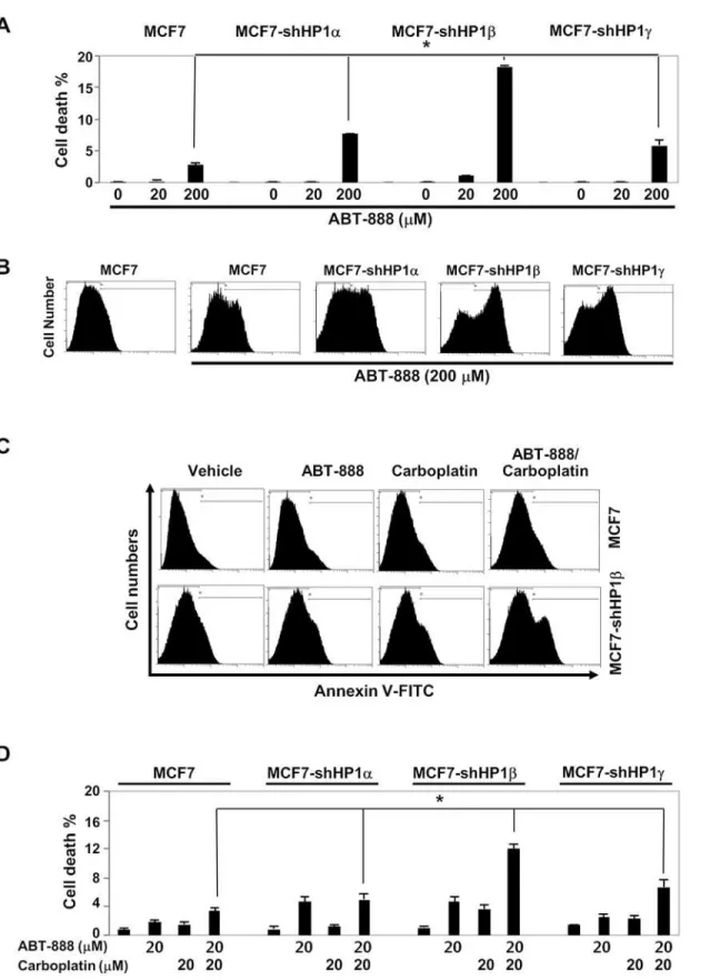
Previously, we reported that HP1 family is required for DNA damage response primarily through the regulation of BRCA1 function [17]. HP1-depleted cells showed defective BRCA1 foci formation, homologous recombination DNA repair and G2/M cell cycle checkpoint con-trol in response to irradiation. As this study showed that significant populations of breast can-cer patients have low or no expression of at least one HP1 subtype (Fig. 2), we tested the effect of individual HP1 on PARP inhibitor therapy. To achieve this goal, MCF7 cells and individual-ly HP1-depleted MCF7 cells (S2 Fig.) were treated with ABT-888 (Veliparib), which is one of
subtypes anti-HP1 antibodies, anti-HP1α, HP1βor HP1γ(Abcam antibodies: ab77256, ab10478, ab10480). IHC staining patterns were compared and IHC staining scores were determined as shown inS1 Fig. Scale bars: 100μm. HP1-High indicates the group of tumors with abundant expression of all three HP1 subtypes. HP1-Low is the group with no or low expression of all three HP1 subtypes. HP1-Mixed group of cancer samples are high level expression with one or two HP1 subtypes. Representative images are shown.
the PARP inhibitors currently undergoing clinical evaluation [25]. MCF7 breast cancer cells were chosen for our experimental paradigm because MCF7 harbors wild-type tumor suppres-sor BRCA1 in addition to wild type p53, ER, and PR [26]. MCF7 cells and individually HP1-depleted MCF7 cells were treated with vehicle or ABT-888 (20 or 200μM) for 72 hours. The
cells were collected and stained with Annexin V and propidium iodide (PI) and analyzed by
Table 1. Correlation analyses of HP1βexpression level with several molecular and pathological cancer markers.
Characteristics Total number of patients HP1β-Low (N = 74) HP1β-High (N = 113) p-value
Median age Age<49 years 83 (43.9%) 32 (43.2%) 50 (44.2%) 0.3141
Age>49 years 107 (56.1%) 42 (56.8%) 63 (56.8%)
Tumor stages T0–T1 58 (31.5%) 24 (36.4%) 32 (28.6%) 0.2818
T2–T3 123 (68.5%) 42 (63.6%) 80 (71.4%)
Lymph node N2 negative 98 (54.0%) 45 (60.1%) 56 (49.6%) 0.1301
N2 positive 92 (46.0%) 29 (39.2%) 57 (50.4%)
ER ER negative 61 (40.7%) 24 (43.6%) 37 (39.0%) 0.5737
ER positive 91 (59.3%) 31 (56.4%) 58 (61.0%)
PR PR negative 71 (47.4%) 22 (40.7%) 50 (51.0%) 0.2234
PR positive 83 (52.6%) 32 (59.3%) 48 (49.0%)
p53 p53 negative 92 (57.6%) 40 (66.7%) 51 (52.0%) 0.0693
p53 positive 67 (42.4%) 20 (33.3%) 47 (48.0%)
Ki-67 Ki-67 negative 72 (44.5%) 39 (60.9%) 34 (34.0%) 0.0007
Ki-67 positive 94 (55.5%) 25 (39.1%) 66 (66.0%)
HER2 HER2 negative 129 (82.2%) 45 (83.3%) 80 (81.6%) 0.069
HER2 positive 27 (17.8%) 9 (16.7%) 18 (18.4%)
Molecular type Luminal A 50 (35.5%) 23 (47.9%) 26 (28.9%) 0.1179
Luminal B 52 (37.0%) 15 (31.3%) 36 (40.0%)
TNBC 26 (18.8%) 8 (16.7%) 18 (20.0%)
HER2+ 12 (8.7%) 2 (4.2%) 10 (11.1%)
doi:10.1371/journal.pone.0121207.t001
Fig 3. Positive correlation of HP1 and Ki-67 levels in breast cancer.Levels of HP1α, HP1βand HP1γIHC signal were positively correlated with Ki-67 levels in breast cancer patients. 0 indicates no or low expression and 1 denotes high expression of respective HP1 subtype, as shown inFig. 2, and Ki-67 level, respectively.
flow cytometry. MCF7 cells with wild type BRCA1 were relatively resistant to PARP inhibitor treatment (Fig. 4A). However, treatment of ABT-888 (20μM) induced high level of apoptosis
in HP1β-depleted MCF7 cells. Although treatment ABT-888 (200μM) barely increased the
Annexin V-positive MCF7 population, it markedly increased Annexin V-positive, presumably apoptotic, HP1α-,β- orγ-knockdown MCF7 cells (Fig. 4B). Notably, the cell death (double stained population by PI and Annexin V) in HP1β-depleted cells was 10 times higher than that of MCF7 cells (Fig. 4A). HP1α- orγ- depleted MCF7 cells were also hypersensitive to ABT-888. This suggests that PARP inhibitor ABT-888 can effectively target HP1-deficient, especially HP1β-deficient, breast cancer cells.
We then examined the combination effects of ABT-888 and carboplatin on apoptosis of MCF7 cells and individually HP1-depleted MCF7 cells. Carboplatin is an alkylating agent that exhibits a cytotoxic effect on cancer cells by binding to DNA and forming interstrand cross-links that block DNA replication. Previously, the synthetic lethality of ABT-888 and carbopla-tin in breast cancer cells with respect to BRCA status was reportedin vitroandin vivo[27]. To test the effect of HP1 status on the synthetic lethality of these two drugs, MCF7 cells and indi-vidually HP1-depleted MCF7 cells were treated with a combination of ABT-888 (20μM) and
carboplatin (20μM). As shown inFig. 4C, neither ABT-888 alone, carboplatin alone nor
com-bination had marked effect on rendering Annexin V-positive in MCF7 cells. However, same amounts of ABT-888 or carboplatin induced cell death of HP1β-depleted MCF7 cells (Fig. 4D). Notably, combination of ABT-888 and carboplatin resulted in marked cytotoxic effects in HP1β-depleted MCF7 cells. These results showed that PARP inhibitors and/or carboplatin can be an effective therapy regimen for patients with breast cancer of no or low HP1βexpressors. Conceivably, HP1αor HP1γdeficiency in tumor tissues can be translated as a predictive mark-er for breast cancmark-er PARP inhibitor thmark-erapy. While HP1αand HP1γcompromised MCF7 cells showed 2~3 fold higher sensitivity to PARP inhibitor treatment, HP1βdeficient cells were much more sensitive to PARP inhibitor (Fig. 4). In other words, HP1 levels, especially HP1β
deficiency, could be a useful predicative marker for BRCAness for the effective use of PARP therapy.
Discussion
HP1 is a potential prognostic marker for breast cancer
Identification of novel biomarkers for breast cancer is crucial for predicting cancer prognosis and therapeutic outcomes [28]. The diverse genetic variations and mutations found in breast cancers make it difficult to classify those tumors into groups to improve therapeutic guidance. Therefore, identification of additional molecular signatures of breast cancers will provide a bet-ter basis for targeted therapy and personalized medicine. Herein, results presented in this study suggest that high levels of HP1βare a poor prognostic marker for breast cancer outcome (Fig. 1). Moreover, high HP1 expressors may indicate a group of patients harboring actively growing breast cancer cells, since all HP1α,βandγexpression correlated with Ki-67, a surro-gate marker for cell proliferation (Fig. 3). Lastly, lack-of-HP1β-expression could serve as a pre-dictive marker to define a breast cancer therapeutic option (Fig. 4).
and IHC study. Here we show that the mRNA and protein expression levels of HP1 are fre-quently altered and diverse among breast cancer biospecimens.HP1βmRNA levels are inverse-ly correlated with survival (OS and DFS) of breast cancer patients (Fig. 1). HP1αprotein levels showed a correlation with several cancer markers including age, p53 status, ER status and Ki-67 (Table 1andFig. 3). However, expressions of all three subtypes of HP1 are frequently regu-lated in similar manner in cancer cells (Fig. 2). Our results reveal that all three HP1 subtypes are potentially useful markers for breast cancer prognosis. Notably, expression levels of HP1 showed strong correlation with Ki-67 level in breast cancer samples (Fig. 3). Ki-67 is used as an indicator to further classify triple negative breast cancers [32]. Analysis of HP1 expression in cancer patients may also be useful for further analyzing breast cancer molecular subtypes. Pre-viously other groups showed that breast cancer cells with high HP1αare more prone to cell cycle progression [31]. This is consistent with our finding showing a positive correlation of HP1αand cell proliferation marker Ki-67. Furthermore, our study shows that there is a strong correlation of Ki-67 expression with other HP1 subtypes. Further investigation of the relation between expression of HP1 subtypes and Ki-67 in other cancers including prostate cancer could also be worthwhile [33]. Our results together with other reports suggest the potential sig-nificance of HP1 in breast cancer prognosis and thus this warrants additional studies.
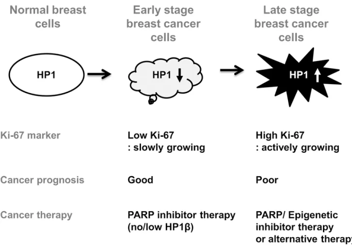
Potential roles of HP1 in carcinogenesis
While the complicated HP1 levels and pattern in breast cancer biospecimens could also reflect the heterogeneity of cancer cells in human breast tumors [6,34], it is intriguing that expression levels of three HP1 subtypes were comparably regulated in some breast cancer cells. These al-tered and heterogeneous staining patterns also implicate that HP1 family plays diverse roles in breast cancers. As HP1 subtypes elicit multiple functions in cells, we surmise that the expres-sion levels and subcellular location of HP1 are dynamically regulated during tumorigenesis (Fig. 5). Previously we showed that HP1 is required for homologous recombination repair and cell cycle control through the regulation of BRCA1 [17]. HP1 is also involved in the other cellular functions, such as transcription and cell proliferation. Thereby, we speculate that the lack-of-HP1-expression in some breast cancer tumors can deregulate their BRCA1 functions in homologous recombination repair and cell cycle checkpoint control. Conceivably, genomic mutations could accumulate in cancer cells with low HP1 levels. This could explain why some cancer patients exhibited lack-of-HP1-expression phenotypes in cancer cells.
However, it is not clear how high HP1 expression contributes to tumorigenesis. High levels of HP1 may deregulate the expression of genes involved in tumorigenesis, thereby promoting the growth and proliferation of cancer cells. This possibility is supported by the observation showing a significant correlation of HP1 expression with Ki-67 level (Fig. 3,Table 1,S1,S2 Ta-bles). Ki-67 is a nuclear protein that correlates with cell cycle progression through S-phase [35]. It is widely held that Ki-67 exists at low levels in normal and resting (G0 phase) cells. This is why Ki-67 is considered to be a surrogate marker for cell proliferation and also a poor prog-nostic marker for several cancers, including breast cancer [36–40]. More recently, HP1γand Ki-67 levels in prostate cancer cases were correlated [33]. We propose that high HP1 expres-sion can be used as a breast cancer marker like Ki-67, indicating actively growing cancer cells, as does Ki-67. This possibility is supported by several reports demonstrating that HP1 forms a
ABT-888 (20μM), carboplatin (20μM) or ABT-888/carboplatin combination (20μM/20μM) for 72 hours. A representative Annexin V staining of MCF7 and HP1βdepleted MCF7 cells.D. Percentage of apoptotic MCF7 and HP1-depleted MCF7 cells were determined by double staining with Annexin V and propidium iodide followed by analysis using flow cytometry.*:p<0.05 from analysis of three independent assays, Student's t-test.
complex with Ki-67 through the C-terminal domain of Ki-67 [41,42]. It is likely that the HP1 and Ki-67 complex is regulated simultaneously and plays critical roles in tumorigenesis.
HP1
β
is a potential predictive marker for PARP inhibitor therapy
Importantly, our results shown inFig. 4clearly suggest that ABT-888, a PARP inhibitor, is more effective in removing low HP1-expressing, especially low HP1β-expressing, breast cancer cells by apoptosis. Conceivably, we propose that PARP inhibitor therapy could be an effective therapy not only for patients with BRCA1/2 mutations but also for patients with no/low HP1β
expressions. However, it is not clear what is the therapeutic recommendation for breast cancer groups with high HP1 expression. It is possible that HP1-high patient group could benefit from either combination therapy of PARP inhibitor/epigenetic drugs or alternative therapy (Fig. 5). Alternative therapeutic strategies could be a better option for breast cancer patients
Fig 5. HP1βis a biomarker for breast cancer prognosis and PARP inhibitor therapy.Respective HP1 expression level is frequently altered in breast cancer cells, suggesting the diverse role of each HP1 subtype in breast cancers. This model shows that the expression level of HP1 subtype in breast cancer cells may be either decreased or increased according to cancer stage, grade, cancer cell proliferation (Ki-67) and aggressiveness. PARP inhibitor therapy may be an effective therapy for patients with no/low HP1βexpression. Combination therapy with epigenetic drugs (including H3K9 methylation inhibitors) or alternative therapy is necessary for patients with breast cancers of high HP1 abundance.
with high HP1 expression. Since HP1 plays critical roles in heterochromatin maintenance, we further speculate that the effects of high HP1 abundance in cancer cells to be overcome by drugs affecting chromatin structure including HDAC (histone deacetylase) inhibitors or H3K9 (histone H2 Lysine 9) methylation inhibitors.
One of the caveats of PARP inhibitor therapy is the selectivity of the drug in killing particu-lar cancer cells [12,13]. PARP inhibitor can selectively kill BRCA1-deficient and HR-repair de-ficient cancer cells [10]. PARP therapy could be an important therapeutic option for breast cancer, ovarian cancer and other cancers and clinical trials of PARP inhibitor are currently in progress [14–16]. One of the limitations of PARP therapy is that there are limited numbers of cancer patients with BRCA1 or BRCA2 mutation. If this experimental finding holds in pre-clinical or pre-clinical studies, many more breast cancer patients could benefit from PARP inhibitor therapy, because HR repair is deficient in many cancers without BRCA1 or BRCA2 mutations. This so-called BRCAness phenomenon was reported previously in breast, ovarian and other cancer cases [9,15,43–45]. Impaired homologous recombination repair can be caused by epi-genetic DNA methylation of promoters or by mutations of DNA damage response regulators [9]. Since we showed that HP1-deficiency impaired homologous recombination repair and ren-dered BRCAness phenotype in breast cancer cells [17], we confirmed the cytotoxicity of PARP inhibitor for HP1-deficient breast cancer cells (Fig. 4). To the best of our knowledge, there is no standard assay to detect BRCAness [15]. This study indicates that analysis of HP1β expres-sion level can be an informative predictive biomarker for BRCAness and for inducing synthetic lethality of breast cancer cells by PARP inhibition. Thus, analysis of HP1βlevel in breast tu-mors not only provides a breast cancer prognosis biomarker but also a predictor for PARP inhibitor therapy.
Supporting Information
S1 Fig. IHC scoring standard for breast cancer samples.Breast cancer samples were stained with an anti-HP1βantibody. IHC scores of each breast cancer samples were scored according to the intensity of staining. This standard staining 0 shows no staining by HP1β. Standard 3 shows the strong staining. IHC scoring was performed according to this staining standard. (TIF)
S2 Fig. Western blot analysis for MCF7 and HP1-depleted MCF7 cells by HP1-specific anti-bodies.MCF7 cells were infected with lentiviral vectors harboring shRNAs for each HP1 sub-types [17]. Knockdown efficiency of HP1 in MCF7 cells are analyzed by Western blot with specific HP1 antibodies.
(TIF)
S1 Table. Contingency analysis of HP1αin breast cancer samples. (DOCX)
S2 Table. Contingency analysis of HP1γin breast cancer samples. (DOCX)
Acknowledgments
Author Contributions
Conceived and designed the experiments: YL DA. Performed the experiments: YL XL. Ana-lyzed the data: YL XL DA. Contributed reagents/materials/analysis tools: FQ YY TO. Wrote the paper: YL DA. Validated clinical outcomes: FQ YY.
References
1. Arranz EE, Vara JA, Gamez-Pozo A, Zamora P. Gene signatures in breast cancer: current and future uses. Translational oncology. 2012; 5(6):398–403. PMID:23323153
2. Weigelt B, Peterse JL, van 't Veer LJ. Breast cancer metastasis: markers and models. Nature reviews Cancer. 2005; 5(8):591–602. PMID:16056258
3. van 't Veer LJ, Dai H, van de Vijver MJ, He YD, Hart AA, Mao M, et al. Gene expression profiling pre-dicts clinical outcome of breast cancer. Nature. 2002; 415(6871):530–6. PMID:11823860
4. Perou CM, Sorlie T, Eisen MB, van de Rijn M, Jeffrey SS, Rees CA, et al. Molecular portraits of human breast tumours. Nature. 2000; 406(6797):747–52. PMID:10963602
5. Weigelt B, Baehner FL, Reis-Filho JS. The contribution of gene expression profiling to breast cancer classification, prognostication and prediction: a retrospective of the last decade. The Journal of patholo-gy. 2010; 220(2):263–80. doi:10.1002/path.2648PMID:19927298
6. Cancer Genome Atlas N. Comprehensive molecular portraits of human breast tumours. Nature. 2012; 490(7418):61–70. doi:10.1038/nature11412PMID:23000897
7. Adamo B, Anders CK. Stratifying triple-negative breast cancer: which definition(s) to use? Breast can-cer research: BCR. 2011; 13(2):105. doi:10.1186/bcr2852PMID:21457488
8. O'Donovan PJ, Livingston DM. BRCA1 and BRCA2: breast/ovarian cancer susceptibility gene products and participants in DNA double-strand break repair. Carcinogenesis. 2010; 31(6):961–7. doi:10.1093/ carcin/bgq069PMID:20400477
9. Turner N, Tutt A, Ashworth A. Hallmarks of 'BRCAness' in sporadic cancers. Nature reviews Cancer. 2004; 4(10):814–9. PMID:15510162
10. McCabe N, Turner NC, Lord CJ, Kluzek K, Bialkowska A, Swift S, et al. Deficiency in the repair of DNA damage by homologous recombination and sensitivity to poly(ADP-ribose) polymerase inhibition. Can-cer research. 2006; 66(16):8109–15. PMID:16912188
11. Dedes KJ, Wilkerson PM, Wetterskog D, Weigelt B, Ashworth A, Reis-Filho JS. Synthetic lethality of PARP inhibition in cancers lacking BRCA1 and BRCA2 mutations. Cell cycle. 2011; 10(8):1192–9. PMID:21487248
12. Bryant HE, Schultz N, Thomas HD, Parker KM, Flower D, Lopez E, et al. Specific killing of BRCA2-deficient tumours with inhibitors of poly(ADP-ribose) polymerase. Nature. 2005; 434(7035):913–7. PMID:15829966
13. Farmer H, McCabe N, Lord CJ, Tutt AN, Johnson DA, Richardson TB, et al. Targeting the DNA repair defect in BRCA mutant cells as a therapeutic strategy. Nature. 2005; 434(7035):917–21. PMID: 15829967
14. Fong PC, Boss DS, Yap TA, Tutt A, Wu P, Mergui-Roelvink M, et al. Inhibition of poly(ADP-ribose) polymerase in tumors from BRCA mutation carriers. The New England journal of medicine. 2009; 361-(2):123–34. doi:10.1056/NEJMoa0900212PMID:19553641
15. Chalasani P, Livingston R. Differential Chemotherapeutic Sensitivity for Breast Tumors With "BRCA-ness": A Review. The oncologist. 2013.
16. Tutt A, Robson M, Garber JE, Domchek SM, Audeh MW, Weitzel JN, et al. Oral poly(ADP-ribose) poly-merase inhibitor olaparib in patients with BRCA1 or BRCA2 mutations and advanced breast cancer: a proof-of-concept trial. Lancet. 2010; 376(9737):235–44. doi:10.1016/S0140-6736(10)60892-6PMID: 20609467
17. Lee YH, Kuo CY, Stark JM, Shih HM, Ann DK. HP1 promotes tumor suppressor BRCA1 functions dur-ing the DNA damage response. Nucleic acids research. 2013; 41(11):5784–98. doi:10.1093/nar/ gkt231PMID:23589625
18. Eissenberg JC, Elgin SC. The HP1 protein family: getting a grip on chromatin. Current opinion in genet-ics & development. 2000; 10(2):204–10.
19. Hiragami K, Festenstein R. Heterochromatin protein 1: a pervasive controlling influence. Cellular and molecular life sciences: CMLS. 2005; 62(23):2711–26. PMID:16261261
21. Ball AR Jr., Yokomori K. Revisiting the role of heterochromatin protein 1 in DNA repair. The Journal of cell biology. 2009; 185(4):573–5. doi:10.1083/jcb.200904033PMID:19451270
22. Huang E, Cheng SH, Dressman H, Pittman J, Tsou MH, Horng CF, et al. Gene expression predictors of breast cancer outcomes. Lancet. 2003; 361(9369):1590–6. Epub 2003/05/16. PMID:12747878 23. van de Vijver MJ, He YD, van't Veer LJ, Dai H, Hart AA, Voskuil DW, et al. A gene-expression
signature as a predictor of survival in breast cancer. N Engl J Med. 2002; 347(25):1999–2009. PMID: 12490681
24. Chang HY, Nuyten DS, Sneddon JB, Hastie T, Tibshirani R, Sorlie T, et al. Robustness, scalability, and integration of a wound-response gene expression signature in predicting breast cancer survival. Proceedings of the National Academy of Sciences of the United States of America. 2005; 102(10):3738–43. PMID:15701700
25. Penning TD, Zhu GD, Gandhi VB, Gong J, Liu X, Shi Y, et al. Discovery of the Poly(ADP-ribose) poly-merase (PARP) inhibitor 2-[(R)-2-methylpyrrolidin-2-yl]-1H-benzimidazole-4-carboxamide (ABT-888) for the treatment of cancer. Journal of medicinal chemistry. 2009; 52(2):514–23. doi:10.1021/ jm801171jPMID:19143569
26. Neve RM, Chin K, Fridlyand J, Yeh J, Baehner FL, Fevr T, et al. A collection of breast cancer cell lines for the study of functionally distinct cancer subtypes. Cancer Cell. 2006; 10(6):515–27. Epub 2006/12/ 13. PMID:17157791
27. Clark CC, Weitzel JN, O'Connor TR. Enhancement of synthetic lethality via combinations of ABT-888, a PARP inhibitor, and carboplatin in vitro and in vivo using BRCA1 and BRCA2 isogenic models. Molec-ular cancer therapeutics. 2012; 11(9):1948–58. doi:10.1158/1535-7163.MCT-11-0597PMID: 22778154
28. McShane LM, Altman DG, Sauerbrei W, Taube SE, Gion M, Clark GM, et al. Reporting recommenda-tions for tumor marker prognostic studies. Journal of clinical oncology: official journal of the American Society of Clinical Oncology. 2005; 23(36):9067–72.
29. Dialynas GK, Vitalini MW, Wallrath LL. Linking Heterochromatin Protein 1 (HP1) to cancer progression. Mutation research. 2008; 647(1–2):13–20. doi:10.1016/j.mrfmmm.2008.10.008PMID:18983859 30. Kirschmann DA, Lininger RA, Gardner LM, Seftor EA, Odero VA, Ainsztein AM, et al. Down-regulation
of HP1Hsalpha expression is associated with the metastatic phenotype in breast cancer. Cancer re-search. 2000; 60(13):3359–63. PMID:10910038
31. De Koning L, Savignoni A, Boumendil C, Rehman H, Asselain B, Sastre-Garau X, et al. Heterochroma-tin protein 1alpha: a hallmark of cell proliferation relevant to clinical oncology. EMBO molecular medi-cine. 2009; 1(3):178–91. doi:10.1002/emmm.200900022PMID:20049717
32. Keam B, Im SA, Lee KH, Han SW, Oh DY, Kim JH, et al. Ki-67 can be used for further classification of triple negative breast cancer into two subtypes with different response and prognosis. Breast cancer re-search: BCR. 2011; 13(2):R22. doi:10.1186/bcr2834PMID:21366896
33. Slezak J, Truong M, Huang W, Jarrard D. HP1gamma expression is elevated in prostate cancer and is superior to Gleason score as a predictor of biochemical recurrence after radical prostatectomy. BMC cancer. 2013; 13:148. doi:10.1186/1471-2407-13-148PMID:23522301
34. Curtis C, Shah SP, Chin SF, Turashvili G, Rueda OM, Dunning MJ, et al. The genomic and transcrip-tomic architecture of 2,000 breast tumours reveals novel subgroups. Nature. 2012; 486(7403):346–52. doi:10.1038/nature10983PMID:22522925
35. Scholzen T, Gerdes J. The Ki-67 protein: from the known and the unknown. Journal of cellular physiolo-gy. 2000; 182(3):311–22. PMID:10653597
36. Urruticoechea A, Smith IE, Dowsett M. Proliferation marker Ki-67 in early breast cancer. Journal of clini-cal oncology: official journal of the American Society of Cliniclini-cal Oncology. 2005; 23(28):7212–20. PMID:16192605
37. Trihia H, Murray S, Price K, Gelber RD, Golouh R, Goldhirsch A, et al. Ki-67 expression in breast carci-noma: its association with grading systems, clinical parameters, and other prognostic factors—a surro-gate marker? Cancer. 2003; 97(5):1321–31. PMID:12599241
38. Jonat W, Arnold N. Is the Ki-67 labelling index ready for clinical use? Annals of oncology: official journal of the European Society for Medical Oncology / ESMO. 2011; 22(3):500–2.
39. Inwald EC, Klinkhammer-Schalke M, Hofstadter F, Zeman F, Koller M, Gerstenhauer M, et al. Ki-67 is a prognostic parameter in breast cancer patients: results of a large population-based cohort of a cancer registry. Breast cancer research and treatment. 2013; 139(2):539–52. doi: 10.1007/s10549-013-2560-8PMID:23674192
41. Kametaka A, Takagi M, Hayakawa T, Haraguchi T, Hiraoka Y, Yoneda Y. Interaction of the chromatin compaction-inducing domain (LR domain) of Ki-67 antigen with HP1 proteins. Genes Cells. 2002; 7-(12):1231–42. PMID:12485163
42. Scholzen T, Endl E, Wohlenberg C, van der Sar S, Cowell IG, Gerdes J, et al. The Ki-67 protein inter-acts with members of the heterochromatin protein 1 (HP1) family: a potential role in the regulation of higher-order chromatin structure. The Journal of pathology. 2002; 196(2):135–44. PMID:11793364 43. Wysham WZ, Mhawech-Fauceglia P, Li H, Hays L, Syriac S, Skrepnik T, et al. BRCAness profile of
sporadic ovarian cancer predicts disease recurrence. PloS one. 2012; 7(1):e30042. doi:10.1371/ journal.pone.0030042PMID:22253870
44. Rigakos G, Razis E. BRCAness: finding the Achilles heel in ovarian cancer. The oncologist. 2012; 17-(7):956–62. doi:10.1634/theoncologist.2012-0028PMID:22673632