The Expression of miR-375 Is Associated with
Carcinogenesis in Three Subtypes of Lung
Cancer
Yi Jin1,2☯, Yalan Liu3☯, Jin Zhang4, Wei Huang5, Hongni Jiang6, Yingyong Hou3, Chen Xu3, Changwen Zhai3, Xue Gao7, Shuyang Wang1, Ying Wu1, Hongguang Zhu1,7, Shaohua Lu3*
1Department of Pathology, School of Basic Medical Science, Fudan University, Shanghai, China, 2Department of Pathology, the First Affiliated Hospital of Wenzhou Medical University, Wenzhou, China, 3Department of Pathology, Zhongshan Hospital, Fudan University, Shanghai, China,4Key Laboratory of Molecular Medicine, Ministry of Education, Institute of Medical Sciences, Department of Biochemistry and Molecular Biology, Shanghai Medical College, Fudan University, Shanghai, China,5Department of Thoracic Surgery, Shanghai Pulmonary Hospital, School of Medicine, Tongji University, Shanghai, China,
6Department of Pulmonary Medicine, Zhongshan Hospital, Fudan University, Shanghai, China, 7Department of Pathology, Huashan Hospital, Fudan University, Shanghai, China
☯These authors contributed equally to this work. *[email protected]
Abstract
Many studies demonstrated unique microRNA profiles in lung cancer. Nonetheless, the role and related signal pathways of miR-375 in lung cancer are largely unknown. Our study investigated relationships between carcinogenesis and miR-375 in adenocarcinoma, squa-mous cell carcinoma and small cell lung carcinoma to identify new molecular targets for treatment. We evaluated 723 microRNAs in microdissected cancerous cells and adjacent normal cells from 126 snap-frozen lung specimens using microarrays. We validated the expression profiles of miR-375 and its 22 putative target mRNAs in an independent cohort of 78 snap-frozen lung cancer tissues using quantitative reverse-transcriptase PCR. More-over, we performed dual luciferase reporter assay and Western blot on 6 targeted genes (FZD8,ITGA10,ITPKB,LRP5,PIAS1andRUNX1) in small cell lung carcinoma cell line
NCI-H82. We also detected the effect of miR-375 on cell proliferation in NCI-NCI-H82. We found that miR-375 expression was significantly up-regulated in adenocarcinoma and small cell lung carcinoma but down-regulated in squamous cell carcinoma. Among the 22 putative target genes, 11 showed significantly different expression levels in at least 2 of 3 pair-wise com-parisons (adenocarcinoma vs. normal, squamous cell carcinoma vs. normal or small cell lung carcinoma vs. normal). Six targeted genes had strong negative correlation with the expression level of miR-375 in small cell lung carcinoma. Further investigation revealed that miR-375 directly targeted the 3’UTR ofITPKBmRNA and over-expression of miR-375 led to significantly decreased ITPKB protein level and promoted cell growth. Thus, our study demonstrates the differential expression profiles of miR-375 in 3 subtypes of lung carcino-mas and finds thatmiR-375 directly targetsITPKBand promoted cell growth in SCLC cell line.
OPEN ACCESS
Citation:Jin Y, Liu Y, Zhang J, Huang W, Jiang H, Hou Y, et al. (2015) The Expression of miR-375 Is Associated with Carcinogenesis in Three Subtypes of Lung Cancer. PLoS ONE 10(12): e0144187. doi:10.1371/journal.pone.0144187
Editor:Rajvir Dahiya, UCSF / VA Medical Center, UNITED STATES
Received:June 19, 2015
Accepted:November 13, 2015
Published:December 7, 2015
Copyright:© 2015 Jin et al. This is an open access article distributed under the terms of theCreative Commons Attribution License, which permits unrestricted use, distribution, and reproduction in any medium, provided the original author and source are credited.
Data Availability Statement:All relevant data are within the paper and its Supporting Information files.
Funding:This work was supported by National Natural Science Foundation of China (No. 81472174 and 81401879), Science and Technology Commission of Shanghai Municipality (No. 14411965900 and 14ZR1406100). The funders had no role in study design, data collection and analysis, decision to publish, or preparation of the manuscript.
Introduction
Lung cancer has long been the leading cause of cancer-related death in males worldwide[1]. His-tologically, lung cancer is classified into 2 major classes, non-small cell lung cancer (NSCLC) and small cell lung cancer (SCLC). NSCLC is a heterogeneous group comprised of 2 most common subtypes, i.e. squamous cell carcinoma (SQ) and adenocarcinoma (AC)[2].Despite the improve-ments in early diagnosis and recent breakthrough in chemo/targeted therapies, the overall 5-year survival rate of lung cancer remains low and the recurrence rate is high[3].Poor prognosis is due to late disease presentation, heterogeneities, and relatively limited understanding of tumor biol-ogy. Therefore, discovery of new molecular markers and targets for the diagnosis and treatment of lung cancer would play pivotal roles in improving prognosis.
MicroRNA (miRNA) is a class of endogenously expressed, noncoding small RNA with around 22 nucleotides. It has been shown that miRNA scan regulate gene expression at the post-transcriptional level through imperfect base pairing with the 3’-untranslated region (3’UTR) of target mRNAs[4].Growing evidence suggests that deregulation of miRNAs may contribute to certain cancer types including lung cancer.Many studies have demonstrated unique miRNA pro-files in lung cancer [5–7].In our previous study on the investigation of miRNA biomarkers in 3 subtypes of lung carcinomas, we found significant up-regulation of the microRNA-375(miR-375) expression levels in AC and SCLC but down-regulation of miR-375 in SQ [8]. We hypothe-size that miR-375 may be a candidate oncogene in AC and SCLC but a tumor suppressor in SQ. In fact, the phenomena of a single miRNA that plays opposite roles during tumor pathogenesis, either as an oncogene or as a tumor suppressor, has been reported in different cancers[9–11].In addition, up-regulation of miR-375 in SCLC was reported recently[12,13]. However, signal path-ways regulated by miR-375 and the role of miR-375 in lung cancer is still largely unknown.
We investigated the role of miR-375 in the carcinogenesis of 3 lung carcinoma subtypes to identify new molecular targets for diagnosis and therapy of lung cancer. In this paper, we show successful validation of distinct miR-375 expression profiles in the 3 subtypes. Furthermore, we present expression profiles of 22 putative target mRNAs of miR-375 in 3 lung carcer sub-types. In addition, we demonstrate that miR-375 promotes cell growth in SCLC cell line and inhibits ITPKB expression at the posttranscriptional level by directly targeting the 3’UTR of
ITPKBmRNA.
Materials and Methods
Clinical specimens
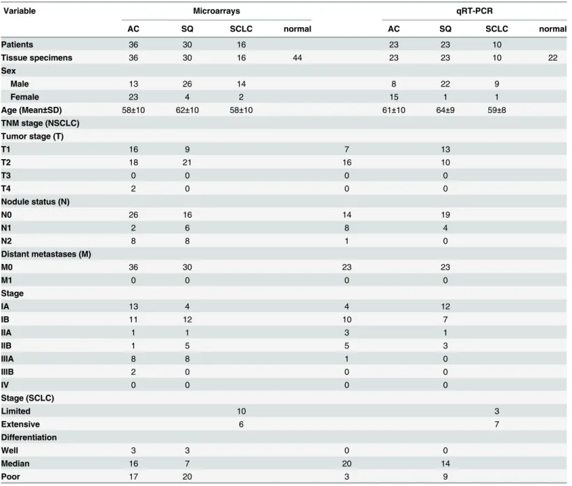
The local Ethics Committee approved the study and written informed consent was obtained from all the patients. For sample selection, routine histological classification was used following World Health Organization Classification of lung tumors[2].The diagnosis of AC was con-firmed by absence of keratinization or intercellular bridges and positive TTF1 staining. SQ diagnosis was confirmed by intercellular bridges or positive P63 staining (at least 40% of tumor cells). For all the SCLC cases, there was complete agreement on neuroendocrine morphology, small cells and neuroendocrine positive staining. If there was a disagreement, the consensus was reached by the detection of TTF1/P63. All cases were independently reviewed by 2 experi-enced lung cancer pathologists. Patients who had received pre-operative radiotherapy or che-motherapy were excluded. The clinical characteristics are presented inTable 1.
RNA isolation
concentration was quantified by NanoDrop 1000 Spectrophotometer (NanoDrop Technolo-gies, Waltham, MA). The quality control of RNA was performed by a 2100 Bioanalyzer using the RNA 6000 Pico LabChip kit (Agilent Technologies, Santa Clara, CA). The quality was mea-sured using RNA integrity number (RIN). A RNA sample was discarded if the RIN score was less than 5.0.
qRT-PCR
Quantitative reverse-transcriptase-polymerase-chain-reaction (qRT-PCR) was performed on 78 macrodissected frozen lung tissues to validate the expression profiles of miR-375 and its putative targeted genes in 3 subtypes of lung carcinomas. In the validation of miR-375,
Table 1. Characteristics of patients and tumors in this study.
Variable Microarrays qRT-PCR
AC SQ SCLC normal AC SQ SCLC normal
Patients 36 30 16 23 23 10
Tissue specimens 36 30 16 44 23 23 10 22
Sex
Male 13 26 14 8 22 9
Female 23 4 2 15 1 1
Age (Mean±SD) 58±10 62±10 58±10 61±10 64±9 59±8 TNM stage (NSCLC)
Tumor stage (T)
T1 16 9 7 13
T2 18 21 16 10
T3 0 0 0 0
T4 2 0 0 0
Nodule status (N)
N0 26 16 14 19
N1 2 6 8 4
N2 8 8 1 0
Distant metastases (M)
M0 36 30 23 23
M1 0 0 0 0
Stage
IA 13 4 4 12
IB 11 12 10 7
IIA 1 1 3 1
IIB 1 5 5 3
IIIA 8 8 1 0
IIIB 2 0 0 0
IV 0 0 0 0
Stage (SCLC)
Limited 10 3
Extensive 6 7
Differentiation
Well 3 3 0 0
Median 16 7 20 14
Poor 17 20 3 9
qRT-PCR was performed using Taqman microRNA assays (Applied Biosystems, Foster City, CA) according to the manufacturer’s instructions. The U47 small nuclear RNA was used as an endogenous control. All assays were carried out in triplicate.
For 22 predicted target genes of miR-375 (ACSL3,CACNG2,CCDC6,CSNK2A1,FZD8,
ITGA10,ITPKB,JAK2,JUND,LAMC1,LRP5,MAP3K5,NLK,PAK7,PDGFC,PDPK1,PIAS1,
RUNX1,RYR2,SOCS5,SP1andWNT5A), qRT-PCR using SYBR Green PCR Master Mix kit (Applied Biosystems, Foster City, CA) was performed according to the manufacturer’s instruc-tions.GAPDHwas used as an endogenous control. All assays were carried out in triplicate. Primer sequences are available upon request. Exclusion criteria for statistical analysis are as fol-lows: 1) A target gene that showed cycle threshold (CT) values above 35 cycles in>20% of the
tested samples and 2) A sample that showed CT values above 35 cycles in>20% of the tested
genes.
Target prediction and pathway analysis
The targets of miR-375 were predicted through the gateway miRecords (http://mirecords. biolead.org/). To increase the prediction accuracy, the genes that were predicted by at least 4 of 11 databases (Diana, microinspector, miranda, mirtarget2, mitarget, nbmirtar, pictar, pita, rna22, rnahybrid and targetscan) were selected as targets.
The KEGG database (http://www.genome.jp/kegg/tool/search_pathway.html) was used to map the predicted targets to lung cancer-associated pathways.
Cells
A SCLC cell line NCI-H82 was kindly provided by Dr. Xueliang Zhu (Institute of Biochemistry and Cell Biology, Shanghai Institutes for Biological Sciences, Chinese Academy of Sciences, Shanghai, China)[13]. NCI-H82 stably transduced with miR-375-expressing (H82-miR-375) or empty (H82-NC) lentiviruses were maintained in RPMI-1640 with 10% fetal bovine serum (FBS). SV40-transformed embryonic kidney cell line 293T, obtained from the American Type Culture Collection, was maintained in DMEM with 10%FBS.
Vector construction
Six putative targeted genes (ITPKB,RUNX1,LRP5,PIAS1,FZD8andITGA10) that had strong negative correlation with miR-375expression level in SCLC were selected for functional studies. Of the 6 targeted genes, the 3’UTR ofITPKBmRNA contained 3 putative target sites for miR-375 (target site 1: seed 1667–1673, target site 2: seed 2463–2469 and target site 3: seed 2513–
2519). Two different segments of theITPKB3’UTR, designated ITPKB-Δ1 (target site 1) and ITPKB-Δ2 (target site 2 and 3), were cloned, respectively.Schematic representation of the
ITPKB3’UTR and the putative binding sites for miR-375 is shown inS2 Fig.
carried the mutated sequence in the complementary site for the seed region of miR-375, were generated based on wild-type 3’-UTR pGL3 reporters by site-specific mutagenesis. All cloned products were verified by sequencing. Primers for cloning miR-375 and oligonucleotides for target 3’UTR construction are presented inS1 Table.
Luciferase reporter assay
293T cells were seeded in 96-well plate. The cells were co-transfected with 2 ng of an internal control vector pRL-renilla (Promega, Madison, WI), 40 ng of different 3’-UTR pGL3-promoter reporters, 160 ng of miR-375 expression vector (pS-miR-375) or empty control (pS-negative). Forty-eight hours after transfection, the firefly and renilla luciferase activities were assayed using Dual-Glo Luciferase assay system (Promega, Madison, WI). The luciferase activity of each sample was normalized by renilla luciferase activity. The normalized luciferase activity for the pS-negative was set as relative luciferase activity 1. All experiments were performed in trip-licate and independently repeated3 times.
Western blot
NCI-H82 cells were infected with miR-375-expressing (H82-miR-375) or empty (H82-NC) lentivirus at a MOI of 80. The infection efficiency was about 100% as assessed by microscopy of GFP fluorescence. The cells were harvested after 72 hours. Total protein of the cells or fresh tissues (2 AC, 2 SQ, 3 SCLC and 1adjacent normal tissue) were prepared using RIPA lysis buffer. Cell protein lysates were separated in 10% SDS-polyacrylamide gels, electrophoretically transferred to polyvinylidenedifluoride membranes (Roche, Pleasanton, CA), then detected with antibodies including anti-ITPKB (for cell line: ab171984, Abcam; for tissue: NBP1- 81589, Novus Biologicals), anti-RUNX1(ab54869, Abcam), anti-LRP5 (ab38311, Abcam), anti-PIAS1 (ab77231, Abcam), FZD8 (ab155650, Abcam), ITGA10 (ab118099, Abcam), anti-GAPDH (Santa Cruz) and peroxidise-conjugated secondary antibodies (Santa Cruz). The intensity of protein fragments was quantified using Quantity One software and was normalized by GAPDH.
CCK-8 assay
NCI-H82 cells were infected with miR-375-expressing (H82-miR-375) or empty (H82-NC) lentivirus as mentioned above. H82-miR-375 or H82-NC cells were seeded in 96-well plates. The cells were incubated for 24h, 48h, and 72h, respectively. CCK-8 reagent (DOJINDO) was added to each well 3h before the end of incubation. Optical density value (OD) of each sample was measured at a wavelength of 450nm. All experiments were performed in triplicate and independently repeated3 times.
Statistical analysis
For microarray data, one-way analysis of variance (ANOVA) with Benjamini-Hochberg correc-tion (p0.001) was performed to determine differentially expressed miRNAs among normal, AC, SQ and SCLC tissues. Hierarchical clustering was performed with Pearson correlation using the differentially expressed miRNAs.
For qRT-PCR data, an unpaired, unequal variancet-test with Benjamini-Hochberg correc-tion (p<0.05) was applied to miR-375 and differentially expressed targeted genes in the
com-parisons of AC vs. normal, SQ vs. normal and SCLC vs. normal tissue. The targeted genes with corrected p<0.05 and fold expression change>2 were selected as candidates for further
Additional methods including laser capture microdissection (LCM), macrodissection and microarray hybridization are described inS2 File.
Results
The miR-375 expression profiles in three subtypes of lung carcinomas
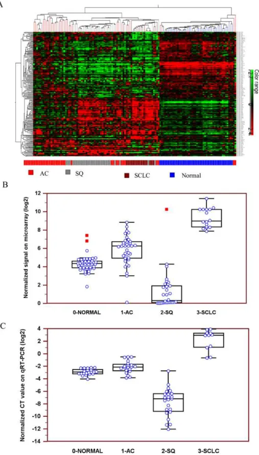
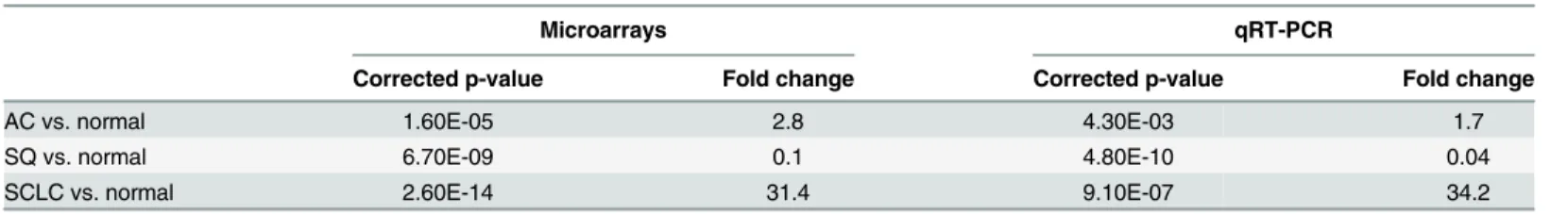
In a microarray analysis, expression profiles of 723 human miRNAs were investigated in selected LCM cancerous cell populations and normal cells derived from 36 AC, 30 SQ, 16 SCLC and 44 adjacent normal tissues.Fig 1Aillustrates the hierarchical clustering of the differ-ential expression levels of miRNAs in 3 subtypes of lung carcinomas and normal lung tissue. The overall miRNA expression profiles clearly divided the samples into four major groups: AC, SQ, SCLC and normal tissue. Up-regulation of miR-375 expression level was observed slightly in AC and significantly in SCLC, while marked down-regulation of miR-375 expression was observed in SQ (Fig 1BandTable 2).The expression profiles of miR-375 in 3 lung carcinoma subtypes were further validated in an independent cohort of 78 snap-frozen surgical lung tissues using qRT-PCR. Similarly, miR-375 expression was significantly up-regulated in AC and SCLC but down-regulated in SQ (Fig 1CandTable 2).
The association of miR-375 expression with lung carcinogenesis
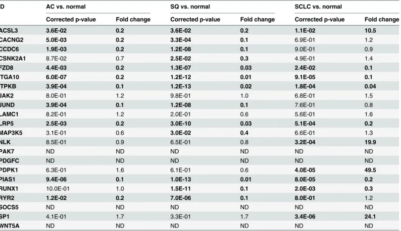
We identified 191 genes through gateway miRecords to be associated with miR-375. After mapping to the KEGG pathway database, 22 genes were identified to be involved in important signaling pathways in lung cancer (S2 Table.).The expression levels of the 22 putative target genes were next determined in 78 macrodis-sected frozen lung tissues. The expression profiles of the putative target mRNAs in 3 subtypes of lung carcinomas are presented inTable 3. Of the 22genes, 11 (ACSL3,CACNG2,CCDC6,
FZD8,ITGA10,ITPKB,JUND,LRP5,PIAS1,RUNX1andRYR2) had markedly differential expression levels in at least 2 of 3 pair-wise comparisons of AC vs. normal, SQ vs. normal or SCLC vs. normal. ExceptACSL3, all the 11 putative target genes showed significant down-regu-lation in all the 3 lung carcinomasubtypes. Conversely,ACSL3showed over 10 fold up-regula-tion when compareits expression level in SCLC to that in normal tissue (p = 0.011). Two genes (CSNK2A1andMAP3K5) showed differential expression levels in SQ vs. normal comparison only. Three genes (NLK,PDPK1andSP1) showed differential expression in SCLC vs. normal control only. Two genes (JAK2andLAMC1) showed comparable expression levels in the 3 comparisons, while other 4 genes (PAK7,PDGFC,SOCS5andWNT5A) did not pass the qual-ity control in which the target gene showed CT values above 35 cycles in>20% of the tested
samples.
Correlation of miR-375 level with target mRNA expression
In the comparison of AC vs. normal (Fig 2A), strong negative correlations were observed between the expression level of miR-375 and that of 10 putative target mRNAs (ACSL3,
CACNG2,CCDC6,FZD8,ITGA10,ITPKB,JUND,LRP5,PIAS1andRYR2). In the comparison of SQ vs. normal (Fig 2B), strong positive correlations were detected between the expression level of miR-375 and that of 13 putative target mRNAs (ACSL3,CACNG2,CCDC6,CSNK2A1,
andRUNX1), while strong positive correlations were observed between the expression of miR-375 and that of 4 putative target mRNAs (ACSL3,NLK,PDPK1andSP1).
ITPKB: a direct target of miR-375
Dual luciferase reporter assay was performed on 6 targeted genes (FZD8,ITGA10,ITPKB,
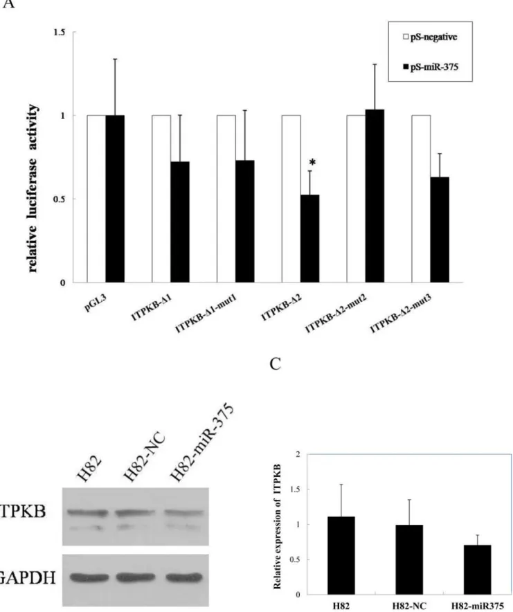
LRP5,PIAS1andRUNX1) that had strong negative correlation with the expression level of miR-375 in SCLC. For the 3 putative target sites at the 3’UTR ofITPKBmRNA, the luciferase activity of the reporter carrying the site 2 and 3 wild-type (ITPKB-Δ2) was significantly sup-pressed by miR-375, while the luciferase activity of the site 1 wild-type (ITPKB-Δ1) was unaf-fected. Importantly, the site 2 mutant (ITPKB-Δ2-mut2) was not targeted by miR-375, while the site 3 mutant (ITPKB-Δ2-mut3) was still inhibited by miR-375 (Fig 3A). Taken together, the results revealed that miR-375 targeted the 3’UTR of theITPKBmRNA primarily through the target site 2 (seed: 2463–2469).
The effect of miR-375 on the endogenous expression of ITPKB was further examined by Western blot. The ectopic expression of miR-375 significantly suppressed the ITPKB protein in the miR-375-transfected H82 cells (H82-miR-375) compared to the vector control trans-fected H82 cells (H82-NC) (Fig 3B and 3C). The data indicated that miR-375 inhibited the expression of ITPKB at the posttranscriptional level by directly targeting the 3’UTR (primarily target site2) ofITPKBmRNA.
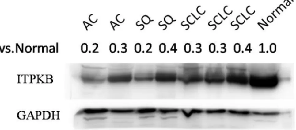
In addition, we observed ITPKB protein expression was significantly down-regulated in lung cancer tissues compared with adjacent normal tissue, which are in consonance with the expression pattern ofITPKBmRNA mentioned above (Fig 4andTable 3).
For other 5 putative targeted genes (RUNX1,LRP5,PIAS1,FZD8andITGA10), minimal effects on miR-375 were observed in their luciferase activities (S3 Fig). Furthermore, the ectopic expression levels of miR-375 were not significantly suppressed by the 5 putative target proteins in H82-miR-375 compared to H82-NC (S4 Fig).
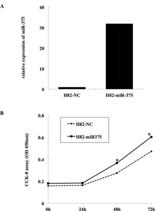
Effect of miR-375 overexpression on SCLC cell growth
The significant up-regulation of miR-375 expression in SCLC samples and its inhibitory effect onITPKBprompted us to investigate its possible biological role in SCLC cells. After infected, miR-375 expression of both H82-NC and H82-miR-375 was detected using real-time
qRT-PCR (Fig 5A). Then we evaluated the cell growth rate by CCK-8 assay. The results shown inFig 5Bsuggested that exogenous expression of miR-375 could promote cell growth of H82 cells in a time-dependence manner. After 24h incubation, no difference in cell viability between the two groups was observed(p>0.05).However, significant difference between the two groups diagram of miR-375 normalized CT values on qRT-PCR. AC: adenocarcinoma, SQ: squamous cell carcinoma, SCLC: small cell lung carcinoma.
doi:10.1371/journal.pone.0144187.g001
Table 2. Expression profiles of miR-375 in 3 subtypes of lung carcinomas.
Microarrays qRT-PCR
Corrected p-value Fold change Corrected p-value Fold change
AC vs. normal 1.60E-05 2.8 4.30E-03 1.7
SQ vs. normal 6.70E-09 0.1 4.80E-10 0.04
SCLC vs. normal 2.60E-14 31.4 9.10E-07 34.2
was observed after 48h and 72h incubation, and the promotion efficiencies were 33.1% and 35.9%, respectively(p<0.01).
Discussion
MiR-375 was first identified as a pancreatic islet-specific miRNA regulating insulin secretion [14–16]. However, further studies revealed that miR-375 is a multifunctional miRNA partici-pating in pancreatic islet development, glucose homeostasis, mucosal immunity, lung surfac-tant secretion and more imporsurfac-tantly, tumorigenesis. Deregulation of miR-375 has been observed in different types of cancers. For instance, Kong et al reported that miR-375 expres-sion was strongly downregulated in oesophageal squamous cell carcinoma (ESCC), and miR-375 inhibited tumor growth and metastasis through repressing insulin-like growth factor 1 receptor (IGF1R)[17]. Another study convinced that miR-375 was frequently downregulated in ESCC cell lines and tissues, and miR-375 was significantly associated with advanced stage, metastasis and poor outcome of ESCC [18]. Leidner et al found that miR-375 was associated with the progression to invasive carcinoma in Barrett’s esophagus [19].Chang et al showed that downregulation of miR-375 in glioma was correlated with unfavorable clinical outcome [20]. Low expression of miR-375 was found in the colorectal cancer [21,22], and also correlated with poor outcome and metastasis in head and neck squamous cell carcinomas [23,24]. He
Table 3. Expression profiles of putative target mRNAs in miR-375.
ID AC vs. normal SQ vs. normal SCLC vs. normal
Corrected p-value Fold change Corrected p-value Fold change Corrected p-value Fold change
ACSL3 3.6E-02 0.2 3.6E-02 0.2 1.1E-02 10.5
CACNG2 5.0E-03 0.2 3.3E-04 0.1 6.9E-01 1.2
CCDC6 1.9E-03 0.2 1.2E-08 0.1 9.0E-01 0.9
CSNK2A1 8.7E-02 0.7 2.5E-02 0.3 4.9E-01 1.4
FZD8 4.4E-03 0.2 1.3E-07 0.03 2.4E-02 0.1
ITGA10 6.0E-07 0.2 1.2E-12 0.01 9.1E-05 0.1
ITPKB 3.9E-04 0.1 1.2E-13 0.02 1.8E-04 0.04
JAK2 8.0E-01 1.2 9.8E-01 1.0 6.8E-01 1.5
JUND 3.9E-04 0.1 1.2E-08 0.1 7.6E-01 0.8
LAMC1 8.2E-01 1.2 2.0E-01 0.6 5.6E-01 1.6
LRP5 2.5E-03 0.2 3.0E-10 0.03 5.1E-04 0.2
MAP3K5 3.1E-01 0.6 3.0E-02 0.4 6.6E-01 1.3
NLK 8.5E-01 0.9 6.5E-01 0.8 3.2E-04 19.9
PAK7 ND ND ND ND ND ND
PDGFC ND ND ND ND ND ND
PDPK1 6.3E-01 1.6 6.1E-01 0.6 4.0E-05 49.5
PIAS1 9.4E-06 0.1 1.0E-13 0.01 8.0E-05 0.2
RUNX1 10.0E-01 1.0 1.5E-11 0.1 2.0E-03 0.3
RYR2 1.2E-02 0.2 7.0E-06 0.1 8.0E-01 1.2
SOCS5 ND ND ND ND ND ND
SP1 4.1E-01 1.7 3.3E-01 1.7 3.4E-06 24.1
WNT5A ND ND ND ND ND ND
AC: adenocarcinoma, SQ: squamous carcinoma, SCLC: small celllungcarcinoma.
ND: not determined, the target gene did not pass the quality control process in which the target gene showed CT values above 35 cycles in>20% of the tested samples.
et al found that miR-375, downregulated in hepatocellular carcinoma (HCC) tissues and cell lines, targets AEG-1 in HCC and suppresses cancer cell growthin vitroandin vivo[25]. In lung cancer, decreased miR-375 expression level in NSCLC tissue samples significantly correlated with advanced stage and lymphatic metastasis[26].Reduced expression of miR-375 was observed in all cancer types mentioned above, while miR-375 upregulation was noticed in breast cancer and prostate carcinoma [27,28]. These findings emphasize a fundamental role of miR-375 in tumorigenesis. Our results were consistent with previous findings. We discovered and validated significant miR-375 up-regulation in AC and SCLC but significant down-regula-tion in SQ.
The current two reports on miR-375 expression in SCLC were consistent with our results [12,13]. Compared to them, our study is unique for the following reasons: First, Zhao et al examined miRNA expression patterns only in cell lines (four SCLC cell lines (H209, H69, H82, and H345), one NSCLC cell line CRL 5908 and one immortalized human lung epithelial cell line Beas-2B), they found that miR-375 was consistently upregulated in all four SCLC cell lines. The other one also drew the conclusion by cultured cells, and used small samples of tissue (7 NSCLC vs. 8SCLC) for verification without normal control. Our study not only included cell lines but also a more comprehensive scanning of clinical samples, enabling us to better identify potential diagnostic miRNA markers. Underscoring the miRNA expression profiles in subtyp-ing SCLC and NSCLC is due to the difficulties in obtainsubtyp-ing primary tissue specimens, as most SCLC tumors are not surgically resected. Second, we discovered miRNA biomarkers on LCM selected cancer cells and adjacent normal cells derived from AC, SQ and SCLC. This approach assures the purity of target cells, reducing background signals from non-cancerous cells and improving the reliability of the discovered biomarker candidates[29].And interestingly, we observed that overexpression of miR-375 can drastically promote cell growth in SCLC cell line NCI-H82.
MiRNAs are negative post-transcriptional regulatory elements of gene expression. Single miRNA can regulate multiple mRNAs and are possibly better drug targets. To better under-stand the molecular mechanism of miR-375 in the 3 subtypes of lung cancer, we further pre-dicted the target genes of miR-375 and filtered the putative target genes through KEGG pathways. We observed that22 target genes are the key members of established signalling path-ways in lung cancer including calcium, insulin, Jak-STAT, MAPK, mTOR, PPAR, TGF-beta and Wnt pathways. Among these genes, we found that 6 target genes (FZD8,ITGA10,ITPKB,
LRP5,PIAS1andRUNX1) had strong negative correlation with miR-375 expression level in the comparison of SCLC vs. normal tissue. The association between lung cancer and 4 target genes (FZD8,LRP5,PIAS1andRUNX1) has been previously reported [30–33].It has been reported thatFZD8is highly expressed in A549 cell line and the dysregulation ofFZD8might play key roles in human cancer through activation of beta-catenin-TCF pathway [30]. Lee et al demon-strated a significant down-expression ofLRP5gene in 6 of 17 lung squamous carcinoma sam-ples [31]. In H1299 cell line,PIAS1inhibited STAT1-mediated gene activation and the DNA binding activity [32]. In NSCLC patients,RUNXwas more frequently methylated in tumor tis-sues than in noncancerous tistis-sues [33].However, the functions ofITGA10andITPKBin lung
Fig 2. Correlation of miR-375 with target mRNA expression. A,Correlation of miR-375 with target mRNA expression in the comparison of AC vs. normal.B,Correlation of miR-375 with target mRNA expression in the comparison of SQ vs. normal.C,Correlation of miR-375 with target mRNA expression in the comparison of SCLC vs. normal. Green circle: down-regulation. Red circle: up-regulation. White circle: the gene did not show differential expression in the pair-wise comparison of AC vs. normal, SQ vs. normal or SCLC vs. normal. Blue circle: the gene did not pass the quality control. AC: adenocarcinoma, SQ: squamous cell carcinoma, SCLC: small cell lung carcinoma.
cancer are still unknown. In our study,ITPKBmRNA and protein expression was significantly down-regulated in lung cancer tissues. The luciferase assay revealed thatITPKBis the direct target gene of miR-375. The over-expression of miR-375 led to significantly decreased ITPKB protein level. The results confirmed that miR-375 negatively regulatedITPKBat the translation level. ForITGA10gene, we did not observe any effects on miR-375 in the luciferase activity and the suppression of the ITGA10 protein expression by miR-375.
Recently, miR-375 has been found to suppress core hallmarks of cancer by targeting several important genes likeAEG-1,YAP1,IGF1RandPDK1[16].The target genes regulated by miR-375 may function spatiotemporally or in cooperation with each other in different cellular pro-cesses. Our identification of ITPKB gene as a direct target of miR-375 provides new insights into the mechanisms underlying tumorigenesis. ITPKB, a member of [InsP3] 3-kinases family, was mapped to the telomeric end of human chromosome 1 and associated with calcium signal-ing pathway. It has been identified as a candidate gene in the pathogenesis of immune disor-ders, multiple sclerosis, Alzheimer disease, and malignant melanoma [34,35]. Since little work has been reported onITPKBfunction in lung cancer pathogenesis, further studiesin vitroand
in vivoare needed to explore the potential effect ofITPKBin lung cancer, which will be helpful for understanding the role of miR-375 in lung cancer and if miR-375 upregulation is a cause or effect of cancer pathogenesis. It is well accepted that the expression levels of miRNAs and their direct mRNA targets may be negatively correlated[36,37].Intriguingly, we did observe signifi-cant positive correlation in the expression level of miR-375 and its 13 putative targets in SQ and its 4 putative targets in SCLC. It has been demonstrated that a miRNA can function as a switch from repression to activation of its target genes [38,39].Further functional studies are needed to investigate the role of the putative targets showing positive correlation with miR-375 expression level.
In summary, we discovered and validated thatmiR-375 expression level is significantly up-regulated in AC and SCLC but down-up-regulated in SQ. Furthermore, we found that miR-375 directly downregulatesITPKBin SCLC and promotes cell growth in SCLC cell line, which were
H82, H82-NC and H82-miR-375 cells were evaluated by western blot. After the transfection of miR-375, the expression level of ITPKB protein was significantly suppressed. C, Quantitative analysis of Western blot. The ITPKB protein intensities were normalized by GAPDH.
doi:10.1371/journal.pone.0144187.g003
Fig 4. Expression of ITPKB protein in 3 lung carcinoma subtypes.Western blot was used to detect the expression of ITPKB protein in lung carcinoma tissues (2AC samples, 2SQ samples, 3SCLC samples) and adjacent normal lung tissue. The ITPKB protein intensities were normalized by GAPDH.The fold changes were 0.2and0.3 in the comparisons of 2AC vs. normal, 0.2 and 0.4of 2SQ vs. normal, 0.3, 0.3 and 0.4of 3SCLC vs. normal, respectively.
not performed in previous studies. This study may be a first step. Further studies in the mecha-nisms of pathogenesis are needed to explore the potential clinical value of miR-375.
Supporting Information
S1 Fig. Laser capture microdissection of lung squamous cell carcinoma.A, H&E-stained slide (X 20). B, Hematoxylin stained slide before LCM (X 20). C, Hematoxylin stained slide after LCM (X 20). D, Cap showing adherent cells (X 20).
(TIF)
Fig 5. Effect of miR-375 on cell growth of NCI-H82. A,miR-375 expression of both H82-NC and H82-miR-375.NCI-H82 cells were infected with miR-375-expressing (H82-miR-375) or empty (H82-NC) lentivirus prior to the proliferation assay.B,miR-375 could promote cell growth of H82 cells. The infected cells were seeded to 96-wells plates. After incubation for several days, the cell growth was measured by CCK-8 assay.*: p< 0.01, compared with H82-NC.
S2 Fig. Schematic representation of the ITPKB 3’UTR and putative binding sites for miR-375.The 3’UTR of the ITPKB mRNA contains 3 putative target sites for miR-375 (site1: seed 1667–1673, site 2: seed2463-2469, site 3: seed2513-2519). Two different segments of the ITPKB 3’UTR, designated ITPKB-Δ1 (target site 1) and ITPKB-Δ2 (target site 2 and 3), were cloned, respectively. The mutation was generated in the complementary site for the seed region of miR-375, as indicated.
(TIF)
S3 Fig. Luciferase assay of 5 putative target genes for miR-375.A, RUNX1. B, LRP5.C, PIAS1. D, FZD8. E, ITGA10.
(TIF)
S4 Fig. Western blot analysis of 5 putative target genes. A,RUNX.B,LRP5.C,PIAS1.D,
FZD8.E,ITGA10. (TIF)
S1 File. Abbreviations.
(DOCX)
S2 File. Supplementary materials and methods.
(DOCX)
S1 Table. Primers for cloning miR-375 and oligonucleotides for target 3’UTR construction
(DOC)
S2 Table. Putative targets of miR-375 in lung cancer signaling pathways.
(DOC)
Author Contributions
Conceived and designed the experiments: YW HZ SL. Performed the experiments: YJ YL WH CX. Analyzed the data: YL WH. Contributed reagents/materials/analysis tools: CZ XG SW YH HJ. Wrote the paper: YJ YL JZ.
References
1. Jemal A, Bray F, Center MM, Ferlay J, Ward E, Forman D. Global cancer statistics. CA: a cancer journal for clinicians. 2011; 61(2):69–90. doi:10.3322/caac.20107PMID:21296855.
2. Beasley MB, Brambilla E, Travis WD. The 2004 World Health Organization classification of lung tumors. Seminars in roentgenology. 2005; 40(2):90–7. PMID:15898407.
3. Miller YE. Pathogenesis of lung cancer: 100 year report. American journal of respiratory cell and molec-ular biology. 2005; 33(3):216–23. doi:10.1165/rcmb.2005-0158OEPMID:16107574; PubMed Central PMCID: PMC2715312.
4. Bartel DP. MicroRNAs: genomics, biogenesis, mechanism, and function. Cell. 2004; 116(2):281–97. PMID:14744438.
5. Yanaihara N, Caplen N, Bowman E, Seike M, Kumamoto K, Yi M, et al. Unique microRNA molecular profiles in lung cancer diagnosis and prognosis. Cancer cell. 2006; 9(3):189–98. doi:10.1016/j.ccr. 2006.01.025PMID:16530703.
6. Mascaux C, Laes JF, Anthoine G, Haller A, Ninane V, Burny A, et al. Evolution of microRNA expression during human bronchial squamous carcinogenesis. The European respiratory journal. 2009; 33 (2):352–9. doi:10.1183/09031936.00084108PMID:19010987.
7. Yang Y, Li X, Yang Q, Wang X, Zhou Y, Jiang T, et al. The role of microRNA in human lung squamous cell carcinoma. Cancer genetics and cytogenetics. 2010; 200(2):127–33. doi:10.1016/j.
8. Huang W, Hu J, Yang DW, Fan XT, Jin Y, Hou YY, et al. Two microRNA panels to discriminate three subtypes of lung carcinoma in bronchial brushing specimens. American journal of respiratory and criti-cal care medicine. 2012; 186(11):1160–7. doi:10.1164/rccm.201203-0534OCPMID:23043084. 9. Wu H, Zhu S, Mo YY. Suppression of cell growth and invasion by miR-205 in breast cancer. Cell
research. 2009; 19(4):439–48. doi:10.1038/cr.2009.18PMID:19238171; PubMed Central PMCID: PMC2664859.
10. Kimura S, Naganuma S, Susuki D, Hirono Y, Yamaguchi A, Fujieda S, et al. Expression of microRNAs in squamous cell carcinoma of human head and neck and the esophagus: miR-205 and miR-21 are specific markers for HNSCC and ESCC. Oncology reports. 2010; 23(6):1625–33. PMID:20428818. 11. Majid S, Dar AA, Saini S, Yamamura S, Hirata H, Tanaka Y, et al. MicroRNA-205-directed transcrip-tional activation of tumor suppressor genes in prostate cancer. Cancer. 2010; 116(24):5637–49. doi: 10.1002/cncr.25488PMID:20737563; PubMed Central PMCID: PMC3940365.
12. Nishikawa E, Osada H, Okazaki Y, Arima C, Tomida S, Tatematsu Y, et al. miR-375 is activated by ASH1 and inhibits YAP1 in a lineage-dependent manner in lung cancer. Cancer research. 2011; 71 (19):6165–73. doi:10.1158/0008-5472.CAN-11-1020PMID:21856745.
13. Zhao H, Zhu L, Jin Y, Ji H, Yan X, Zhu X. miR-375 is highly expressed and possibly transactivated by achaete-scute complex homolog 1 in small-cell lung cancer cells. Acta biochimica et biophysica Sinica. 2012; 44(2):177–82. doi:10.1093/abbs/gmr110PMID:22172490.
14. Kloosterman WP, Lagendijk AK, Ketting RF, Moulton JD, Plasterk RH. Targeted inhibition of miRNA maturation with morpholinos reveals a role for miR-375 in pancreatic islet development. PLoS biology. 2007; 5(8):e203. doi:10.1371/journal.pbio.0050203PMID:17676975; PubMed Central PMCID: PMC1925136.
15. Avnit-Sagi T, Kantorovich L, Kredo-Russo S, Hornstein E, Walker MD. The promoter of the pri-miR-375 gene directs expression selectively to the endocrine pancreas. PloS one. 2009; 4(4):e5033. doi:10. 1371/journal.pone.0005033PMID:19343226; PubMed Central PMCID: PMC2660411.
16. Yan JW, Lin JS, He XX. The emerging role of miR-375 in cancer. International journal of cancer Journal international du cancer. 2014; 135(5):1011–8. doi:10.1002/ijc.28563PMID:24166096.
17. Kong KL, Kwong DL, Chan TH, Law SY, Chen L, Li Y, et al. MicroRNA-375 inhibits tumour growth and metastasis in oesophageal squamous cell carcinoma through repressing insulin-like growth factor 1 receptor. Gut. 2012; 61(1):33–42. doi:10.1136/gutjnl-2011-300178PMID:21813472.
18. Li J, Li X, Li Y, Yang H, Wang L, Qin Y, et al. Cell-specific detection of miR-375 downregulation for pre-dicting the prognosis of esophageal squamous cell carcinoma by miRNA in situ hybridization. PloS one. 2013; 8(1):e53582. doi:10.1371/journal.pone.0053582PMID:23301089; PubMed Central PMCID: PMC3536738.
19. Leidner RS, Ravi L, Leahy P, Chen Y, Bednarchik B, Streppel M, et al. The microRNAs, MiR-31 and MiR-375, as candidate markers in Barrett's esophageal carcinogenesis. Genes, chromosomes & can-cer. 2012; 51(5):473–9. doi:10.1002/gcc.21934PMID:22302717; PubMed Central PMCID:
PMC3547654.
20. Chang C, Shi H, Wang C, Wang J, Geng N, Jiang X, et al. Correlation of microRNA-375 downregulation with unfavorable clinical outcome of patients with glioma. Neuroscience letters. 2012; 531(2):204–8. doi:10.1016/j.neulet.2012.10.021PMID:23103713.
21. Dai X, Chiang Y, Wang Z, Song Y, Lu C, Gao P, et al. Expression levels of microRNA-375 in colorectal carcinoma. Molecular medicine reports. 2012; 5(5):1299–304. doi:10.3892/mmr.2012.815PMID: 22377847.
22. Wang S, Wang L, Bayaxi N, Li J, Verhaegh W, Janevski A, et al. A microRNA panel to discriminate car-cinomas from high-grade intraepithelial neoplasms in colonoscopy biopsy tissue. Gut. 2013; 62 (2):280–9. doi:10.1136/gutjnl-2011-301554PMID:22535378.
23. Hui AB, Bruce JP, Alajez NM, Shi W, Yue S, Perez-Ordonez B, et al. Significance of dysregulated metadherin and microRNA-375 in head and neck cancer. Clinical cancer research: an official journal of the American Association for Cancer Research. 2011; 17(24):7539–50. doi: 10.1158/1078-0432.CCR-11-2102PMID:22031094.
24. Harris T, Jimenez L, Kawachi N, Fan JB, Chen J, Belbin T, et al. Low-level expression of miR-375 corre-lates with poor outcome and metastasis while altering the invasive properties of head and neck squa-mous cell carcinomas. The American journal of pathology. 2012; 180(3):917–28. doi:10.1016/j.ajpath. 2011.12.004PMID:22234174; PubMed Central PMCID: PMC3349885.
26. Li Y, Jiang Q, Xia N, Yang H, Hu C. Decreased expression of microRNA-375 in nonsmall cell lung can-cer and its clinical significance. The Journal of international medical research. 2012; 40(5):1662–9. PMID:23206448.
27. de Souza Rocha Simonini P, Breiling A, Gupta N, Malekpour M, Youns M, Omranipour R, et al. Epige-netically deregulated microRNA-375 is involved in a positive feedback loop with estrogen receptor alpha in breast cancer cells. Cancer research. 2010; 70(22):9175–84. doi: 10.1158/0008-5472.CAN-10-1318PMID:20978187.
28. Szczyrba J, Nolte E, Wach S, Kremmer E, Stohr R, Hartmann A, et al. Downregulation of Sec23A pro-tein by miRNA-375 in prostate carcinoma. Molecular cancer research: MCR. 2011; 9(6):791–800. doi: 10.1158/1541-7786.MCR-10-0573PMID:21593139.
29. Wang S, Wang L, Zhu T, Gao X, Li J, Wu Y, et al. Improvement of tissue preparation for laser capture microdissection: application for cell type-specific miRNA expression profiling in colorectal tumors. BMC genomics. 2010; 11:163. doi:10.1186/1471-2164-11-163PMID:20219115; PubMed Central PMCID: PMC2853520.
30. Saitoh T, Hirai M, Katoh M. Molecular cloning and characterization of human Frizzled-8 gene on chro-mosome 10p11.2. International journal of oncology. 2001; 18(5):991–6. PMID:11295046.
31. Lee EH, Chari R, Lam A, Ng RT, Yee J, English J, et al. Disruption of the non-canonical WNT pathway in lung squamous cell carcinoma. Clinical medicine Oncology. 2008; 2008(2):169–79. PMID: 20401333; PubMed Central PMCID: PMC2855195.
32. Megidish T, Xu JH, Xu CW. Activation of p53 by protein inhibitor of activated Stat1 (PIAS1). The Journal of biological chemistry. 2002; 277(10):8255–9. doi:10.1074/jbc.C200001200PMID:11788578. 33. Feng Q, Hawes SE, Stern JE, Wiens L, Lu H, Dong ZM, et al. DNA methylation in tumor and matched
normal tissues from non-small cell lung cancer patients. Cancer epidemiology, biomarkers & preven-tion: a publication of the American Association for Cancer Research, cosponsored by the American Society of Preventive Oncology. 2008; 17(3):645–54. doi:10.1158/1055-9965.EPI-07-2518PMID: 18349282; PubMed Central PMCID: PMC2798850.
34. Nalaskowski MM, Fliegert R, Ernst O, Brehm MA, Fanick W, Windhorst S, et al. Human inositol 1,4,5-tri-sphosphate 3-kinase isoform B (IP3KB) is a nucleocytoplasmic shuttling protein specifically enriched at cortical actin filaments and at invaginations of the nuclear envelope. The Journal of biological chemis-try. 2011; 286(6):4500–10. doi:10.1074/jbc.M110.173062PMID:21148483; PubMed Central PMCID: PMC3039344.
35. Tajouri L, Mellick AS, Tourtellotte A, Nagra RM, Griffiths LR. An examination of MS candidate genes identified as differentially regulated in multiple sclerosis plaque tissue, using absolute and comparative real-time Q-PCR analysis. Brain research Brain research protocols. 2005; 15(2):79–91. doi:10.1016/j. brainresprot.2005.04.003PMID:15905117.
36. Kim S, Choi M, Cho KH. Identifying the target mRNAs of microRNAs in colorectal cancer. Computa-tional biology and chemistry. 2009; 33(1):94–9. doi:10.1016/j.compbiolchem.2008.07.016PMID: 18723399.
37. Wang YP, Li KB. Correlation of expression profiles between microRNAs and mRNA targets using NCI-60 data. BMC genomics. 2009; 10:218. doi:10.1186/1471-2164-10-218PMID:19435500; PubMed Central PMCID: PMC2686738.
38. Vasudevan S, Tong Y, Steitz JA. Switching from repression to activation: microRNAs can up-regulate translation. Science. 2007; 318(5858):1931–4. doi:10.1126/science.1149460PMID:18048652. 39. Nunez-Iglesias J, Liu CC, Morgan TE, Finch CE, Zhou XJ. Joint genome-wide profiling of miRNA and
mRNA expression in Alzheimer's disease cortex reveals altered miRNA regulation. PloS one. 2010; 5 (2):e8898. doi:10.1371/journal.pone.0008898PMID:20126538; PubMed Central PMCID: