INTRODUCTION
The Metropolitan Area of Buenos Aires (MABA) is comprises Buenos Aires city and 24 districts, with an estimated population of 14 million, being the 10th largest megacity in the world and the 3rd in Latin America. By its 200 km2, Buenos Aires is the largest city in Argentina, with a population of approximately 3 million people.
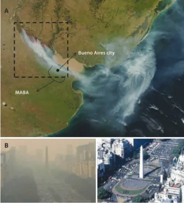
In the Delta Islands of the Paraná River, approximately 112 miles a way from the city of Buenos Aires, pasture burning for grazing li-vestock is a fairly common practice. Between April 16th and April 24th 2008, there was a series of wildires that afected over 70,000 hectares. The extent of the ire, the drought, and the wind direction generated a thick cloud of smoke that caused the most severe episode of acute air pollution in the MABA ever recorded. The northern and northeas-ABSTRACT
Purpose: To evaluate the acute impact of the wildfire smoke episode in 2008 on the ocular surface of subjects living in the Metropolitan Area of Buenos Aires (MABA). Methods: A total of 86 subjects were evaluated: Group 1 comprised patients from a public ophthalmology hospital (N=35) and Group 2 comprised healthy volunteers (N=51). All subjects answered a questionnaire on ocular symptoms and underwent ophthalmologic examination [bulbar conjunctival hyperemia, corneal fluorescein staining, rose bengal vital staining, tear break-up time (TBUT ), Schirmer I test, tear lysozyme, and impression cytology] during and after the acute episode. Concentrations of carbon monoxide (CO), nitrogen dioxide (NO2), and particulate matter (PM) were measured before, during, and after the acute episode. Results: Both groups showed a statically significant increase in ocular symptoms and bulbar conjunctival hyperemia and a statically significant decrease in tear break-up time during the acute episode. Group 1 showed more severe symptoms and a statistically significant increase in fluorescein and rose bengal staining intensities during the acute episode. We found a significant negative correlation between ocular symptoms and tear break-up time. During the episode, the levels of CO, NO2, and particulate matter in MABA were four times higher than the usual average levels for the same period in 2007 and 2009.
Conclusions: Increased air pollution from the burning of biomass is associated with a decrease in the stability of the tear film (TBUT ), generating areas of ocular surface exposure that may be the cause of the increased feeling of irritation. Group 1 was more affected by not having a healthy ocular surface, and thus consulted an ophthalmologist. Cytological changes in the conjunctiva were not observed, which could be due to the short duration of the episode.
Keywords: Urban fires; Conjunctiva/cytology; Cytodiagnosis; Air pollution; Tears/ physiology; Fluorescein/diagnostic use; Rose bengal/diagnostic use; Hyperemia; Argentina
RESUMO
Objetivo: Avaliar os efeitos agudos da fumaça do episódio de incêndio violento ocor-rido em 2008, sobre a superfície ocular de sujeitos que vivem na Região Metropolitana de Buenos Aires (MABA).
Métodos: Um total de 86 indivíduos foram avaliados: Grupo 1: pacientes de um hospital público de oftalmologia (N=35) e Grupo 2: voluntários saudáveis (N=51). Todos os participantes responderam a um questionário sobre os sintomas oculares e foram submetidos a exame oftalmológico (hiperemia conjuntival bulbar, teste de fluoresceína, corante rosa bengala, tempo de ruptura do filme lacrimal (TBUT), teste de Schirmer I, lisozima lacrimal e citologia de impressão) durante e após o episódio agudo. As concentrações de monóxido de carbono, dióxido de nitrogênio e partículas (PM) foram medidas antes, durante e após o episódio agudo.
Resultados: Ambos os grupos apresentaram aumento estatisticamente significativo dos sintomas oculares, hiperemia conjuntival bulbar, e diminuição estatisticamente significativa no tempo de ruptura do filme lacrimal durante o episódio agudo. Grupo 1 apresentou maior intensidade dos sintomas e aumento estatisticamente significativo no teste de fluoresceína e rosa bengala durante o episódio agudo. Encontramos uma correlação negativa significativa entre os sintomas oculares e tempo de ruptura do filme lacrimal. Durante o episódio agudo de 2008, os níveis de CO, NO2 e PM na Região
Metropolitana de Buenos Aires foram 4 vezes maiores do que os níveis médios habituais para o mesmo período de 2007 e 2009.
Conclusões: O aumento da poluição do ar a partir da queima de biomassa está associado a uma diminuição da estabilidade do filme lacrimal (TBUT) gerando zonas da exposição da superfície ocular, que podem ser a causa do aumento da sensação de irritação. Grupo 1 foi mais afetado por não ter superfície ocular saudável e, portanto, consultaram um oftalmologista. Mudanças citológicas da conjuntiva não foram observadas e isso poderia ser devido à curta duração do episódio.
Descritores: Incêndios urbanos; Conjuntiva/citologia; Citodiagnóstico; Poluição do ar; Lágrimas/fisiologia; Fluoresceína/uso diagnóstico; Rosa bengala/uso diagnóstico; Hiperemia; Argentina
tern areas of the province of Buenos Aires, including MABA, had been swathed in thick smoke (Figure 1 A, B). It is estimated that the wildires of the Delta of the Paraná River afected approximately 20 million people. The smoke was so dense in the city of Buenos Aires that on April 18th, the airports and highways were closed, and classes in all schools were suspended (Figure 1 B, C). The levels of carbon mo noxide (CO), nitrogen dioxide (NO2), and particulate matter (PM) registered during the episode were the highest ever registered in the region of the River de la Plata.
Most of the previous studies on the impact of wildires were con-ducted focusing on short-term outcomes related to hospital admittan-ces or speciic cohorts, particularly susceptible to respiratory distress, such as patients with chronic obstructive pulmonary disease or asthma or those who are directly exposed to the ires, such as iremen(1-3).
Impact of wildfire smoke in Buenos Aires, Argentina, on ocular surface
Efeitos da fumaça de incêndios na superfície ocular em Buenos Aires, Argentina
Martin Berra1,2, Gustavo Galperín1,2, laura DawiDowski3, Julia tau2, isaBel Márquez4, aleJanDro Berra2,4
Submitted for publication: September 8, 2014 Accepted for publication: January 26, 2015
1 Hospital de Oftalmología Pedro Lagleyze, Buenos Aires, Argentina.
2 Laboratorio de Investigaciones Oculares, Departamento de Patología, Facultad de Medicina, Uni versidad de Buenos Aires, Buenos Aires, Argentina.
3 Grupo de Monitoreo Ambiental, Comisión Nacional de Energía Atómica, Buenos Aires, Argentina. 4 Laboratorio BioFundus, Buenos Aires, Argentina.
Funding: No specific financial support was available for this study.
Disclosure of potential conflicts of interest: None of the authors have any potential conflict of interest to disclose.
The ocular surface is daily in contact with air, and epidemiolo-gical studies have indicated increasing incidence of dry eye disease (DED) worldwide(4-5). This common ocular condition has multiple causes that are not entirely understood. The emerging awareness that environmental factors can contribute to DED is supported by recent studies(6-8). Moreover, impact of the environment on the pathophysiology of DED has been studied and conirmed in animal models of DED(8-11).
There are few studies that show the ocular impact of wildires(12-13). The wildire episode in the northern area of the province of Buenos Aires was a unique opportunity to study the ocular impact of the exposure to the smoke produced by pasture burning on patients and healthy volunteers in a megacity.
METHODS
P
OPULATIONOFSTUDYThe study population consisted of two groups, each with two sub groups (Table 1).
Group 1 (G1) comprised 35 patients who visited the Emergency Unit of Hospital Oftalmologico Pedro Lagleyze on April 19th due to ocular complaints; Hospital Oftalmologico Pedro Lagleyze is the largest Ophthalmology Hospital in Argentina and located in the study area.
Group 2 (G2) included 51 healthy volunteers who accompanied the patients to the hospital on April 19th. Both groups were evaluated on a second appointment on May 22nd, when the air pollutant levels had recovered to normal values in the city of Buenos Aires (Table 1).
We included 35/67 patients and 51/72 healthy volunteers becau-se only 35 patients and 51 healthy volunteers visited the hospital on both April 19th and May 22nd.
To be included in G1 and G2, subjects had to give their informed consent before being enrolled in the study, had to be between 18 and 65 years old, and had to have lived and worked in MABA between March and May 2008, far from areas close to stationary sources of signiicant emissions, such as factories. The exclusion criteria for G2 included having a previous history of ocular surface disease or using contact lenses or any systemic or topical ocular drugs.
This study was conducted in accordance with the Declaration of Helsinki and was approved by the Ethical Committee of the Hospital Oftalmologico Pedro Lagleyze, Buenos Aires, Argentina.
O
CULARSYMPTOMSQUESTIONNAIREAll subjects answered a symptoms questionnaire composed by ques tions that assessed the presence of ocular burning, dryness, foreign body sensation, irritation, and itching. Subjects were asked to grade the intensity of the symptoms on a scale of 1 to 3 (1 = minor, 2 = mild, and 3 = severe). We also investigated if any factor worsened the symptoms, such as the time of the day of exposure, staying in- or outdoors, if it was the irst time they experienced the symptoms, and if they were using any eye drops to improve the ocular symptoms.
O
PHTHALMOLOGICEXAMINATIONThe usual ocular surface tests available in the hospital and our standard of care in our institution were used to evaluate the subjects.
Bulbar conjunctival hyperemia
Bulbar conjunctival hyperemia was recorded using ive levels of severity from grade 0 (normal) to grade 4 (severe)(14) with a slit lamp (SL-8Z; Topcon Corp., Tokyo, Japan). The inter-subject variation in gra-ding was determined to be 1.0 scale units. Interpretation of gragra-ding levels was as follows: 0 = Normal, 1 = Trace, 2 = Mild, 3 = Moderate, and 4 = Severe.
Corneal luorescein staining
Fluorescein staining with cobalt blue light was used to assess the presence or absence of corneal keratitis. Corneal luorescein staining was evaluated with luorescein strips (Diagnóstico Ocular, Buenos Aires, Argentina), which were wetted with 0.9% sodium chloride and gently applied to the inferior fornix. The cornea was divided into ive regions (central, superior, inferior, nasal, and temporal), and each re-gion was graded by a scale, with values ranging from grade 0 (normal) to grade 4 (severe)(14). Grades of each area were added to produce a inal score.
Conjunctival rose bengal vital staining
Vital staining with rose bengal was performed by instilling one drop of 1% rose bengal solution (Farmacia Magister, Buenos Aires, Argen-tina) into the inferior fornix. The staining pattern was evaluated and graded according to the Van Bijsterveld scoring system on a scale that ranges from 0 to 9. Scores higher than 4 were considered abnormal(15).
Tear break-up time
Tear break-up time (TBUT) was measured by instilling one drop of 1% luorescein solution (Farmacia Magister, Buenos Aires, Argentina) into the inferior fornix. The subject was asked to blink several times Figure 1. A) Satellite image (MODIS/Aqua) of the wildires in the Delta of the Parana River
on April 18th, 2008. Red boxes indicate wildire areas. Scale: 1 px=250 m (NASA. Fires and smoke over Argentina, seen from MODIS on the Aqua satellite at 2008/18/04 at 17:50 UTC. Scale: 1 px=250 m) http://rapidire.sci.gsfc.nasa.gov/gallery/?20081090418/Argentina. A2008109.1750.250 m.jpg, 2008). B and C) Images of the same location in the city of Buenos Aires at two diferent moments: (B) April 18th, 2008, during the wildires in the Delta of the Paraná River (http://nidodecaranchos.blogspot.com/2008_04_01_archive. html). (C) May 20th, 2008 (http://cliobuenosaires.blogspot.com/2010/05/el-obelisco-un- simbolo-que-deine.html).
A
B C
Table 1. Study population
Group Subgroup Subjects N
Time of examination relative to the acute episode
G1 G1A Patients 35 During (19th April)
G1B The same as G1A 35 After (22nd May) G2 G2A Healthy volunteers 51 During (19th April)
and then stop, at which point TBUT was assessed by monitoring the time (in seconds) elapsing to the appearance of the initial black spot on the cornea under slit lamp examination with a cobalt blue ilter(16). Values of 10 seconds or below were considered abnormal(17).
Schirmer’s I test
Subjects were evaluated by Schirmer’s I test without topical anes-thesia. One graded sterile Schirmer strip (Opthalmos™, Brazil) was pla-ced in the lateral canthus of the inferior lid margin of both eyes, and the subjects were instructed to keep their eyes closed during the test. After 5 minutes, the length of wetting was measured in millimeters, and values of 10 mm or less were considered abnormal(17).
Tear lysozyme concentration
Tears were collected by gently applying a 5-mm diameter ilter paper disc in the inferior conjunctival cul-de-sac of both eyes for one minute with eyes closed. Samples were kept at -20°C until processed. To determine tear lysozyme concentration, we used the Micrococcus
lysodeikticus (ATCC 4698, M3770; Sigma-Aldrich, St. Louis, MO) agar
difusion assay(18) in Mueller Hinton agar plates (Bio Merieux, Marcy l’Etoile, France). Each disc was placed in the plate with the M.
lyso-deik ticus (2 × 10⁶ CFU/mL) suspension gel, and the inhibition halo was
measured after 24 hours. To calculate the lysozyme concentration, a standard curve was obtained using identical discs wetted with 10,000, 1,000, 100, and 10 µg/mL of lysozyme (ATCC 4698, L6876; Sigma-Aldrich) diluted in phosphate-bufered saline (Invitrogen Corp., Carlsbad, CA). Values of 1000 µg/mL or below were considered abnormal(19-20).
Impression cytology
Semicircular ilters, approximately 15 mm diameter, (Polyvinylide-ne Fluoride -PVDF-, 22-μm pore size; Millipore Corp., Bedford, MA, USA) were applied to the inferior tarsal and bulbar conjunctiva after instillation of one drop of topical anesthetic (tetracaine) in each eye, and any excess luid was wiped away. The paper fragments were gently pressured with the blunt end of the forceps, and the fragments were peeled of and immediately immersed in tubes containing absolute ethanol. After ixation, specimens were rehydrated in 70% ethyl alcohol, then placed successively in periodic acid-Schif reagent, sodium metabisulite, Gill’s hematoxylin, and Scott’s tap water. Speci-mens were then rinsed with 95% alcohol and absolute alcohol. Xylene was used to make the ilter paper transparent. Slides were examined under a conventional light microscope, using 400× magniication. Mor phometric analysis was performed using a point-counting techni-que, PAS positive areas were counted across 10 ran dom microscopic high-power ields (HPF) on a 100-point, and 50 lines grid on a video system coupled to the microscope. The result for each subject was calcu-lated as the average of the 10 HPF counts. A single investigator perfor-med all the observations and was blinded to the origin of the samples(18).
Ocular damage was based on the scale of Nelson, comprising degrees of severity. This scale is based on the density of goblet cells and appearance of epithelial cells (grade squamous metaplasia). Level 0 is considered normal; 1, slightly altered; 2, moderately altered; and 3, severely altered(21).
Assessment of air pollutants during and after the acute episode Average CO and NO2 concentrations over cumulative periods of three minutes were measured. The calibration of the equipment was made with high quality certiied synthetic air. Nondispersive infrared absorptiometry was used to measure CO with an HORIBA monitor, model APMA-360, using a gas standard of 12 ppm to ix the span scale calibration. The HORIBA monitor APNA-360 was used for measuring NO2 on the basis of the chemiluminescence method. The equipment was calibrated with a NO gas standard of 99.7 ppm diluted with a 1:1000 ratio by means of an HORIBA device, SGGU.514. These measures were performed before, during, and after the acute episode. Data from particulate matter (PM) were obtained from the ambient agency of the city of Buenos Aires.
S
TATISTICALANALYSISResults are expressed as the mean ± standard deviation. When a test was performed in both eyes, the mean of the two measurements was used in the statistical analysis. Statistical analysis was performed using the T-test for independent samples to compare data between groups and the T-test for paired samples to compare data within groups. Linear regression was used to analyze the association between symptom levels and tear break-up time values. All tests were per-formed with a signiicance level of 0.05 using SPSS Statistics 17.0. (Copyright© 2007 Sun Microsystems, Inc., 4150 Network Circle, Santa Clara, California 95054, USA).
RESULTS
O
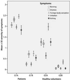
CULARSYMPTOMSQUESTIONNAIREThe individuals of group G1 were patients who went sponta-neously to the Emergency Unit of Hospital Oftalmologico Pedro Lagleyze with ocular complaints on April 19th, 2008. The 35 patients in G1 included 25 women and 10 men, with an average age of 42.5 ± 10.1 years. A total of 31/35 patients had a history of ocular surface pathology: Eighteen sufered from dry eyes (seven had Sjögren’s syndrome), 10 from allergic conjunctivitis, and three sufered from ocular pemphigoid. Four patients had no history of ocular surface pathology but were included in this group because their ocular surfa-ce complaints were of a severity suicient to attend hospital. During the acute episode, all patients (G1A) experienced at least three of the symptoms evaluated (burning, dryness, foreign body sensation, itchi-ness, and irritation). Burning, foreign body sensation, and irritation were the most frequent and severe symptoms (100%) referred during the acute episode. A total of 83% experienced the symptoms during the whole day; 66% reported worsening of the symptoms when outdoors; and only 11% experienced these symptoms for the irst time. There was a signiicant diference in the frequency and severity of the symptoms when the patients were compared over time, i.e., during vs.
after the acute episode (G1A vs. G1B, p<0.01; Figure 2).
Table 2. Test results expressed as means ± standard deviation obtained during and after the acute episode for each group
Groups
Bulbar conjuctival hyperemia
Cornea luorescein staining
Rose bengal
vital staining TBUT (seconds)
Schirmer’s I test (mm/5 min)
Tear lyzozyme (ug/ml)
Impression cytology (10 HPF)
G1A 1.8 ± 1.0 3.7 ± 1.2 6.4 ± 2.1 4.4 ± 1.1 11.3 ± 9.5 1533 ± 987 202 ± 145
G1B 1.1 ± 1.0 1.9 ± 0.7 3.7 ± 1.8 6.2 ± 1.3 10.1 ± 8.2 1612 ± 1072 186 ± 120
G2A 0.8 ± 0.3 0.7 ± 0.3 1.9 ± 1.2 4.9 ± 1.2 17.6 ± 9.7 2489 ± 412 298 ± 132
G2B 0.3 ± 0.1 0.6 ± 0.2 1.7 ± 0.8 7.8 ± 1.2 15.3 ± 7.5 2570 ± 426 315 ± 151
*p<0.05 among groups (G1A vs. G1B and G2A vs. G2B). All the diferences between groups (G1A vs. G2A and G1B vs. G2B) were statistically signiicant.
* * * *
* *
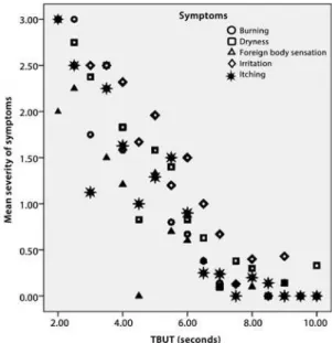
Figure 3. Correlation between severity of symptoms and tear break-up time (TBUT). There was a statistically signiicant correlation between the reported severity of ocular burning, dry eye, foreign body sensation, itching and irritation, and mean TBUT values. Values are expressed as means ± 0.95 conidence intervals.
A total of 51 healthy volunteers were evaluated, including 23 women and 28 men, with an average age of 39.1 ± 11.9 years. During the acute episode, 79% of healthy volunteers (G2A) experienced at least three of the symptoms evaluated. The most frequent and severe symptom was ocular irritation (80%). Most people reported sustaining ocular irritation during the whole day (55%) or at the end of the day (41%). In addition, 65% reported worsening of the symptoms when outdoors and 57% experienced these symptoms for the irst time. There was a signiicant diference in the frequency and severity of the symptoms when the healthy volunteers were compared over time, i.e., during vs. after the acute episode (G2A vs.
G2B, p<0.01; Figure 2).
O
PHTHALMOLOGICEXAMINATIONTable 2 shows the values obtained from G1A-B (N=35) and G2A-B (N=51) for all tests performed.
The patients (G1) showed a statistically signiicant increase in bulbar conjunctival hyperemia (G1A vs. G1B, p=0.0061), corneal luorescein staining (G1A vs. G1B, p<0.0001), and rose bengal vital staining (G1A vs. G1B, p<0.0001) during the acute episode.
The healthy volunteers (G2) only showed a statistically signiicant increase in bulbar conjunctival hyperemia (G2A vs. G2B, p=0.0001). No statistically signiicant diferences were found in corneal luorescein staining (G2A vs. G2B, p>0.05) or rose bengal vital staining (G2A vs. G2B,
p>0.05) during the acute episode.
No statistically signiicant diferences within the groups were found when analyzing the values obtained by Schirmer’s I test, tear lysozyme, and impression cytology.
The healthy volunteers showed statistically signiicant diferences when compared with patients for the entire test performed during and after the acute episode (G1A vs. G2A and G1B vs. G2B, p<0.05).
Both patients (G1) and healthy volunteers (G2) showed a statisti-cally signiicant (p<0.0001) decrease in TBUT values during the acute episode. We also observed a negative correlation between severity of symptoms and TBUT values (Figure 3), signiicant (p<0.05) for the symptoms dryness and irritation (Table 3).
P
OLLUTANTLEVELSDuring April 16th to April 20th, the measured mean levels of CO, NO2, and PM concentrations were four times higher than the usual average levels for this time of the year. Figure 4 depicts the concen-trations of these substances before, during, and after the episode. Hourly measured values of CO reached 17 ppm on April 17th, which is 8.5 times as much as the average value (approximately 2 ppm) registered in the area. Moreover, the 8 hours average value was grea-ter than the standard value recommended by the WHO Air Quality Guidelines (AQG) established by the World Health Organization(22) in 29 opportunities. For NO2 and PM, average levels are usually not exceeding 50% of the maximum levels recommended by the WHO, but during the acute episode, the concentrations were twice as high as those recommended in the WHO guidelines(23-24).
DISCUSSION
In this study, we assessed the impact of acute exposure to high levels of pollutants produced by pasture burning on the ocular sur-face of subjects living in an afected megacity.
Previous studies have demonstrated that ocular symptoms are a frequent inding in people exposed to the smoke generated by ires, including wildires(12-13,24).
During the acute episode, the patients who attended the hos-pital emergency room and the healthy volunteers experienced ocu lar symptoms, an increase in bulbar conjunctival hyperemia, and a reduc-tion in TBUT values.
These results indicate that the patients showed more severe symp-toms and a greater reduction in TBUT values. Meanwhile, Schirmer´s I test, tear lysozyme, and impression cytology showed no dife rences.
Low TBUT values and high concentrations of air pollutants (PM10, NO2, and CO) measured during the acute episode indicated an in-luence of high concentrations of air pollutants on tear ilm stability; this result is similar to a previous study(25).
Table 3. Data obtained from linear regression analysis. Dependent variable = TBUT
Constant variables
Unstandardized coeicients
Standardized coeicients
B Std. error Beta t p
Burning -0.195 0.132 -0.126 -1.478 <0.141
Dryness -0.388 0.128 -0.261 -3.029 <0.003 Foreign body
sensation
-0.108 0.102 -0.064 -1.061 <0.290
Irritation -0.524 0.113 -0.370 -4.622 <0.001 Itching -0.072 0.096 -0.052 -0.745 <0.457
toxicity or as a consequence of ocular dryness caused by tear ilm ins tability.
CONCLUSION
In this study, we demonstrated that acute exposure to high levels of air pollution causes tear ilm instability without remodulation of the ocular surface. Furthermore, the perceptions of symptoms of nor mal subjects were not severe enough to trigger a visit to the emer -gency unit. In contrast, patients who sufered from dry eyes, allergic conjunctivitis, Sjögren’s syndrome, and ocular pemphigoid experien-ced these symptoms with a higher degree of severity, trigge ring a
visit to the emergency unit. This indicates that this last group of pa-tients is more susceptible to developing and/or sufering from these ocular symptoms.
REFERENCES
1. Lipsett M, Waller K, Shusterman D, Thollaug S, Brunner W. The respiratory health impact of a large urban ire. Am J Public Health. 1994;84(3):434-8.
2. Mott JA, Meyer P, Mannino D, Redd SC, Smith EM, Gotway-Crawford C, et al. Wildland forest ire smoke: health efects and intervention evaluation, Hoopa, California, 1999. West J Med. 2002;176(3):157-62. Comment in: West J Med. 2002;176(3):162-3. 3. Vedal S, Dutton SJ. Wildire air pollution and daily mortality in a large urban area. Environ
Res. 2006;102(1):29-35. Comment in: Environ Res. 2008;106(3):423-4; discussion 425. 4. Moss SE, Klein R, Klein BE. Prevalence of and risk factors for dry eye syndrome. Arch
Ophthalmol. 2000;118(9):1264-8.
5. Moss SE, Klein R, Klein BE. Incidence of dry eye in an older population. Arch Ophthal-mol. 2004;122(3):369-73.
6. Iyer JV, Lee SY, Tong L. The dry eye disease activity log study. Scientiic World J. 2012; 2012:589875.
7. Wolkof P, Skov P, Franck C, Petersen LN. Eye irritation and environmental factors in the oice environment-hypotheses, causes and a physiological model. Scand J Work Environ Health. 2003;29(6):411-30. Comment in: Scand J Work Environ Health. 2003; 29(6):407-9.
8. Alves M, Novaes P, Morraye Mde A, Reinach PS, Rocha EM. Is dry eye an environmental disease? Arq Bras Oftalmol. 2014;77(3):193-200.
9. Barabino S, Dana MR. Animal models of dry eye: a critical assessment of opportunities and limitations. Invest Ophthalmol Vis Sci. 2004;45(6):1641-6.
10. Nakamura S, Shibuya M, Nakashima H, Hisamura R, Masuda N, Imagawa T, et al. Invol-vement of oxidative stress on corneal epithelial alterations in a blink-suppressed dry eye. Invest Ophthalmol Vis Sci. 2007;48(4):1552-8.
11. Stern ME, Schaumburg CS, Siemasko KF, Gao J, Wheeler LA, Grupe DA, et al. Autoan-tibodies contribute to the immunopathogenesis of experimental dry eye disease. Invest Ophthalmol Vis Sci. 2012;53(4):2062-75.
12. Kunzli N, Avol E, Wu J, Gauderman WJ, Rappaport E, Millstein J, et al. Health efects of the 2003 Southern California wildires on children. Am J Respir Crit Care Med. 2006;174(11):1221-8. Comment in: Am J Respir Crit Care Med. 2007;175(6):629; author reply 629; Am J Respir Crit Care Med. 2006;174(11):1168-9.
13. Viswanathan S, Eria L, Diunugala N, Johnson J, McClean C. An analysis of efects of San Diego wildire on ambient air quality. J Air Waste Manag Assoc. 2006;56(1):56-67. 14. Efron N. Grading scales for contact lens complications. Ophthalmic Physiol Opt. 1998;
18(2):182-6.
15. Lemp MA. Report of the National Eye Institute/Industry workshop on Clinical Trials in Dry Eyes. CLAO J. 1995;21(4):221-32.
16. van Bijsterveld OP. Diagnostic tests in the Sicca syndrome. Arch Ophthalmol. 1969; 82(1):10-4.
17. The deinition and classiication of dry eye disease: report of the Deinition and Classi ication Subcommittee of the International Dry Eye WorkShop (2007). Ocul Surf. 2007;5(2):75-92.
18. Novaes P, do Nascimento Saldiva PH, Kara-Jose N, Macchione M, Matsuda M, et al. Ambient levels of air pollution induce goblet-cell hyperplasia in human conjunctival epithelium. Environ Health Perspect. 2007;115(12):1753-6.
19. Gonzalez-Garcia MJ, Gonzalez-Saiz A, de la Fuente B, Morilla-Grasa A, Mayo-Iscar A, San-José J, et al. Exposure to a controlled adverse environment impairs the ocular surface of subjects with minimally symptomatic dry eye. Invest Ophthalmol Vis Sci. 2007;48(9):4026-32.
20. Jones LT. The lacrimal secretory system and its treatment. Am J Ophthalmol. 1966; 62(1):47-60.
21. Nelson JD. Impression cytology. Cornea. 1988;7(1):71-81.
22. World Health Organization. Carbon Monoxide. 2nd ed. Geneva: WHO; 1999. (Environ-mental Health Criteria 213)
23. World health Organization. WHO Air quality guidelines for particulate matter, ozone, nitrogen, dioxide and sulfur dioxide. Global update 2005. Summary of risk assessment [Internet]. Geneva: WHO; 2005. (WHO/SDE/PHE/OEH/06.02.) [cited 2008 Jul 27]. Avai-lable from: http://whqlibdoc.who.int/hq/2006/WHO_SDE_PHE_OEH_06.02_eng.pdf 24. Morgan O, Verlander NQ, Kennedy F, Moore M, Birch S, Kearney J, et al. Exposures and reported symptoms associated with occupational deployment to the Bunceield fuel depot ire, England 2005. Occup Environ Med. 2008;65(6):404-11.
25. Novaes P, Saldiva PH, Matsuda M, Macchione M, Rangel MP, Kara-Jose N, et al. The efects of chronic exposure to traic derived air pollution on the ocular surface. En-viron Res. 2010;110(4):372-4.
26. Zelikof JT, Chen LC, Cohen MD, Schlesinger RB. The toxicology of inhaled woodsmoke. J Toxicol Environ Health B Crit Rev. 2002;5(3):269-82.
Figure 4. A) Carbon Monoxide (CO) and nitrogen dioxide (NO2) hourly measured levels before, during, and after the acute episode.B)Levels of total particulate matter (PM) before, during, and after the acute episode. Data were obtained from the Envi-ronmental Protection Agency of Buenos Aires.
A