Phospholipase C
b
1: Translin-Associated Factor X
Omozuanvbo R. Aisiku1, Loren W. Runnels2, Suzanne Scarlata1*
1Department of Physiology and Biophysics, Stony Brook University, Stony Brook, New York, United States of America,2Department of Pharmacology, University of Medicine and Dentistry of New Jersey-Robert Wood Johnson Medical School, New Brunswick, New Jersey, United States of America
Abstract
Mammalian phospholipase Cb1 (PLCb1) is activated by the ubiquitous Gaqfamily of G proteins on the surface of the inner
leaflet of plasma membrane where it catalyzes the hydrolysis of phosphatidylinositol 4,5 bisphosphate. In general, PLCb1 is mainly localized on the cytosolic plasma membrane surface, although a substantial fraction is also found in the cytosol and, under some conditions, in the nucleus. The factors that localize PLCb1in these other compartments are unknown. Here, we identified a novel binding partner, translin-associated factor X (TRAX). TRAX is a cytosolic protein that can transit into the nucleus. In purified form, PLCb1 binds strongly to TRAX with an affinity that is only ten-fold weaker than its affinity for its functional partner, Gaq. In solution, TRAX has little effect on the membrane association or the catalytic activity of PLCb1.
However, TRAX directly competes with Gaqfor PLCb1 binding, and excess TRAX reverses Gaqactivation of PLCb1. In C6 glia
cells, endogenous PLCb1 and TRAX colocalize in the cytosol and the nucleus, but not on the plasma membrane where TRAX is absent. In Neuro2A cells expressing enhanced yellow and cyano fluorescent proteins (i.e., eYFP- PLCb1 and eCFP-TRAX), Fo¨rster resonance energy transfer (FRET) is observed mostly in the cytosol and a small amount is seen in the nucleus. FRET does not occur at the plasma membrane where TRAX is not found. Our studies show that TRAX, localized in the cytosol and nucleus, competes with plasma-membrane bound Gaq for PLCb1 binding thus stabilizing PLCb1 in other cellular
compartments.
Citation:Aisiku OR, Runnels LW, Scarlata S (2010) Identification of a Novel Binding Partner of Phospholipase Cb1: Translin-Associated Factor X. PLoS ONE 5(11): e15001. doi:10.1371/journal.pone.0015001
Editor:Vladimir N. Uversky, University of South Florida College of Medicine, United States of America
ReceivedAugust 13, 2010;AcceptedOctober 5, 2010;PublishedNovember 29, 2010
Copyright:ß2010 Aisiku et al. This is an open-access article distributed under the terms of the Creative Commons Attribution License, which permits unrestricted use, distribution, and reproduction in any medium, provided the original author and source are credited.
Funding:This work was supported by the National Institutes of Health, NIH053132. The funders had no role in the study design, data collection and analysis, decision to publish, or preparation of the manuscript.
Competing Interests:The authors have declared that no competing interests exist.
* E-mail: [email protected]
Introduction
Inositide-specific mammalian phospholipase Cbeta (PLCb) enzymes are the main effectors of the Gaqfamily of G proteins and are coupled to agents such as angiotension, dopamine, serotonin, bradykinin, etc. (for review see [1,2,3,4]). PLCbcatalyzes the hydrolysis of the signaling lipid phosphatidylinositol 4,5 bisphosphate (PI(4,5)P2) to produce the second messengers inositol 1,4,5 trisphosphate and diacylglycerol that in turn stimulate the release of Ca2+
from intracellular stores and activate protein kinase C, respectively. Both the Gaqfamily of G proteins and the PLCb enzymes they activate are found in all mammalian cells lines.
In cultured cells, PLCb1 resides mainly on the surface of the plasma membrane where it associates with its membrane-bound activator Gaqand can access its PI(4,5)P2substrate. In addition to this plasma membrane population, a significant population of PLCb1 resides in the cytosol, and under some circumstances, in the nucleus. The factors that localize PLCb1 to these alternate compartments are unknown, especially since PLCb1 is expected to have a high propensity to localize to the plasma membrane due to its strong, non-specific lipid binding behavior [5]. Additionally, the basal activity of PLCb1 is very low and it is unclear how it can be activated in these alternate compartments since Gaq appears to only reside at the plasma membrane (see [6,7]).
There are several possible mechanisms that may underlie the cytosolic localization of PLCb1. The first might be a saturation of
binding sites on the plasma membrane. While we lack the knowledge to accurately quantify binding sites and the local cellular concentration of competing proteins, we note that the cellular concentration of Gaqappears to be higher than PLCb1 allowing Gaqto interact with its other effectors, phosphatidylino-sitol 3-kinase and RhoGEF (see [8]). Another possibility is that one or more cytosolic proteins might promote the plasma member localization of PLCb1. With this idea in mind, we searched for alternate protein partners of PLCb1 using a yeast two hybrid approach and identified the protein TRAX (translin-associated factor X). TRAX forms strong complexes with its only known partner, translin [9]. Translin is a single-stranded DNA and RNA-binding protein with proposed functions in chromosomal translo-cations in lymphoid cells and mRNA transport and storage in brain and testis [10]. Both translin and TRAX are part of the RNA-induced silencing complex (RISC) where they help guide double stranded RNA into the silencing machinery [11]. Additionally, TRAX has been implicated to function as a localization factor for translin. When the cellular level of translin exceeds TRAX, it remains in the cytosol [12]. However, when the cellular level of translin is reduced, translin can partition into the nucleus through the nuclear localization signal (NLS) of TRAX.
TRAX and PLCb1 interact strongly in solution and form complexes in living cells. We find that TRAX competes with Gaq for PLCb1 binding and activation. Our studies show that TRAX stabilizes the cytosolic and nuclear localization of PLCb1.
Methods
Sample Preparation
Purified proteins were used in all in vitro experiments. His-tagged PLCb1 from rat and Gaqfrom rat were expressed in Sf9 cells and purified based on previously described methods (see [13,14]). Preparation of C-terminal truncated PLCb1 has been described [5]. Large, unilamellar vesicles (LUVs), 100nm in diameter, were prepared by extrusion. All lipids (1-palmitoyl-2-oleoyl phosphatidylethanolamine (POPE), 1 -palmitoyl-2-(1 -palmitoyl-2-oleoyl-phosphatidylserine (POPS), 1-palmitoyl-2-oleoylphosphatidylcho-line (POPC)) were purchased from Avanti Polar Lipids (Alabaster, AL) with the exception of the tritiated PI(4,5)P2 which was purchased from Perkin Elmer. Human TRAX cDNA purchased from Open Biosystems was cloned into pET32a expression vectors purchased from Novagen. His-tagged TRAX was expressed in Rosetta cells and purified on a Ni-NTA column. The integrity of the TRAX preparation was determined by western blot analysis and circular dichroism spectra (see results).
Protein Labeling and Reconstitution
In vitro binding studies were carried out by labeling either the PLCb1 or Gaq constructs with the thiol reactive probe 7-diethylamino-3- (49-maleimidylphenyl)-4-methylcoumarin (CPM, Invitrogen, Inc) as previously described to yield fluorescent proteins with an ,1:1 probe:protein ratio (see [15]). We note
that labeling by this method does not affect the activity of PLCb1 or its ability to be stimulated by Gaq(see [14]). To prevent protein aggregation, Gaqwas labeled in the presence of storage detergents (0.1% lubrol) and was reconstituted into preformed extruded lipid vesicles by simple addition.
Optical Measurements and Data Analysis
CD spectra were carried out on an Olis RSM 1000 CD spectrophotometer (On-line Instrument Systems, Inc. Bogart, GA). TRAX concentration was 20mM in 20 mM Hepes, 0.16 M NaCl, pH 7.4. Binding affinity measurements were performed on an ISS spectrofluorometer (Urbana, IL) using a 3 mm cuvette (see [15]). CPM-labeled proteins were excited at 380 nm and scanned 420 to 560 nm. Binding affinity was determined as a function of the increase in CPM fluorescent intensity as non-fluorescent protein was incrementally added. Briefly, the sample spectra were corrected by subtracting out identical spectra in control cuvettes that substituted buffer for non-fluorescent protein. The area under the emission peaks of the corrected spectra were calculated using ISS Vinci software. These area were then plotted as a function of concentration of added protein, and the resulting curves were fit to a bimolecular dissociation constant using Sigma Plot (Jandel, Inc.). Membrane binding studies were carried out by measuring the change in intensity of a 20mM solution of CPM- PLCb1 as freshly extruded large unilamellar vesicles were incrementally added. The data were corrected for background by carrying out an identical titration where buffer was substituted for CPM- PLCb1, The corrected data were them fit to a hyperbolic curve using Sigmaplot (Jandel, Inc.).
Activity Measurements
Measurements were made using full length and truncated His-tagged PLCb1, Gaq, and TRAX and small, unilamellar vesicles
consisting of PI(4,5)P2, POPE, POPS (1:1:1) doped with 3 H-PI(4,5)P2prepared by sonication (see [13]). Briefly, 2mM of lipid were incubated at 37uC with PLCb1 in the linear range of the activity as determined for each by running a time course experiment (usually between 2 and 5 minutes). The reaction was initiated by the addition of Ca2+. Activities are reported as the
percent of radioactive PI(4,5)P2hydrolysis that occurred.
Cell Culture
Neuro2A cells from American Type Culture Collection (www. ATCC.org) were cultured in 50/50 DMEM/F12 media supple-mented 10% FBS. C6 glioma cells were cultured in DMEM containing 10% FBS, 100mM sodium pyruvate and 1% PenStrep at 37uC with 5% CO2.
DNA was transfected into cells by electroporation using a protocol adapted from Maniatis. Cells were grown to near 100% confluence and washed with sterile PBS. The cells were then trypsinized, centrifuged 5 minutes at 15006g and resuspended in 10mL of fresh growth medium. 800ml of cells were pipeted into a 0.4cm BioRad cuvette and placed in an electroporator (BioRad Gene Pulser Xcell). Cells were then plated and covered with fresh medium.
Immunofluorescence
Cells were fixed and stained with primary antibodies to TRAX and PLCb1 (Santa Cruz Biochemicals, Inc.). In certain cases as noted in the text, eYFP-tagged PLCb1was transfected via electroporation prior to fixing. Cells were initially washed with PBS+ 1mM Ca2+
and 2 mM Mg2+
. Cells were then fixed with 3.7% formaldehyde in PBS and permeabilized with a solution of 0.2% NP40 in PBS. After, the cells were blocked in 4% goat serum in 1X TBS, washed and a primary antibody added. Cells were incubated for 1 hour, washed and treated with a secondary antibody. After another wash, the cells were viewed on either a Zeiss Axiovert 200M with an AxioCam MRm camera, or an Olympus Fluoview FV1000 laser confocal microscope. Data were analyzed using either Olympus (Fluoview) software or Image J (NIH).
FRET analysis
A fully functional eYFP-tagged PLCb1 was cotransfected with TRAX fused with eCFP. Cells were allowed to incubate for 48 hours. Afterwards the live cells were viewed through the CFP, YFP and FRET channels and FRET measurements on regions of interest were made using sensitized emission on an Olympus Fluoview FV1000 laser confocal microscope (for details, see [16]). Analysis was done using Fluoview software (Olympus, Inc.).
Results
Identification of TRAX as a PLCb1 binding partner
A yeast two-hybrid (Y2H) screen of a rat brain library with the C2 domain-containing COOH-terminus of rat PLCb1 (a.a. 643-1216) was performed to identify novel interacting partners important to the functional regulation of PLCb1 and uncovered the TRPM7 ion channel as a binding partner [17,18]. Among the 69 rat Y2H clones from the screen that were sequenced based on selected growth and b-galactosidase activity, three overlapping clones were identified as translin-associated factor X (TRAX). Since TRAX has a cytosolic and nuclear localization, we carried out further tests to determine whether it is a PLCb1 binding partner.
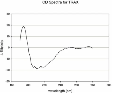
purified PLCb1 is readily assessed by its catalytic activity and its ability to be activated by Gaq, TRAX does not have a catalytic function. Thus, we determined whether TRAX was folded properly by circular dichroism (CD) spectroscopy. In Fig. 1we show the CD spectrum of a solution of 20mM TRAX in buffer. We find that the TRAX CD spectrum displays a high degree of secondary structure that corresponds to ,80% helical content
(http://www.embl.de/˜andrade/k2d). This high helical content is expected from its sequence where it is predicted to have over 50% helical structure with the remainder being loops (www. predictprotein.org).
We measured the association between TRAX and PLCb1 using fluorescence methods. In these experiments His6- PLCb1 was expressed in Sf9 cells and purified, and then labeled with the fluorescent probe CPM (diethylamino-3- (49 -maleimidylphenyl)-4-methylcoumarin). We then monitored changes in the fluorescence spectrum of CPM- PLCb1 with the addition of purified TRAX (see methods and [15] for details). We find that the fluorescence intensity increased systematically with the addition of TRAX without a significant shift in the emission spectrum. Fitting this increase (an 80% increase relative to control samples that did not contain TRAX) to a bimolecular dissociation curve gives an apparent dissociation constant of Kd= 8+/21 nM (Fig. 2a). Interestingly, this apparentKd is only,10 fold weaker than the
one measured for the interaction between PLCb1 and activated Gaq[14]. We repeated the titration using an initial concentration of PLCb1 that is above the Kd, 10 nM, to verify that the association was shifted to the stoichiometric regime, and a midpoint of 10 nM was obtained (data not shown).
The COOH-terminal region of PLCb1 was used as bait to identify TRAX in the yeast 2-hybrid screen suggesting that this region binds TRAX. Therefore, we repeated the fluorescence binding study using a COOH-terminal deletion mutant of PLCb1 (PLCb1–DC). We find that the loss of the C-terminus results in a
drastic reduction in its affinity for TRAX (Fig. 2b). This result confirms that the primary interaction site between TRAX and PLCb1 is within C-terminal region. Thus, all further experiments used the full length enzyme.
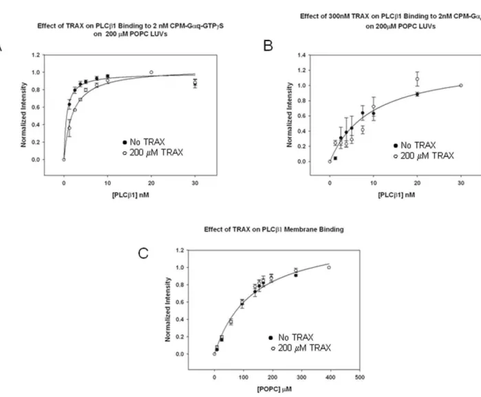
TRAX competes with Gaq-GTP for binding to PLCb1 PLCb1 strongly associates with Gaq-GTP on membrane surfaces (Kd= 0.67 nM) through sites in its C2 and COOH-terminal regions [19,20]. Since both TRAX and Gaqbind to the COOH-terminal region, we asked whether TRAX would compete with binding of Gaq to full length PLCb1. For these studies, purified Gaqactivated with non-hydrolyzable GTPcS was labeled with CPM and reconstituted onto large unilamellar vesicles (LUVs) composed of POPC:POPS:POPE (1:1:1). We measured the dissociation constant between PLCb1 and Gaq-GTPcS in the absence and presence of excess TRAX (300nM). The presence of TRAX resulted in a 3-fold reduction in PLCb1- Gaq-GTPcS binding affinity, showing that TRAX competes with Gaq for binding to PLCb1 (Fig 3a).
We repeated the above study substituting unactivated Gaq(GDP) for activated Gaq (GTPcS). It is worth noting that in the deactivated state, the affinity between Gaq and PLCb1 is reduced by a factor of,50 [14] giving rise to large experimental
error. As expected, we find that TRAX does not appear to compete with deactivated Gaqfor binding to PLCb1. This result may suggest that deactivation of Gaq (GDP) alters its binding interaction with PLCb1, consistent with previous work [6], to a site that is less competitive for TRAX association (Fig. 3b).
PLCb1 binds strongly to lipid membranes where it associates with Gaqand accesses its substrate. Membrane binding of PLCb1 has been found to be primarily mediated through the N-terminal PH domain of PLCb1 and to a lesser extent, its C-terminal region [5]. Since TRAX might affect the association between PLCb1 and Gaq by altering its membrane binding affinity, we determined
whether TRAX would interfere with PLCb1’s association with membranes. We find it does not (Fig. 3c). A lack of effect of TRAX on the membrane binding of PLCb1 correlates well with the finding that TRAX associates with the C-terminal region and not the N-terminal membrane binding region.
TRAX interferes with the activation of PLCb1 by Gaq subunits
We next determined whether TRAX has the ability to modulate the enzymatic activity of PLCb1 or its activation by Gaq. These studies were carried out by monitoring the amount of PI(4,5)P2 hydrolysis catalyzed by 2 nM PLCb1 in the presence and absence of TRAX. We find that a 300 molar excess of TRAX does not greatly affect the initial velocity of the reaction catalyzed by PLCb1, but reduces the velocity at later times, suggesting that TRAX causes a small reduction in the maximum rate (Fig. 4a). We then tested whether TRAX could block PLCb1 activation by Gaq. Again, a large excess of TRAX was used to allow it to compete with Gaq, which binds PLCb1 much more strongly. We find that TRAX prevents the activation of PLCb1 by Gaq (Fig.4b), which is consistent with the ability of TRAX to disrupt the association between PLCb1 and Gaq(GTPcS) (Fig.3a).
TRAX and PLCb1 are associated in cultured cells
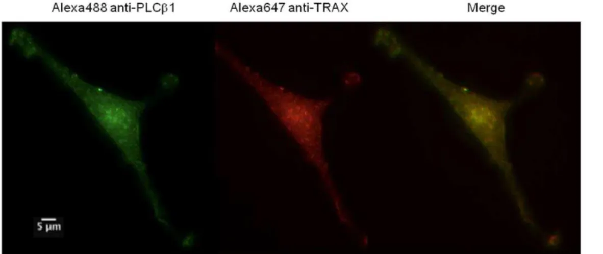
We verified that TRAX and PLCb1 associate in cells using fluorescence microscopy. First, we used immunofluorescence to determine whether endogenous TRAX and endogenous PLCb1 are colocalized in C6 glia cells. The images inFig. 5show almost complete colocalization of the proteins throughout the cell suggesting association between the two proteins.
Since colocalization will only indicate whether the proteins reside in the same region of the cell, we measured the physical association of TRAX and PLCb1 by Fo¨rster resonance energy transfer (FRET) using attached ‘‘donor’’ and ‘‘acceptor’’
fluores-cent probes. For these studies, we linked enhanced yellow fluorescence proteins (eYFP) to PLCb1 and enhanced cyano fluorescence protein (eCFP) to TRAX. The observation of FRET from eCYP donors to eYFP acceptors indicates that the probes are at least within 30 A˚ of each other [21], implying that the two labeled proteins are physically associated.
We transfected Neuro2A cells with eYFP- PLCb1 and eCFP-TRAX and measured the amount of FRET by sensitized emission (see methods) using a confocal microscope. FRET values were compared to a positive control consisting of eYFP-X12-eCFP and a negative control consisting of free eYFP and eCFP (see [6]). An example of a FRET study is shown inFig. 6. We find that a large degree of FRET occurs between the proteins in the cytosol while only a small amount is seen in the nucleus and none on the plasma membrane compartments since TRAX does not localize to this compartment and cannot provide donor energy for FRET (Fig. 6). Averaging the amount of FRET over the entire cell gives a value of 40.1+0.6% (n = 5). The lack of FRET in the nucleus contrasts with the coimmunofluorescence results in C6 glia cells and can be explained by a lack of PLCb1 in the nuclear compartment. Thus, these studies show that TRAX and PLCb1 associate in the cytosol of cells.
Discussion
In this study, we have identified TRAX as a novel cellular binding partner of PLCb1. The potential interaction between PLCb1 and TRAX was found in a yeast 2-hybrid experiment using the 574 residue C2-domain containing COOH-terminal region of PLCb1 as bait, and subsequent binding studies between the purified proteins verified this interaction.
The binding of TRAX to the COOH-terminal region of PLCb1 suggests that this interaction is specific to the PLCbfamily rather than the other known mammalian families of PLCs (i.e.d,c,eand f) since this region of PLCb is not found in other PLCs. This Figure 2. TRAX binds strongly to PLCb1. A– Binding of TRAX to 2 nM CPM- PLCb1 as monitored by the increase in CPM intensity where the normalized fluorescence intensity is shown as a function of TRAX concentration. In these studies, an 80% increase in intensity was observed as compared to control samples that substituted buffer for TRAX. Also shown is the fitted curve to a bimolecular dissociation constant whereKd= 861 nM (n = 6 and S.D. is shown).B– Identical study as2Aexcept that the COOH-terminal deletion mutant of PLCb1 (PLCb1-DC) was used instead of the full length enzyme (n = 3 and S.D. is shown). While a binding curve is shown to guide the eye, the affinity between the proteins was too weak to be accurately fit to a bimolecular dissociation constant. We note that the total change in CPM intensity was also,80% at the end of the titration.
region contains the only identified nuclear localization signal of PLCb1 (a nuclear export signal on PLCb has never been identified), as well as mitogen-activation protein kinase and protein kinase C phosphorylation sites (we note that unpublished studies by our group suggest that PKC phosphorylation does not affect the interaction between TRAX and PLCb1: Aisiku, Dowal & Scarlata,unpublished). Importantly, the COOH-terminal region of PLCb1 is necessary for Gaq activation, and has GTPase-promoting activity [19,22]. Therefore, it is not surprising that TRAX competes with Gaq for binding to PLCb1. The crystal structure of the isolated COOH-terminal region of PLCb1 has been solved and is found to be an intertwined helical dimer [23], although it is not clear whether PLCb1 itself is dimeric. While it is impossible to speculate the mode of interaction between TRAX and PLCb1, it is notable that translin, TRAX’s known binding partner, is comprised of a network of helixes [24].
We assessed the association between endogenous TRAX and PLCb1 in C6 glial cells, which express these proteins at high levels. We speculate that the high level of expression of PLCb1 in these cells is responsible for the large cytosolic population since all of the
plasma membrane localized Gaqbinding sites may be saturated. We find that the two proteins co-localize throughout the cell. We then visualized the associated proteins using FRET by co-expressing fluorescent tagged proteins in a cell line where their endogenous expression is more limited (i.e. Neuro2A cells). We find that the proteins complexes largely reside in the cytosol, since little PLCb1 localizes to the nucleus and since TRAX is not found on the plasma membrane in these cells. Interestingly, the FRET values that have been reported for PLCb1 and Gaq[6] are close to the FRET values between PLCb1 and TRAX observed here. PLCb1 is known to have a high plasma membrane population due to its strong interaction with Gaqboth in the basal and stimulated states [6]. The ability of TRAX to hold PLCb1 in the cytosol despite the higher affinity of the enzyme for Gaq and the concentrating effect of the membrane in promoting protein association correlates well with the strong association between TRAX and PLCb1. It is interesting to note that recent biochemical studies suggest that Gaq and PLCb1 bind to a protein scaffold on the plasma membrane which stabilizes their interactions [25].
Figure 3. TRAX competes with Gaqfor PLCb1 binding but not membranes. A– Binding of PLCb1 to 2 nM activated CPM- Gaq(GTPcS) in the
absence (
N
) and presence (#) of 200 nM TRAX right panel showing the loss in Gaqaffinity when TRAX is present, where n = 6 and S.D. is shown. Wenote that an,20% increase in CPM intensity was seen both without and with TRAX.B-Binding of PLCb1 to 2 nM deactivated CPM- Gaq(GDP) in the
absence (
N
) and presence (#) of 200 nM TRAX, where n = 3 and S.D. is shown.C- Binding of PLCb1 to PC:PS:PE (1:1:1) large, unilamellar vesicles in the absence (N
) (Kp= 132mM) and presence (#) (Kp= 120mM) of 200 nM TRAX as measured by the increase in CPM intensity as LUVs are titrated into theFigure 4. TRAX affects the activation of PLCb1 by Gaq. A– The effect of 300 nM TRAX on the rate of PI(4,5)P2hydrolysis catalyzed by 25nM
PLCb1 (n = 3 and S.D. is shown). As can be seen, TRAX does not affect the initial velocity of the curve.B-Prevention of activation of 5 nM PLCb1 by 5nM Gaqby 300 nM TRAX, where n = 8 and S.D. is shown.
doi:10.1371/journal.pone.0015001.g004
Figure 5. TRAX and PLCb1 co-localize in C6 glial cells.Example of a co-immunofluorescence study of endogenous PLCb1 (left panel) as visualized by Alexa488-labeled antibody, TRAX (middle panel) visualized by Alexa647-labeled antibody and the resulting merged image (right panel) in C6 glial cells. The scale bar is 5mm.
While the majority of PLCb1 resides on the plasma membrane, a significant amount is also seen in the cytosol. Additionally, we can only speculate about the function of PLCb1 in the cytosol. While its activity is expected to be too low to impact the level of PI(4,5)P2 levels in internal membranes, it might be sufficient to keep internal PI(4,5)P2at basal levels.
In C6 glia cells, TRAX and PLCb1 appear to be co-localized in the nucleus as well as the cytosol. Since both proteins have a nuclear localization sequence, it is just as likely that either partner could be responsible for nuclear transit. Nuclear transit might occur through exposure of either nuclear localization signal upon protein association or upon saturation of PLCb1’s plasma membrane and cytosolic sites. Although PLCb1 is not considered a nuclear protein, it has been found in the nucleus under some conditions (see [26,27]). We observe large amount of the enzyme in undifferentiated PC12 cells (Dowal & Scarlata, unpublished). Cocco and colleagues find PLCb1 travels to the nucleus in Swiss 3T3 cells upon stimulation with insulin-like growth factor [28]. The role of PLCb1 in the nucleus is unknown, but is assumed to be involved in the nuclear phosphadylinositol signaling pathway [29]. It is notable that PLCb1 does not appear to be associated with the nuclear membrane.
The identification of TRAX as a potential binding partner of PLCb1 is surprising since TRAX is not known to be associated with inositol phosphate signaling. One study did report a link between TRAX and activation of a G protein coupled receptor linked to cAMP [30] giving rise to the possibility that TRAX may modulate additional aspects of G protein signaling. To date, TRAX has only been known to bind to translin and modulate its cellular localization [12]. It has recently been found that TRAX and translin are part of the RNA-induced silencing complex, and their presence helps to guide RNA insertion into the complex to
induce gene silencing [11]. PLCb1 has been shown to reside in nuclear speckles [29,31] which are storage deposits for splicing factors and other transcription machinery (for review see [32]). The localization of PLCb1 in speckles suggests a possible role in RNA splicing and/or in mRNA processing.
While the strong binding constant and large FRET value measured for cytosolic PLCb1 and TRAX suggest a specific cellular role, the functional consequence of these proteins’ association is not yet clear. Unfortunately, we do not yet have the ability to accurately measure the concentrations of these proteins in the cell, but realize that these numbers will change will cell type, localization, cycle and differentiation. Therefore, we cannot determine the importance of TRAX in regulating the localization of PLCb1 at this time. One possibility is that TRAX buffers PLCb1 in the cytosol until Gaqbecomes activated to high enough levels to displace TRAX from PLCb1. TRAX may also regulate the entry of PLCb1 into the nucleus so that it may participate in RNA processing. Alternately, PLCb1 may regulate entry of TRAX into the nucleus or its association to translin. These functional studies are presently underway.
Acknowledgments
The authors are grateful to Dr. Steven Smith (Stony Brook University) for the use of his circular dichoism instrument, and to Drs. Guo and Rebecchi (Stony Brook University) for their helpful comments.
Author Contributions
Conceived and designed the experiments: OA SS. Performed the experiments: OA LR. Analyzed the data: OA LR SS. Contributed reagents/materials/analysis tools: OA LR SS. Wrote the paper: OA LR SS.
References
1. Suh P, Park J, Manzoli L, Cocco L, Peak J, et al. (2008) Multiple roles of phosphoinositide-specific phospholipase C isozymes. BMB reports 41: 415–434. 2. Rebecchi MJ, Pentyala SN (2000) Structure, function, and control of
phosphoinositide-specific phospholipase C. PhysiolRev 80: 1291–1335. 3. Exton JH (1996) Regulation of phosphoinositide phospholipases by hormones,
neurotransmitters, and other agonists linked to G proteins. Annual Review of Pharmacology & Toxicology 36: 481–509.
4. Rhee SG (2001) Regulation of phosphoinositide-specific phospholipase C. AnnRevBiochem 70: 281–312.
5. Runnels LW, Jenco J, Morris A, Scarlata S (1996) Membrane binding of phospholipases C-b1and C-b2is independent of phosphatidylinositol 4,5-bispho-sphate and the a and bg subunits of G proteins. Biochemistry 35: 16824–16832.
6. Dowal L, Provitera P, Scarlata S (2006) Stable association between G alpha(q) and phospholipase C beta 1 in living cells. J Biol Chem 281: 23999–24014. 7. Hughes TE, Zhang H, Logothetis D, Berlot CH (2001) Visualization of a
functional Gaq-green fluorescent protein fusion in living cells. JBiolChem 276: 4227–4235.
8. Philip F, Sengupta P, Scarlata S (2007) Signaling through a G Protein Coupled Receptor and its corresponding G protein follows a stoichometrically limited model. JBiolChem 282: 19203–19216.
9. Yang S, Cho YS, Chennathukuzhi VM, Underkoffler LA, Loomes K, et al. (2004) Translin-associated Factor X Is Post-transcriptionally Regulated by Its Partner Protein TB-RBP, and Both Are Essential for Normal Cell Proliferation. J Biol Chem 279: 12605–12614.
Figure 6. TRAX associates with PLCb1 in N2A cells.Example of a FRET study showing the raw images of eYFP- PLCb1 (left panel), eCFP-TRAX (middle panel) and the normalized FRET image (right panel) in transfected Neuro2A cells where amount of FRET is determined by the sensitized emission (see [6]. The scale bar is 5mm.
10. Wang J, Boja ES, Oubrahim H, Chock PB (2004) Testis Brain Ribonucleic Acid-Binding Protein/Translin Possesses both Single-Stranded and Double-Stranded Ribonuclease Activities. Biochemistry 43: 13424–13431.
11. Liu Y, Ye X, Jiang F, Liang C, Chen D, et al. (2009) C3PO, an Endoribonuclease That Promotes RNAi by Facilitating RISC Activation. Science 325: 750–753.
12. Cho YS, Chennathukuzhi VM, Handel MA, Eppig J, Hecht NB (2004) The Relative Levels of Translin-associated Factor X (TRAX) and Testis Brain RNA-binding Protein Determine Their Nucleocytoplasmic Distribution in Male Germ Cells. J Biol Chem 279: 31514–31523.
13. Runnels LW, Scarlata SF (1998) Regulation of the rate and extent of phospholipase C beta 2 effector activation by the beta gamma subunits of heterotrimeric G proteins. Biochemistry 37: 15563–15574.
14. Runnels LW, Scarlata S (1999) Determination of the affinities between heterotrimeric G protein subunits and their phospholipase C- b effectors. Biochemistry 38: 1488–1496.
15. Philip F, Scarlata S (2006) Real time measurements of protein affinities on membrane surfaces by fluorescence spectroscopy. ScienceSTKE. pp 15–25. 16. Golebiewska U, Scarlata S (2008) G{alpha}q Binds Two Effectors Separately in
Cells: Evidence for Predetermined Signaling Pathways. Biophys J 95: 2575–2582.
17. Runnels LW, Yue L, Clapham DE (2001) TRP-PLIK, a Bifunctional Protein with Kinase and Ion Channel Activities. Science 291: 1043–1047.
18. Runnels LW, Yue L, Clapham DE (2002) The TRPM7 channel is inactivated by PIP2 hydrolysis. Nat Cell Biol 4: 329–336.
19. Park D, Hhon D-Y, Lee C-W, Ryu SH, Rhee SG (1993) Removal of the carboxyl-terminal region of phospholipase C-b1 by calpain abolishes activation by Gaq. Journal of Biological Chemistry 268: 3710–3714.
20. Wang T, Pentyala S, Elliott JT, Dowal L, Gupta E, et al. (1999) Selective interaction of the C2 domains of phospholipase C-beta1 and - beta2 with activated Galphaq subunits: an alternative function for C2- signaling modules. Proc Natl Acad Sci U S A 96: 7843–7846.
21. Patterson GH, Piston DW, Barisas BG (2000) Forster distances between green fluorescent protein pairs. Anal Biochem 284. pp 438–440.
22. Berstein G, Blank JL, Jhon D-Y, Exton JH, Rhee SG, et al. (1992) Phospholipase C-b1 is a GTPase-Activating Protein for Gq/11, Its Physiologic Regulator. Cell 70: 411–418.
23. Singer AU, Waldo GL, Harden TK, Sondek J (2002) A unique fold of phospholipase C-beta mediates dimerization and interaction with G alpha q. Nat Struct Biol 9: 32–36.
24. Sugiura I, Sasaki C, Hasegawa T, Kohno T, Sugio S, et al. (2004) Structure of human translin at 2.2 Angstrom resolution. Acta Crystallographica Section D 60: 674–679.
25. Garcı´a-Hoz C, Sa´nchez-Ferna´ndez G, Dı´az-Meco MT, Moscat J, Mayor F, et al. (2010) Gaq Acts as an Adaptor Protein in Protein Kinase Cf(PKCf )-mediated ERK5 Activation by G Protein-coupled Receptors (GPCR). Journal of Biological Chemistry 285: 13480–13489.
26. Manzoli L, Billi AM, Faenza I, Matteucci A, Martelli AM, et al. (1999) Nuclear phospholipase C: a novel aspect of phosphoinositide signalling. Anticancer Res 19: 3753–3756.
27. Cocco L, Martelli AM, Mazzotti G, Barnabei O, Manzoli FA (2000) Inositides and the nucleus: phospholipase Cbeta family localization and signaling activity. Adv Enzyme Regul 40: 83–95.
28. Manzoli L, Billi AM, Rubbini S, Bavelloni A, Faenza I, et al. (1997) Essential role for nuclear phospholipase C beta1 in insulin-like growth factor I-induced mitogenesis. Cancer Research 57: 2137–2139.
29. Cocco L, Faenza I, Fiume RM, Gilmour SR, Manzoli FA (2006) Phosphoino-sitide-specific phospholipase C (PI-PLC) [beta]1 and nuclear lipid-dependent signaling. Biochem Biophys Acta 1761: 509–521.
30. Sun C-N, Cheng H-C, Chou J-l, Lee S-Y, Lin Y-W, et al. (2006) Rescue of p53 Blockage by the A2A Adenosine Receptor via a Novel Interacting Protein, Translin-Associated Protein X. Molecular Pharmacology 70: 454–466. 31. Cocco L, Martelli AM, Vitale M, Falconi M, Barnabei O, et al. (2002) Inositides
in the nucleus: regulation of nuclear PI-PLCbeta1. Adv Enzyme Regul 42: 181–193.