The
Aspergillus fumigatus pkcA
Mutant
Is Defective in the Activation of the Cell Wall
Integrity Pathway but Is Dispensable for
Virulence in a Neutropenic Mouse Infection
Model
Marina Campos Rocha1, Krissia Franco de Godoy1, Patrícia Alves de Castro2, Juliana Issa Hori3, Vinícius Leite Pedro Bom2, Neil Andrew Brown4, Anderson Ferreira da Cunha1,
Gustavo Henrique Goldman2,5, Iran Malavazi1*
1Departamento de Genética e Evolução, Centro de Ciências Biológicas e da Saúde, Universidade Federal
de São Carlos, São Carlos, São Paulo, Brazil,2Departamento de Ciências Farmacêuticas, Faculdade de
Ciências Farmacêuticas de Ribeirão Preto, Universidade de São Paulo, Ribeirão Preto, São Paulo, Brazil, 3Departamento de Farmacologia, Faculdade de Medicina de Ribeirão Preto, Universidade de São Paulo,
Ribeirão Preto, São Paulo, Brazil,4Department of Plant Biology and Crop Science, Rothamsted Research,
Harpenden, Herts, United Kingdom,5Laboratório Nacional de Ciência e Tecnologia do Bioetanol,
Campinas, São Paulo, Brazil
*imalavazi@ufscar.br
Abstract
Aspergillus fumigatusis an opportunistic human pathogen, which causes the life-threaten-ing disease, invasive pulmonary aspergillosis. In fungi, cell wall homeostasis is controlled by the conserved Cell Wall Integrity (CWI) pathway. InA.fumigatusthis signaling cascade is partially characterized, but the mechanisms by which it is activated are not fully eluci-dated. In this study we investigated the role of protein kinase C (PkcA) in this signaling cas-cade. Our results suggest thatpkcAis an essential gene and is activated in response to cell wall stress. Subsequently, we constructed and analyzed a non-essentialA.fumigatus pkcAG579Rmutant, carrying a Gly579Arg substitution in the PkcA C1B regulatory domain. ThepkcAG579Rmutation has a reduced activation of the downstream Mitogen-Activated Protein Kinase, MpkA, resulting in the altered expression of genes encoding cell wall-related proteins, markers of endoplasmic reticulum stress and the unfolded protein response. Furthermore, PkcAG579Ris involved in the formation of proper conidial architec-ture and protection to oxidative damage. ThepkcAG579Rmutant elicits increased production of TNF-αand phagocytosis but it has no impact on virulence in a murine model of invasive pulmonary aspergillosis. These results highlight the importance of PkcA to the CWI pathway but also indicated that additional regulatory circuits may be involved in the biosynthesis and/or reinforcement of theA.fumigatuscell wall during infection.
OPEN ACCESS
Citation:Rocha MC, Godoy KFd, de Castro PA, Hori JI, Bom VLP, Brown NA, et al. (2015) TheAspergillus fumigatus pkcAG579RMutant Is Defective in the Activation of the Cell Wall Integrity Pathway but Is Dispensable for Virulence in a Neutropenic Mouse Infection Model. PLoS ONE 10(8): e0135195. doi:10.1371/journal.pone.0135195
Editor:Jae-Hyuk Yu, The University of Wisconsin—
Madison, UNITED STATES
Received:May 13, 2015
Accepted:July 19, 2015
Published:August 21, 2015
Copyright:© 2015 Rocha et al. This is an open access article distributed under the terms of the
Creative Commons Attribution License, which permits unrestricted use, distribution, and reproduction in any medium, provided the original author and source are credited.
Data Availability Statement:All relevant data are within the paper and its Supporting Information files.
Funding:This work was supported by Fundação de Amparo à Pesquisa do Estado de São Paulo—
FAPESP (http://www.fapesp.br/en/), grant number: 2009/53546-5 to IM; Conselho Nacional de Desenvolvimento Científico e Tecnológico—CNPq
Introduction
Aspergillus fumigatusis a ubiquitous mold and opportunistic human pathogen that causes a number of clinical diseases including the life-threatening disease, invasive pulmonary aspergil-losis (IA). Immunocompromised individuals such as those with prolonged neutropenia, recipi-ents of hematopoietic stem-cell transplants or solid-organ transplants, and patirecipi-ents with advanced acquired immunodeficiency syndrome or chronic granulomatous diseases have a high incidence of IA [1]. Despite the impact of this pathogen on human health, the multitude of virulence determinants deployed by this organism and the complex interplay between them throughout the infection process are not fully understood. Such topics have been under enhanced scrutiny since the completion of theA.fumigatusgenome sequence [2]. Described virulence determinants include the abundant production of highly dispersive conidia, thermo-tolerance, nutritional versatility and the secretion of secondary metabolites [3–5]. Additionally, the fungal cell wall has been shown to perform multiple roles in virulence including its struc-tural and protective functions, its role in cell-to-cell adhesion, and finally its role in the preven-tion of non-self recognipreven-tion by the host’s immune system [6,7].
The fungal cell wall is a rigid and highly dynamic structure, which varies in composition among different fungal species. InSaccharomyces cerevisiae, the Cell Wall Integrity (CWI) pathway is the main signaling cascade governing thede novosynthesis of the cell wall respond-ing to cell wall stress that arise durrespond-ing normal growth conditions or through environmental pressures [8]. Stimuli that activate the CWI pathway are sensed by mechanosensors located on the plasma membrane, such as Mid2 and Mtl1, and members of the Wsc protein family [9,10]. These sensors transmit intracellular signals to the small Rho1 GTPase via the activation of two guanine nucleotide exchange factors (GEFs), Rom1-2 [8]. Once activated, Rho1p promotes the activation of Protein Kinase C (Pkc1). PKC is the apical kinase in the CWI pathway, which activates a MAPK (Mitogen-activated protein kinase) signaling cascade. The MAPK core of the
S.cerevisiaeCWI comprises of Bck1, the paralogues Mkk1/Mkk2 and the terminal MAPK, Mpk1 [11,12]. The phosphorylation and activation of Mpk1 controls the function of two tran-scription factors, Rlm1 and SBF (Swi4/Swi6), which are responsible for regulating the expres-sion of genes involved in cell wall biosynthesis and cell cycle control, respectively [13–15]. The PKC-CWI signaling circuit is functionally conserved among eukaryotes and has been charac-terized in many fungal species includingA.fumigatus[16–21].
Alignment of fungal PKC sequences indicated that the proteins have a serine/threonine kinase domain and a regulatory domain comprising of the sub-domains HR1, C2 and two cys-teine-rich repeats (C1A and C1B) located between the pseudosubstrate region and the fungal specific Q/A/P-rich region [21,22]. Classical and novel PKCs contain twin C1 domains occur-ring in the same molecule, which are designated as C1A and C1B [23]. The C1 domain of con-ventional PKC enzymes was first defined due to its ability to bind phorbol esters, which are non-metabolizable structural mimics of diacylglycerol (DAG) that bind to, and activate, pro-teins containing C1 domains, such as conventional PKCs [24,25]. C1 domains play a crucial role in the translocation of PKCs and other molecules from the cytosol to membranes, in response to phorbol esters, or DAG, upon receptor activation [26]. Sequence alignments and closer inspections of the yeast C1 repeats show that Pkc1p does not bind DAG [23,27]. The same is not true for other members of the fungal family of PKCs. For example, inCryptococcus neoformans, the DAG C1 binding domain of Pkc1 is required for the proper localization of lac-cases in the cryptococcal cell wall and for the formation of melanin [28]. InNeurospora crassa
PKC is activated by exogenous DAG and phorbol esters, and translocated to the plasma mem-brane from the cytoplasm [29]. InS.cerevisiae, an interaction with the Rho1 GTPase of the CWI pathway was observed to be mediated by the Pkc1 C1 domain [27,30]. This interaction is Competing Interests:The authors have declared
supported by the cell wall phenotype associated with site-specific mutations in this region of Pkc1 [31].
Previous studies have reported thatpkcAis an essential gene inAspergillus nidulans[22,
32]. In addition, apkcAmutant calledcalC2was isolated in a screen for mutants showing hypersensitivity to the cell wall damaging agent, Calcofluor White (CFW), and was identified as carrying a C2537G mutation in the C1B domain [21,33]. Besides its role in the CWI path-way, theA.nidulans pkcAgene was also described as being involved in penicillin production, morphogenesis, farnesol tolerance and cell death [21,22,32,34].
The CWI pathway inA.fumigatusis partially characterized, but the mechanisms by which it is activated are not fully elucidated. In addition, the functions of other unidentified signaling circuit(s) that may coordinately operate alongside the canonical CWI pathway to promote cell wall homeostasis remain to be determined (reviewed in [35]). The present study aimed to enhance our understanding of the function ofA.fumigatusCWI pathway and specifically the role of PkcA. For that purpose, we constructed and analyzed anA.fumigatus pkcAG579R
mutant, carrying a Gly579Arg substitution in the C1B regulatory domain. We demonstrated thatpkcAtranscript is weakly induced in response to cell wall stress and that thepkcAG579R
mutation compromises the activation of the downstream MAPK, MpkA, resulting in the altered expression of genes encoding cell wall-related proteins, markers of endoplasmic reticu-lum (ER) stress and the Unfolded Protein Response (UPR). Furthermore,pkcAseemed to be involved in the formation of proper conidial architecture and protection to oxidative damage inA.fumigatus.
Materials and Methods
Strains and culture conditions
TheA.fumigatusstrainakuBKU80[36] and the mutant strains used in this study were main-tained in complete medium [YG; glucose 2% (w/w), 0.5% yeast extract (w/w), 1X trace ele-ments) or minimal medium [MM; glucose 1% (w/w), 1x high nitrate salt solution and 1x trace elements, pH 6.5]. The composition of trace elements and high nitrate salt solution was described previously [37]. For solid complete medium (YAG) or solid minimal medium, 2% agar (w/w) was added to YG or MM, respectively. To grow theakuBKU80pyrG-strain, the media was supplemented with 1.2 g/l of uridine and uracil. MM+sorbitol had the same compo-sition as MM, but contained the osmotic stabilizer D-sorbitol (1.2 M). The analysis of growth rate at different temperatures was determined by spotting 1x10⁴conidia into the center of a 90 mm petri dish containing 20 ml of solid medium. The diameter was scored at 24 hours intervals.
To access the germination kinetics, 1x10⁶conidia of each strain were inoculated onto glass coverslips placed within a 35 mm petri dish containing 2 ml of YG medium, which was incu-bated at 37°C or 45°C for 2, 4, 6 and 8 hours. After incubation, coverslips with adherent germl-ings were transferred to fixative solution [PBS 1X; DMSO 5% (v/v) formaldehyde 3.7% (v/v)] for 10 minutes at room temperature. Coverslips were briefly rinsed with PBS buffer, mounted and visualized in a bright field microscope. A conidiospore was counted as germinated if it pos-sessed a germ tube, which is readily detectable as small protuberances on the spherical spore surface.
cylinders. The other half plate of YAG was maintained in order to prevent the agar cylinders form drying out in the incubator. A sterile tip was use to inoculate fresh conidia of the respec-tive strains on the sides of the agar cylinders. A sterile coverslip was put on the top of each cyl-inder. Plates were incubated at 37°C during 24 to 72 hours. After the incubation, the coverslips containing the adherent hyphae and conidiophores were stained with lactophenol cotton blue solution (Fluka) and microscopically inspected.
To induce cell wall stress, 1x10⁷conidia from wild-type andpkcAG579Rstrains were incu-bated in 50 ml liquid YG for 16 hours or MM for 24 hours. After incubation, 300μg/ml of Congo Red (CR) was added to the cultures and incubated for additional 15, 30 and 60 minutes. Control was left untreated. Mycelia from each time point, pre- and post-CR exposure, were col-lected via vacuum filtration, immediately frozen in liquid nitrogen and stored at -80°C until used for either RNA or protein extractions.
Construction of the
A
.
fumigatus pkcA
G579Rmutant and complementing
strain
The gene replacement cassette was constructed byin vivorecombination inS.cerevisiaeas described by [38] and reported in [39]. Briefly, two fragments encompassing thepkcA
(Afu5g11970) gene were PCR-amplified from genomic DNA of the CEA17 strain according to
S1 Fig. Primers used are listed inS1 Table. The fragment amplified by the primers pkcA GC FW and pkcA 4120 REV contained the G!C transversion at the position 2044 and 250 bp of the downstreampkcAregulatory sequence. The 3’pkcAflanking region was also PCR-ampli-fied from genomic DNA. Primers pkcA START SC and Afu5g11970 3R contained a short homologous sequence to the multiple cloning site of the plasmid pRS426 (small letters indi-cated inS1 Table). ThepyrGinserted into the gene replacement cassette was amplified from pCDA21 plasmid [40] and was used to generate a marker for prototrophy in the mutant strain. Gene replacement cassette was generated by transforming the four independent fragment along with theBamHI-EcoRI cut pRS426, intoS.cerevisiaeFGSC 9721 (FY834) strain using the lithium acetate method [39]. Genomic DNA extracted from theS.cerevisiaetransformant cells was used to transformEscherichia colichemocompetent DH5αcells to rescue the recom-bined plasmid pRS426 containing the gene replacement cassette. The presence of the single mutation G2044C was confirmed by fully sequencing thepkcAgene within the pRS426 plasmid harboring the gene replacement cassette. The isolated plasmid was used as template to PCR-amplify the cassette using the outermost primers indicated inS1 Fig. All the PCR amplifica-tions were performed using Phusion Hot Start II High-Fidelity DNA Polymerase (Thermo Sci-entific). The gene replacement cassette was transformed intoA.fumigatusby using the polyethylene glycol mediated protoplast technique, according to the procedures previously described [41] but using Lallzyme MMX (Lallemand, Canada) as the lytic cocktail [39].
To complement thepkcAG579Rstrain, thepkcAgene plus the two 1.0 kb flanking regions was PCR amplified using the genomic DNA from the CEA17 strain as a template and primers cpkcA FW and cpkcA REV (S1 Table). Protoplasts of thepkcAG579Rstrain were transformed with the 5,871 bp PCR product and plated on media containing 300μg/ml of CR. Several rever-tants, which were able to grow under these conditions, were further analyzed by PCR, using primers pkcA GC FW and Afu5g1970 3R and tested for the complementing phenotypes giving the same results. One of these were chosen and named as cpkcAG579R.
ThepkcAgene was also targeted for entire gene deletion. The deletion cassette was gener-ated byin vivorecombination method inS.cerevisiaeas described above for thepkcAG579R
replacement cassette. Briefly, approximately 2.0 kb from the 50-untranslated region (UTR) and
30
primers Afu5g11970 5F and Afu5g11970 3R contained a short sequence homologous to the multiple cloning site of the pRS426 plasmid. The internal primers used for the amplification of the flankingpkcAregions (Afu5g11970 5R and Afu5g11970 3F, respectively for the 5’- and 3’ -flanking regions) contained overhangs forpyrGgene.pyrGgenes was also used as a selectable marker for uridine and uracil prototrophy and was amplified form de pCDA21 plasmid. Each fragment along with theBamHI/EcoRI cut pRS426 was transformed into theS.cerevisiae. The deletion cassette was PCR-amplified from the recombined plasmid as described above and used forA.fumigatustransformation.
DNA manipulation, construction of cassettes and Southern blot analysis
Southern blot analysis was used to demonstrate that the cassettes had integrated homologously at the targetedA.fumigatus pkcAlocus. Genomic DNA fromA.fumigatuswas extracted by grinding frozen mycelia in liquid nitrogen and genomic DNA was extracted as previously described [39]. Standard techniques for manipulation of DNA were carried out as described [42]. For Southern blot analysis,XhoI restricted chromosomal DNA fragments were separated on 1% agarose gel and blotted onto Hybond N+nylon membranes (GE Healthcare). Probe labeling for detection was performed using [α-32P]dCTP using the Random Primers DNA Labeling System (Life Technologies). Labeled membranes were exposed to X-ray films, which were scanned for image processing.Susceptibility assay to cell wall, oxidative and endosplasmic reticulum
stressing agents
To monitor growth under cell wall stress 1x10⁵conidia of each strain were inoculated onto the center of a solid YG plates supplemented with varying concentrations of caffeine (CAF), Calco-fluor White (CFW), anidulafungin [AF; (Ecalta, Pfizer)], CR, Sodium Dodecyl Sulfate (SDS) and ethylenediaminetetraacetic acid (EDTA), as specified in the results section. Alternatively, to assess sensitivity to nikkomycin Z (NKZ), and fluconazole (FLUC), 10 fold serial dilutions of conidia from the wild-type and mutant strains were used. The plates were incubated for 2–3 days at 37°C, and the extent of vegetative growth was used as a relative indicator of sensitivity. For the experiments using solid MM supplemented with 1.2 M sorbitol, serial dilutions of conidia ranging from 1x10⁶to 1x10³were spotted onto agar plates. For the evaluation of the oxidative stress tolerance, 1x10⁵conidia were inoculated in 24-well plates containing 1 ml of liquid MM and varying concentration of menadione or paraquat, as specified in the results sec-tion. The sensitivity to reactive oxygen species (ROS) generated by diamide and H2O2was tested by an inhibition zone assay in MM agar plates, as described by [19]. ER stress in the pres-ence of DTT (dithiothreitol) was likewise tested in liquid YG culture, while brefeldin A (BFA) and tunicamycin (TM) were tested in solid media, as previously described forA.fumigatus
[43].
Survival in the presence of chelerythrine
reduction of MTT and the formation of formanzan salt. Plates were centrifuged (3,600 g for 10 minutes). The content of each well was removed and 100μl of isopropanol containing 5% (v/v) 1 M HCl was added to dissolve the formanzan crystals. Plates were incubated overnight at room temperature in the dark. The optical density was measured spectrophotometrically with a microtiter plate reader at 570 nm (Biorad). The mean MTT determinations were obtained from 12 replicates per plate, with five independent experiments. The results were expressed as mean ± SD and were considered statistically different with ap-value0.05, as determined by the Student’s T test using Graph-Pad Prism software.
Antifungal susceptibility (Etest diffusion assay)
E-test strips (Probac) were used according to the manufacturer’s instructions to determineA.
fumigatussusceptibility to caspofungin, voriconazole and amphotericin B. Briefly, conidial sus-pensions were prepared in sterile MilliQ water and counted using the Neubauer chamber. 1x10⁶conidia were used to inoculate 90 mm plates of RPMI 1640 agar (Sigma R1383) buffered with MOPS (pH 7.0). 100μl of conidial suspension was spread evenly onto the surface of the agar plates using a wetted swab. Plates were allowed to dry for 20 min before Etest strips were applied. The plates were incubated at 37°C and analyzed after 24 and 48 hours before being photographed. The Minimal Inhibitory Concentration (MIC) was defined as the lowest drug concentrations at which the border of the elliptical inhibition zone intercepted the scale on the antifungal strip.
RNA extraction, real time RT-PCR procedures and analysis of
hacA
splicing by RT-PCR
Genes that have well-known or putative functions in cell biosynthesis and reinforcement were chosen for gene expression studies by using real time RT-PCR. Mycelia was disrupted by grind-ing in liquid nitrogen with a pestle and mortar and the total RNA was extracted usgrind-ing the Tri-zol reagent (Life Technologies) according to the manufacturer’s protocol. Samples were treated with Turbo DNase I treatment (Life Technologies) to remove genomic DNA. The DNAse treatment was validated by real time PCR usingA.fumigatusβ-tubulin (tubA) primers using the DNAse-treated RNA as template in the reactions. DNAse-treated RNA quality was con-firmed by denaturing agarose gel (2.2 M formaldehyde; 1.2% (wt/vol) agarose) stained with ethidium bromide, and visualized under UV light to evaluate the presence of intact 25S and 18S rRNA bands. RNA concentration and quality were measured with a nanophotometer (NanoVue, GE HealthCare). RNA integrity was assessed using a 2100 Bioanalyzer (Agilent Technologies). A total of 2μg of DNAse-treated total RNA from eachA.fumigatusstrain was reverse transcribed with High Capacity cDNA Reverse Transcription kit (Life Technologies) using oligo dTV and random primers blend. Real-time RT-PCR was conducted using Power Sybr Green PCR Master Mix (Life Technologies). Primers for the individual cell wall biosyn-thesis genes were designed using Primer Express 3.0 software (Life Technologies) and are listed inS2 Table. Real time RT-PCR was performed in duplicate with three independent biological samples in a StepOne Plus Real Time PCR System (Life Technologies). The concentration of each primer pair was optimized prior to the efficiency curve reaction. Only primers having amplification efficiency ranging from 95%-105% were used according to reference [44]. Non-template controls (NTC) were used to confirm elimination of contaminating DNA in every run. Melt curve analysis was performed after the PCR was complete to confirm the absence of non-specific amplification products. The fold change in mRNA abundance was calculated using 2−ΔΔCt[45] and all values were normalized to the expression of theA.fumigatusβ
ThehacAmRNA splicing analysis was carried out according to the protocol described pre-viously forA.fumigatus[46] with minor modifications. Briefly, RNA was extracted and reverse transcribed exactly as described above and the Afu hacA (u-i) FW and Afu hacA (u-i) REV (S2 Table) were used in the RT-PCR reaction using the first-strand cDNA as a template. This primer pair flanks the unconventional 20 nucleotide intron and yield fragments of 120 bp or 100 bp for thehacAuninduced (non-spliced) andhacAinduced (spliced) transcripts. The cycling conditions were: 98°C, 10 s; 25 cycles of 98°C, 1 s; 52°C, 5 s; 72°C, 10 s; 72°C, 1 min, using Phusion Flash High-Fidelity PCR Master Mix (Thermo Scientifc). Amplicons were load onto a 12% acrylamide/7M urea gel in 1X TBE after heating the samples (95°C; 5 minutes) in RNA loading buffer. The PCR products were stained with ethidium bromide for visualization and image capture. The images generated were subjected to densitometric analysis using the ImageJ software [47]. The cDNA loading for each sample was normalized bytubAamplified with primers tubA FW e tubA REV, described inS2 Table.
Protoplast counting
To assess the ability of thepkcAG579Rstrain to generate protoplasts under standard conditions containing cell wall-degrading enzymes, 2x10⁶conidia from each strain were inoculated in 50 ml liquid YG and incubated for 16 hours at 37°C (180 rpm). Cells were washed twice with ster-ile MilliQ water and 100 mg of mycelium wet weight were incubated in 50 ml of a osmotic sta-bilized protoplasting solution [(0.4 M ammonium sulfate; 50 mM citric acid pH 6.0; yeast extract 0,5% (w/v), sucrose 1% (w/v)] according to reference [39] containing 0,3% of Lallzyme MMX as lytic cocktail (β-glucanase and pectinase blend sourced fromTrichoderma sp. andA.
niger) and 400 mg of BSA at 30°C (90 rpm). The protoplasts yield was analyzed using Neu-bauer chamber after 0, 4 and 6 hours of incubation.
Staining for dectin-1 and chitin
The staining was performed as described previously [48,49]. Briefly,A.fumigatusconidia were grown for 8 hours at 37°C in liquid MM, UV-irradiated, blocked using blocking solution (goat serum 2%, BSA 1%, 0.1% Triton X-100, 0.05% Tween 20, 0.05% NAF and 0.01 M PBS) for 1 hour at room temperature, and stained with conditioned medium containing 1μg/ml of s-dec-tin-hFc followed by DyLight 594-conjugated, goat anti-human IgG1 [50]. For chitin staining, UV-irradiated germlings were treated with CFW 2μg/ml for 5 minutes. After washing, stained cells were visualized under identical imaging conditions for parallel comparison using a Zeiss Observer Z1 fluorescence microscope. Staining was quantified as the averaged amount of stain-ing to the total fungal area usstain-ing ImageJ software.
Biofilm formation assay
Rodlet layer extraction
The hydrophobins were extracted from the dormant spore surface by incubating dry conidia with 48% hydrofluoric acid (HF) for 72 h at 4°C according to reference [52]. Briefly, the con-tents were centrifuged (9,000 g for 10 min) and the supernatant obtained was dried under N2. The dried material was reconstituted in milliQ H2O and an aliquot was subjected to 15% SDS-PAGE gel. Bands corresponding to the 16 kDa and 14.5 kDa RodA protein were visualized by silver nitrate staining following standard protocols. The amount of conidia of each strain subjected to HF extraction was further validated by CFU (colony forming unit) counting onto YG solid medium.
Protein extraction and western blot analysis of phosphorylated MpkA
To assess the phosphorylation status of MpkA, freshly harvested conidia (1x10⁷) of the wild-type andpkcAG579Rstrains were inoculated in 50 ml liquid YG medium at 37°C for 16 hours (180 rpm). After incubation, 300μg/ml of CR was added to the cultures and incubated for an additional 30, 60 and 120 minutes. Control was left untreated. The phosphorylation of MpkA upon pharmacological inhibition of PkcA by chelerythrine was performed by growing the wild-type andpkcAG579Ras mentioned above. Then chelerythrine (25μM) was added or not (control) for 120 minutes.Mycelia were ground in liquid nitrogen with pestle and mortar. For protein extraction, 0.5 ml lysis buffer described in reference [20] containing 10% (v/v) glycerol, 50 mM Tris–HCl pH 7.5, 1% (v/v) Triton X-100, 150 mM NaCl, 0.1% (w/v) SDS, 5 mM EDTA, 50 mM NaF, 5 mM sodium pyrophosphate, 50 mMβ-glycerophosphate, 5 mM sodium orthovanadate, 1 mM PMSF, and 1X Complete Mini protease inhibitor (Roche Applied Science) was added to the ground mycelium. Extracts were centrifuged at 20,000 g for 40 minutes at 4°C. The superna-tants were collected and the protein concentrations were determined using the Hartree method [53]. 50μg of protein from each sample were resolved in a 12% (w/v) SDS–PAGE and trans-ferred to polyvinylidene difluoride (PVDF) membranes (BioRad). The phosphorylation of the MAP kinase MpkA, was examined using phospho p44/42 and anti p44/42 MAPK anti-body (9101 and 9102, respectively; Cell Signaling Technologies) following the manufacturer’s instructions using a 1:1000 dilution in TBST buffer (137 mM NaCl, 20 mM Tris, 0.1% Tween-20) containing 5% BSA and 16 hours incubation at 4°C. Primary antibody was detected using an HRP-conjugated secondary antibody raised in rabbit (A0545; Sigma). Antiγ-tubulin (yN-20; Santa Cruz Biotechnology) were used as loading control in the experiments. Incubation was performed in a 1:2000 dilution in TBST containing 3% skimmed milk and incubated in a rocking platform for 16 hours at 4°C. Antiγ-tubulin antibodies were detected by peroxidase (HRP)-conjugated second antibody (Sigma). Chemoluminescent detection was achieved by using ECL Prime Western Blot detection kit (GE HealthCare). Images were generated by exposing the membranes to the ChemiDoc XRS gel imaging system (BioRad). The images were subjected to densitometric analysis using ImageJ software [47].
BMDMs preparation, phagocytosis index and determination of TNF-
α
levels
The phagocytic assay was performed according to [55]. Briefly, in a 24-well plate containing one 15 mm diameter coverslip per well, 2x10⁴macrophages were incubated with 1 ml of RPMI-FBS at 37°C with 5% CO2for 1 hour. Next, the cells were washed with 1 ml of assay medium to remove non-adherent cells. In each well, 1 ml of RPMI-FBS containing 1x10⁵ conidia (1:5 macrophage/conidia ratio) was added. The samples were incubated at 37°C with 5% CO2for 80 min, then the supernatant was removed and 500μl of 3.7% formaldehyde–PBS was added. After 15 min, the samples were washed with 1 ml of ultrapure water and incubated for additional 20 min with 495μl of water and 5μl of CFW (10 mg/ml). The samples were washed and mounted on slides with 50% glycerol. A Zeiss Observer Z1 fluorescence micro-scope was used to assess the percentage of phagocytized spores. Since macrophage cells were not permeable, only internalized conidia remained unstained by CFW. At least 100 conidia were counted per sample, and a phagocytosis index was calculated.
For TNF-α, macrophages (5x10⁵) were plated in 48-well plates for 16 h at 37°C, 5% CO2in RPMI 140 media containing 10% FBS and 5% of LCCM. For fungal infection, strains were cul-tured for 18 hours up to a hyphal stage at a density of 2x10⁴per well, UV-irradiated and used to stimulate the BMDMs. The cells were centrifuged to synchronize the infection and allowed to infect for 18 h. The supernatant was collected and the cytokine was measured by enzyme-linked immunosorbent assay (ELISA) with a mouse TNF-αkit (R&D Quantikine ELISA) according to the manufacturer's instructions. For positive control, it was used 1μg/ml of LPS fromE.coli(Sigma).
Animal model of invasive pulmonary aspergillosis and ethics statement
Virulence ofA.fumigatusstrains was analyzed using a murine model for invasive aspergillosis, as detailed described by Dinamarcoet al. [56]. Briefly, outbreed female mice (BALB/c strain; body weight, 20 to 22 g) were housed in vented cages containing 5 animals. Mice were immu-nosuppressed with cyclophosphamide at 150 mg/kg of body weight administered intraperito-neally on days -4, -1, and 2 prior to and postinfection. Hydrocortisonacetate (200 mg/kg) was injected subcutaneously on day -3.A.fumigatusconidia used for inoculation were grown on YAG medium for 2 days prior to infection. Conidia were freshly harvested in PBS and filtered using Miracloth (Calbiochem). Conidial suspensions were spun for 5 min at 3,000 x g, washed three times with PBS, counted using a hemocytometer, and resuspended at a concentration of 2.5x10⁶conidia/ml. Viable counts of the administered inocula were determined, following serial dilution, by plating on YAG medium, and the conidia were grown at 37°C. Mice were anesthetized by halothane inhalation and infected by intranasal instillation of 5.0x10⁴conidia in 20μl of PBS. As a negative control, a group of 5 mice received sterile PBS only. Mice were weighed every 24 h from the day of infection and visually inspected twice daily. In the majority of cases, the endpoint for survival experimentation was identified when a 20% reduction in body weight was recorded, at which time the mice were sacrificed. The statistical significance of comparative survival values was calculated using log rank analysis and the Prism statistical analysis package.This study and the protocols herein described involving animal care were approved by the Local Ethics Committee for Animal Experiments from the Federal University of São Carlos—
monitored at least twice daily and humanely sacrificed if moribund (defined by lethargy, dys-pnea, hypothermia and weight loss). All stressed animals were sacrificed by cervical
dislocation.
Results
Construction of the
pkcA
G579Rmutant strain
A BLASTp search of theA.fumigatusgenome identified a single uncharacterized open reading frame (Afu5g11970) as the putative homologue of theS.cerevisiaePKC1 and theA.nidulans
PkcA, which was hence named PkcA, to be consistent with the previous nomenclature for this gene inA.nidulans[22]. TheA.fumigatusPkcA sequence is 1,106 amino acids and the location of each domain was reported previously [21,22]. The genome ofA.fumigatuscontains a single PKC orthologue, which is similar to other fungi except forSchizosaccharomyces pombeand
Sporothrix schenckiiwhere an additional PKC copy is present [57,58]. PkcA protein sequences fromA.nidulansandA.fumigatusdisplayed 84% amino acid identity and 88% protein sequence similarity (e-value 0.0), whileS.cerevisiaePkc1p andA.fumigatusPkcA displayed 66% amino acid identity and 82% protein sequence similarity (e-value 2.1e-255).
As an initial approach to functionally characterize theA.fumigatus pkcAgene, we took advantage of a well-defined mutation inA.nidulansgene encoding PkcA, namedcalC2, and created anA.fumigatusmutant carrying the same mutation within thepkcAgene sequence. This mutation is located at nucleotide position 2044 and consists of a G to C transversion inside the cysteine-rich C1B regulatory domain. This mutation is located beside the C-terminal limit of the C1B domain and introduces a charged arginine residue replacing the original neu-tral glycine, which is highly conserved among fungal and mammals PKCs [21].
The gene replacement cassette for the generation of thepkcAG579Rmutant strain was obtained using anin vivo S.cerevisiaefusion-based approach [39] and consisted of a cassette containing the mutatedpkcAsequence followed by 250 bp of the endogenous terminator region (S1 Fig). The introduction of the G2044C point mutation was confirmed by sequencing the entirepkcAgene from the cassette. No additional mutations were observed (data not shown). Replacement ofpkcAlocus by the substitution cassette was rigorously confirmed by diagnostic PCR (data not shown). Replacement occurred in several transformants, which were further confirmed by Southern blotting analysis of theXhoI-digested genomic DNA of the mutant indicating a correct and single integration of the replacement cassette. One of these transformants was selected for further phenotypic characterization and was named as
pkcAG579R(S1 Fig). The mutant allele was also complemented with the corresponding wild-type gene (cpkcAG579Rstrain) aiming to confirm the occurrence of possible secondary muta-tions during the construction of the deletion strain. Complementation of thepkcAgene in the
pkcAG579Rmutant background was confirmed by PCR (S1 Fig). Complemented strain was indistinguishable from the wild-type strain.
The
pkcA
gene is essential for viability and is required for vegetative
growth in
A
.
fumigatus
A.fumigatusPkcA protein domain organization resembles the structure of novel PKC’s, as reported forA.nidulans[21,22]. Previously, thepkcAorthologue inA.nidulanswas shown to be an essential gene [22,32,59]. Consequently, we began our examination of theA.fumigatus
CWI pathway by attempting to generate apkcAnull mutant. In spite of many attempts, we were unable to obtain any transformants for thepkcAgene deletion, even usingΔakuBKU80
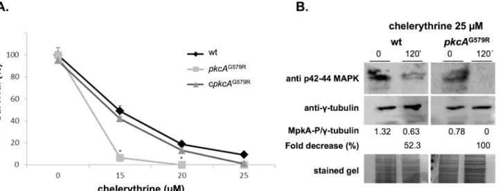
These results suggested thatpkcAinA.fumigatusis also essential for viability. As an attempt to further investigate the effects ofpkcAinactivation inA.fumigatus, we looked for PKC pharma-cological inhibitors that acted directly on the catalytic kinase domain, which is separate from the C1B motif located in the PKC regulatory domain [22]. Clerythrine (CHE) is described as a potent inhibitor of PKC interacting with the catalytic domain of PKC and has been shown to inhibitS.cerevisiaegrowth [60,61]. So, we reasoned that it would be possible to observe a syn-thetic lethality effect derived from the partial loss of function ofpkcAin thepkcAG579Rmutant in the presence of this drug. The MIC determination for CHE inA.fumigatusperformed in YAG medium according to the Clinical and Laboratory Standards Institute (CLSI) M38-A2 protocol was 20μM (data not shown). Accordingly, we exposed conidia form wild-type, mutant and complemented strains to different concentrations of CHE below and above the MIC for 24 hours and then measured the ability of the cells to resume growth in fresh media. Viability was determined after 24 hours of recovery and quantified in a MTT assay. Survival of the mutant strain was reduced in comparison to the wild-type and complemented strains at lower drug concentration (Fig 1A). The wild-type and complementing strains showed about 20% survival at the MIC concentration, while the mutant was not viable (0%). In order to dem-onstrate that this was due to inhibition ofA.fumigatusPkcA by CHE, MpkA phosphorylation was examined by Western blot assay. The results show that there was no phosphorylation of MpkA after 120 minutes of CHE exposure in thepkcAG579Rstrain and a reduction of about 52.3% in the wild-type strain (Fig 1B). These results indicate that the G579R mutation in C1B domain of PkcA combined with the inhibition of the catalytic site of the protein lead to a fungi-cidal effect.
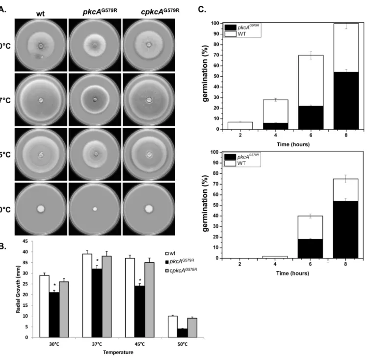
An initial phenotypic analysis ofA.fumigatus pkcAG579Rmutant strain displayed decreased radial hyphal growth at all temperatures tested and this difference was more evident at 45°C and 50°C. The radial growth rate of the complemented strain was similar to the wild-type strain, but not thepkcAG579Rmutant, in both complete (Fig 2A) and minimal medium (data Fig 1. Chelerythrine treatment leads to a fungicidal effect ofA.fumigatus pkcAG579Rmutant.(A) 2x10³
conidia from each strain were grown for 24 hours in liquid MM at 30°C in the presence of the indicated concentration of chelerythrine. After growth, cells were centrifuged, washed in pre-warmed medium and allow to recover in fresh MM medium for an additional 24 hours. After this time, survival was determined via the MTT assay comparing the formanzan salt formation at each time point in comparison to the untreated control.*p0.01. (B) Western blot for MpkA phosphorylation. Wild-type and
pkcAG579Rstrains were grown for 16 h in YG at 37°C and then chelerythrine (25μM) was added, or not (control), to the medium for 120 minutes. Anti-phospho-p44/42 MAPK antibody was used to detect the phosphorylation of MpkA. Theγ-tubulin antibody and Coomassie Brilliant Blue stained gels were used as loading sample controls. Signal intensity was quantified using the ImageJ software by dividing the intensity of phosphorylatred MpkA/γ-tubulin and expressed as arbitrary units.
not shown). At 37°C the radial growth rate of thepkcAG579Rmutant was approximately 10% less than the wild-type and complemented strains, while at 45°C there was a 50% decrease in growth rate (Fig 2B). In addition to the significant defects in vegetative growth, conidia form thepkcAG579Rmutant showed a significant delay in germination. When germination was mon-itored in complete medium, there was a significant decrease (p0.01) in the emergence of the germ-tubes in thepkcAG579Rmutant of 50% at both 37°C and 45°C after 8 hours of growth (Fig 2C). ThepkcAG579Rmutant did not exhibit altered pigmentation or differences in the Fig 2. PkcA contributes to thermotolerant growth and germination inA.fumigatus.1x104conidia of each strain were inoculated onto the center of a YAG medium and radial growth was measured after 3 days at the indicated temperatures (A-B). 1x10⁶conidia of each strain were inoculated in 2 ml liquid YG culture and incubated at 37°C and 45°C during 2, 4, 6 and 8 hours before the percentage of germination was evaluated (C). Experiments were performed in triplicate and the results are the mean±standard deviation.*Statistically significant by Student’s T-test (p0.01).
number of conidia formed either at 37°C or 45°C (data not shown). Taken together, these mor-phological analyses of thepkcAG579Rmutant suggest that the impaired function ofpkcA inter-feres in the hyphal growth and elongation.
The
pkcA
gene is required for the maintenance of cell wall integrity,
proper conidial architecture and oxidative stress tolerance
The involvement of thepkcAG579Rmutation in cell wall biogenesis inA.fumigatuswas further tested by utilizing several known cell wall perturbation agents. ThepkcAG579Rmutant showed increased sensitivity to the cell wall stressing agents Congo Red (CR), Calcofluor White (CFW) and the echinocandin anidulafungin (AF), plus to Sodium Dodecyl Sulfate (SDS), which dis-rupts the plasma membrane and lyses cells with membrane defects, and caffeine (CAF) that has been extensively used to probe signal transduction and cell integrity phenotypes inS. cere-visiae[62] (Fig 3A). Lower concentrations of CR and CFW were also tested (S2 Fig). A subtle increase in the sensitivity of thepkcAG579Rmutant to the chitin synthase inhibitor, nikkomycin Z (NKZ), and to fluconazole (FLUC) was observed in comparison to the wild-type and comple-mented strains (Fig 3B). ThepkcAG579Rmutant also showed decreased tolerance to EDTA (Fig 3A).
CR and CFW displayed the most pronounced effects on thepkcAG579Rmutant. The increased sensitivity to CR and CFW phenotype could be partially rescued on minimal media containing the osmotic stabilizer sorbitol (1.2 M). In addition, this phenotypic rescue was com-plete in the presence of AF, but not evident in the presence of CAF (S3 Fig). We did not observe lysis or swelling phenotype of conidia during germination for thepkcAG579Rmutant in the presence of any cell wall damaging compound (data not shown), although excessively swelling during germination was observed inA.nidulans calc2mutant [21]. As an indirect approach to investigate the cell wall composition and architecture of thepkcAG579Rmutant strain, hyphae of thepkcAG579R, wild-type and complemented strains, which were grown in liquid YG medium, were subjected to enzymatic digestion by Lallzyme MMX. The protoplasts were then harvested and counted. Digestion of thepkcAG579Rmutant yielded about 4 times more proto-plasts than the wild-type and complemented strains (Fig 4A) indicating that thepkcAG579R
mutant cell wall was much more susceptible to the enzymatic degradation, suggesting that it possessed a modified carbohydrate composition or it is more enzymatically accessible. To investigate if this carbohydrate modification affected the cell wall organization, the fungal cell wall chitin andβ-1,3-glucan levels were assessed in these three strains via CFW staining and soluble dectin-1 staining, respectively. The intensity of CFW staining per fungal area was simi-lar inpkcAG579Rmutant, wild-type and complementing strains (Fig 4B). In contrast, the
pkcAG579Rmutant has higher levels ofβ-1,3-glucans (about 2.7- fold) than the wild-type and complementing strains (Fig 4C). Collectively, these results suggest that thepkcAG579Rmutant had altered cell wall organization. In addition to providing structural integrity to the fungal cell, the cell wall is the main structure forming interactions with host tissues. So, we subse-quently investigated the ability of thepkcAG579Rmutant to promote cell-to-cell adhesion mea-suring the initial stages of biofilm formation of mature hyphae in polystyrene plates. Biofilm formation was significantly reduced (60%) in thepkcAG579Rmutant (Fig 4D) when compared to the wild-type and complemented strains. Collectively, these results suggest that the PkcA signaling cascade influences adherence to polystyrene and biofilm formation inA.fumigatus
and complemented strains (Fig 4E). This alteration seems to be an event unrelated to the conidial developmental program in thepkcAG579Rmutant, since no abnormalities in the Fig 3. Growth phenotypes of thepkcAG579Rmutant.(A) 1x10⁵conidia of each strain were inoculated onto the center of a solid YG plates supplemented with different cell wall perturbing agents: Congo Red (CR); Calcofluor White (CFW); caffeine (CAF); anidulafungin (AF); Sodium Dodecyl Sulfate (SDS) and ethylenediaminetetraacetic acid (EDTA). (B) The indicated number of conidia was spotted onto solid YG plates supplemented with nikkomycin Z (NKZ) and fluconazole (FLUC). The plates were incubated for 3 days (A) or 2 days (B) at 37°C.
conidiophore structures were observed when slide cultures were analyzed under lactophenol cotton blue staining (data not shown).
Based on the cell wall-related phenotypes of thepkcAG579Rmutant, we next examined the susceptibility of this strain, plus the wild-type and complemented strains, to the major classes of antifungal compounds in clinical use [63] via the E-test method, including voriconazole, amphotericin B and caspofungin (Fig 5). No differences were observed between the three strains for amphotericin B (data not shown) but there was a larger area of growth inhibition for thepkcAG579Rmutant on voriconazole (endpoints 0.125 and 0.094 for wild-type andpkcAG579R
Fig 4.pkcAG579Rmutant strain presents increased protoplasts production andβ-1,3-glucan content, lower biofilm formation and reduced hydrophobin content.(A) The digestion mixture included the Lallzyme MMX and 100 mg of mycelium into 50 ml of osmotic stabilizer solution at the indicated incubation times. Protoplasts were quantified using a Neubauer chamber. Detection of the exposed chitin (B) andβ-1,3−glucan (C) contents on the cell surface. Conidia were cultured in liquid MM to the hyphal stage, UV-fixed, and stained with CFW or soluble dectin-1to detect the content of exposed chitin andβ-glucan, respectively. The intensity of staining was calculated by averaging the amount of staining to the total area of each fungal cell using ImageJ software. These experiments were performed in triplicate, and the results are displayed as mean values±standard deviation.*Student’s T-test (p0.05). (D) Biofilm formation was evaluated by crystal violet absorbance at 570 nm and expressed as the percentage of adhesion considering 100% for the wild-type strain. Experiments were performed in quintuplicate and the results are the mean±standard deviation. (*p0.01). (E) Silver stained SDS-PAGE profiles of HF acid extracts from resting conidia showing the hydrophobin concentration of the wild-type,pkcAG579Rand complementing strains. The number of conidia subjected to the hydrophobin extraction was validated by CFU counting on solid YG medium. RodAp corresponds to the native RodA and RodAp*to partially degraded or processed RodA.
mutant strain, respectively) and caspofungin (endpoints 0.094 and 0.023 for the wild-type
pkcAG579Rmutant strain, respectively) on the buffered RPMI medium. The caspofungin inhibi-tion zone was clearer in thepkcAG579Rmutant in contrast to the wild-type and complemented strains. Caspofungin is fungistatic toA.fumigatusforming a known incomplete clearing area around the caspofungin strip [43,64,65]. However, the clearer area obtained for thepkcAG579R
mutant exposed to caspofungin may suggest that it can be fungicidal uponpkcApartial loss-of-function.
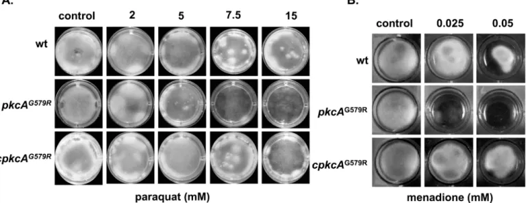
Next, we investigate the response of the mutant to oxidative damage to get information if thepkcAcould be indirectly involved in Reactive Oxygen Species (ROS) tolerance. The
pkcAG579Rmutant showed increased sensitivity to paraquat (PQT) and menadione in compari-son to the wild-type and complemented strains (Fig 6). However, no effect was observed in the presence of H2O2and diamide (data not shown). Taken together, these phenotypic analyses of thepkcAG579Rmutant suggest that theA.fumigatus pkcAaffects the ability of the fungus to Fig 5. ThepkcAG579Rmutant is sensitive to antifungal drugs.Antifungal susceptibility using E-test gradient strips for voriconazole (A) and caspofungin
(B).
form normal cell wall and itsbona fidetolerance to oxidative damage elicited primarily by anion superoxide.
The activation of the MAP kinase MpkA upon cell wall stress is defective
in the
pkcA
G579Rmutant
InS.cerevisiae, the PKC1 CWI pathway is activated by the exposure to cell wall stressing agents culminating with the Mpk1p (Slt2p) phosphorylation [66–68]. Although inA.fumigatus phos-phorylation of the Mpk1p homologue, MpkA is achieved upon cell wall stress caused by Gluca-nex and CFW [16,20] the requirement of PkcA in this signaling cascade was not investigated. Accordingly, the hypothesis that PkcA is acting upstream the MpkA for the activation of the CWI pathway was tested by the evaluation of the phosphorylation of MpkA upon CR-induced cell wall stress. CR was used to induce cell wall stress in the wild-type andpkcAG579Rstrains, for which the latter was highly sensitive to 300μg/ml of this compound (Fig 3AandS2 Fig). The MpkA protein was phosphorylated in response to CR in the time course experiment pre-senting an increase of about 32%, 35% and 31% after 15, 30 and 60 minutes, respectively (Fig 7), indicating that CR can induce the CWI pathway inA.fumigatus. In contrast, the phosphor-ylation of MpkA was lower in thepkcAG579Rmutant showing a decrease of about 10%, 19% and 31% after 15, 30 and 60 minutes post- CR treatment. Interestingly, the phosphorylation of MpkA was 47.3% higher in thepkcAG579Rstrain prior to the CR treatment. These results indi-cate that PkcA signals MpkA phosphorylation in theA.fumigatusCWI pathway.
Loss-of-function of
pkcA
contributes to the Endoplasmic Reticulum (ER)
stress condition during cell wall stress
A connection between the cell wall integrity and the ER stress pathways has been described, since mutants defective in the activation of Unfolded Protein Response (UPR) are more sensi-tive to cell wall stressing agents [43,46] and reviewed in [69,70]. This relationship is supported by the fact that UPR maintains ER functionality via balancing the income of new proteins with processing capacity, which in turn influences secretion, cell wall homeostasis and ultimately Fig 6. Growth phenotypes of thepkcAG579Rmutant strain in the presence of oxidative damage.The strains were grown in liquid MM in 24-well plates
supplemented with in the indicated concentrations of paraquat (A) or menadione (B) during 48 hours at 37°C.
fungal pathogenesis [43,71,72]. InA.fumigatusthe canonical UPR is activated when mis-folded proteins accumulate in the ER lumen leading to the oligomerization and activation of the cytosolic kinase and RNAse domains of the ER stress sensor IreA [46], which excises an unconventional intron (20 nucleotides) from the cytoplasmic mRNA ofhacA(uninduced;
hacAu) [43]. The spliced mRNA ofhacA(induced;hacAi) can be translated in a fully functional bZIP transcription factor, which migrates to the nucleus allowing the transcription of genes related to the ER folding capacity [70]. Based on this information, we wanted to determine how thepkcAmutant is affected by ER-stressing agents such as DTT (dithiothreitol), tunica-mycin (TM) and BFA (brefeldin A), which activate UPR by: unfolding proteins by reducing disulfide bonds, inhibiting N-linked glycosylation and interfering the anterograde protein transport form ER to the Golgi, respectively [73]. ThepkcAG579Rmutant was more sensitive to all ER stressing agents (Fig 8A–8C). In addition, the activation ofhacAtranscription factor was assessed by RT-PCR according to the assay previously described by [46]. Both wild-type and
pkcAG579Rstrains were exposed to 1 mM of DTT during 60 minutes, which can activate UPR Fig 7. ThepkcAG579Rmutant strain is defective in activating the CWI pathway.Conidia of the wild-type and mutant strains were grown overnight in liquid
YG medium and cell wall stress was induced by exposure to CR for 0, 15, 30 and 60 minutes. Samples were collected at the indicated time points for western blot preparation. Phosphorylated and the total fraction of MpkA were detected using anti-phospho p44/42 and anti-p44/42 MAPK antibodies, respectively.γ -tubulin antibody and Coomassie Brilliant Blue stained gel were used as loading sample controls (A). Densitometry analysis of western blots using the ImageJ software presenting the difference in the phosphorylated MpkA/non-phosphorylated MpkA ratio in the wild-type and mutant strain expressed as percentage in the histogram (B).
Fig 8. Cell wall stress caused by loss ofpkcAis connected to UPR signaling inA.FUMIGATUS.(A) The indicated number of conidia of each strain was spotted on solid YG medium supplemented with BFA (brefeldin A) or in a 24 well plate (1x10⁴conidia/well) containing the indicated concentration of TM (tunicamycin) (B); or (C) DTT. (D) Analysis ofhacAmRNA splicing in the wild-type andpkcAG579Rmutant. Overnight liquid cultures of each strain were subjected to cell wall stress by the addition of CR during the indicated amount of time before total RNA was extracted. RT-PCR was used to amplify thehacA
inA.fumigatus[43,46]. Interestingly, in thepkcAG579Rmutant strain, the UPR is activated under a condition without DTT as shown by the 2- fold increase in thehacAitranscript (Fig 8D, upper graph). To evaluate the mRNA abundance ofhacAiduring cell wall stress, the strains were exposed to CR. In the wild-type strain exposed to CR there was an increased accumula-tion of thehacAimRNA (2.4- fold mRNA increase) after 15 minutes and a drop to 1.4- fold after 30 and 60 minutes. ThehacAimRNA accumulation in thepkcAG579Rmutant was 2.2-fold higher than the wild-type strain prior to CR treatment (Fig 8D, lower graph). In addition, levels ofhacAiwere maintained at high levels (2.8-, 2.8- and 2.2- fold increase) post CR treat-ment (Fig 8D). These results suggest that cell wall defects elicited by the PkcA-mediated CWI pathway are directly connected to the ER stress and also provide experimental data reinforcing the hypothesis that the secretion system is fundamental for maintaining cell wall homeostasis. This idea is also supported by the fact that the increased sensitivity of thepkcAG579Rmutant strain to DTT and BFA can be rescued to the wild-type levels when the medium is supple-mented with D-sorbitol as osmotic stabilizer (S4 Fig).
The
pkcA
G579Rmutant shows an altered expression pattern of cell wall
integrity-related genes upon exposure to cell wall stress
TheA.fumigatus pkcAG579Rstrain was shown to be more sensitive to several cell wall damaging compounds, and CR was able to induce a PkcA-dependent CWI pathway leading to the phos-phorylation of MpkA (Fig 7). Hence, we used a transcriptional approach to investigate the cause of the increased sensitivity of thepkcAG579Rmutant to cell wall stressing agents, via mon-itoring the expression of genes known to be involved in the cell wall biosynthesis and reinforce-ment inA.fumigatus. Hyphae from wild-type andpkcAG579Rmutant strains were exposed to CR for 0, 15, 30 and 60 minutes prior to the quantification of mRNA abundance using real time RT-PCR. Accordingly, we examined the mRNA levels ofpkcA,mpkA, and the Afu3g08520 (here namedrlmA) that encodes the putative homologue ofS.cerevisiae RLM1
transcription factor [14,18]. In addition, the expression of the main cell wall biosynthesis genes:α-1,3-glucan synthases (agsA-C);β-1,3-glucan synthase (fksA); 1,3-β-glucanosyl trans-ferases (gelA-C), and the eightA.fumigatuschitin synthases (chsA-GandcsmB), were also investigated (Figs9and10).
Upon CR treatment,pkcAwas slightly induced, whilempkAtranscript levels were increased in the wild-type strain after 30 and 60 minutes CR treatment. Interestingly, there was lower abundance ofmpkAtranscripts in thepkcAG579Rmutant compared to the wild-type strain (0.3- and 1.3- fold increase after 30 minutes, respectively). This correlated with the lower level of MpkA phosphorylation under the same growth conditions (Fig 7). Likewise, levels of the
rlmAtranscription factor were up-regulated in the wild-type strain in a time-dependent man-ner, whilerlmAexpression was significantly lower in thepkcAG579Rmutant in the presence or absence of CR (Fig 9), suggesting that the activation ofrlmAin response to CR depends on the PkcA-MpkA pathway.
Regarding the cell wall biosynthetic enzymes assessed in this study, all the genes showed increased expression in the wild-type strain, at different levels for at least one time point, except for the chitin synthaseschsB,chsDthat were repressed andcsmBthat showed similar expres-sion during CR exposure. InterestinglychsBandchsDwere expressed at a higher level in the
(non-splicedhacAform =hacAu) yielding fragments of 100 and 120 nucleotides, respectively. PCR products were separated in an acrylamide gel and stained. As a control, both wild-type and mutant strain were treated with 1 mM of DTT to induce UPR. Genomic DNA was also used as template in the same reactions yielding only the 120 bp band (upper panel). Images were subjected to densitometric analysis of pixel intensity and normalized by the expression of
tubArun as loading control in each RT-PCR.
pkcAG579Rmutant in the absence of CR (2.6- and 3.3- fold increase, respectively), indicating that these two genes may be critical for the cell to cope with cell wall stress imposed solely by the loss-of-function topkcA(Fig 10). Levels of the catalytic subunit of theβ-1,3-glucan synthasefksAwere also increased in a time dependent-manner in the wild-type strain reaching 2.2- fold induction after 60 minutes of CR exposure. In contrast different values were observed in thepkcAG579Rmutant especially after 30 and 60 minutes of treatment (0.8- and 1.5- fold induction). This was expected due to the increased sensitivity of thepkcAG579Rmutant to cas-pofungin and anidulafungin, which indicated alteredβ-1,3-glucan andβ-1,6-glucan synthesis. However, the induction offksAin thepkcAG579Rmutant after 15 minutes of CR treatment (2.1- fold increase) indicates that it was not completely affected by thepkcAmutation, suggest-ing that either residual activity of PkcAG579Rcan stimulate its activation, orfksAexpression does not depend on PkcA-MpkA signaling cascade.
Relevant differences in the transcript levels were observed in the threeα-1,3-glucan synthase encoding genes,agsA,agsBandagsC. Interestingly,agsAmRNA was maintained at low levels regardless of CR exposure, but the transcription ofagsBandagsCwere increased after CR stress in the wild-type strain. The largest fold change was observed foragsC, which was increased over 4.5-, 8.2- and 9.1- fold after 15, 30 and 60 minutes of CR treatment respectively. On the other hand, the transcription ofagsBandagsCin thepkcAG579Rmutant was markedly reduced to 0.2- and 1.9- fold, respectively after 60 minutes of incubation (Fig 9). These results suggest that the transcription ofα-1,3-glucan synthase depends on the PkcA-MpkA signaling trans-duction inA.fumigatus.
The mRNA levels of the genes encoding cell wall remodeling enzymes,gelA,gelBandgelC, were also analyzed. No significant differences were observed between the wild-type and
pkcAG579Rmutant strains for thegelBandgelC. In contrast, thegelAtranscript levels in the wild-type strain, but not thepkcAG579Rmutant, increased in all time points post CR treatment. This also indicates that the transcription ofgelAdepends on PkcA kinase activity. Chitin synthase encoding genes,chsAandchsG, showed higher induction after CR exposure (6.9- and 5.1- fold, respectively at 60 minutes) and this induction was clearly lower in thepkcAG579R
mutant at the same time point (2.8- and 3.2- fold, respectively). The same was observed for
chsCand chsFin all time points (Fig 10). Interestingly,csmBshowed similar expression profile in thepkcAG579Rmutant in comparison to the wild-type strain, except for the timepoint 30 minutes where there was a 3.9- fold induction. Taken together, these data suggest that PkcA contributes to the transcriptional regulation of several but not all cell wall-related genes inA.
fumigatus.
The
pkcA
G579Rmutant elicits increased TNF
−
α
levels and macrophage
recognition but it is not able to disturb virulence in a low dose
neutropenic murine infection model
ThepkcAG579Rmutant has an impaired response to cell wall stress and alterations to the conidia surface. Thus, we reasoned if these phenotypes would impact on the host immune response. So, we used Bone Marrow Derived Macrophages (BMDMs) to measure the levels of the proinflammatory cytokine Tumor Necrosis Factor alpha (TNF−α) released by these cells Fig 9. Transcriptional analysis of cell wall-related genes.The wild-type, andpkcAG579Rstrains were grown for 24 hours in YG medium and transferred to fresh pre-warmed YG with either 0 or 300μg/ml of CR and grown for a further 15, 30 and 60 minutes. mRNA abundance for each gene was assessed by real time RT-PCR and normalized toβ-tubulin. Fold increase in each strain represents the normalized mRNA abundance relative to the wild-type strain at time point 0 (i. e. prior to CR exposure). Data represent the average value of at least three biological replicates, each repeated in duplicate in the same run and standard deviation.*p0.05.
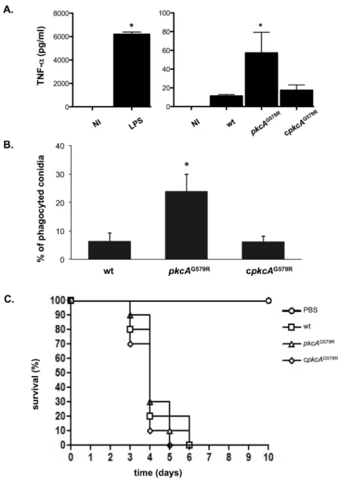
after co-incubation withA.fumigatusconidia. TNF−αis an important inflammatory mediator secreted by macrophages when exposed toA.fumigatus[74,75]. BMDMs co-cultured with
pkcAG579Rstrain showed about 4- fold higher TNF−αproduction than the wild−type or the complemented strain (Fig 11A). We also test the ability of BMDM in internalizing wild-type, Fig 10. Transcriptional analysis of chitin synthase genes.The wild-type, andpkcAG579Rstrains were grown for 24 hours in YG medium and transferred to fresh pre-warmed YG with either 0 or 300μg/ml of CR and grown for a further 15, 30 and 60 minutes. mRNA abundance for each gene was assessed by real time RT-PCR and normalized toβ-tubulin. Fold increase in each strain represents the normalized mRNA abundance relative to the wild-type strain at time point 0 (i. e. prior to CR exposure). Data represent the average value of at least three biological replicates, each repeated in duplicate in the same run and standard deviation.*p0.05.
mutant and completing strain conidia. About 25% of thepkcAG579Rstrain conidia were phago-cytized after the 80 minutes of co-incubation. In contrast, only 6.3% of the wild-type and com-plementing strains conidia were internalized (Fig 11B). These results suggest that the effect caused bypkcAG579Rmutation on theA.fumigatusCWI is important for macrophage recogni-tion and inflammatory responses. To determine the possible influence ofpkcAon virulence, the wild-type,pkcAG579Rmutant and the complemented strains were compared in a murine Fig 11.pkcAG579Rstrain presents no virulence attenuation in a mouse model but activates innate immunity againstA.fumigatus.(A) Secretion of TNF-αfrom bone marrow derived macrophages (BMDM) after co-incubation withA.fumigatushyphae of wild-type,pkcAG579Rand complementing strains. TNF-α levels were quantified by ELISA from the culture supernatant after 18 hours of BMDMs infection. Data show average±standard deviation (*p0.005). NI: Non−infected; LPS: lipopolysaccharide (positive control). (B) Phagocytosis index is increased in thepkcAG579Rmutant strain (average±standard deviation,*p0.01 compared to the wild−type and the complemented strain). (C) Comparative analysis of wild type,pkcAG579R and complemented strains in a neutropenic murine model of invasive pulmonary aspergillosis.
model for invasive aspergillosis [56]. In spite of the higher TNF-αelicited production and the increased phagocytosis levels in thepkcAG579Rmutant, all the strains caused the same absolute mortality and similar survival kinetics after the immunosuppressive regimen and intranasal infection after five to six days (Fig 11C). These results indicate that although PkcAG579R influ-ences macrophage recognition, it does not affect fungal survival and virulence in the immuno-compromised mammalian host.
Discussion
The fungal CWI pathway that orchestrates cell wall biosynthesis and remodeling is evolution-ary conserved in many fungal organisms. InA.fumigatus, this pathway is partially character-ized, but how it is activated is not completely understood yet. Here, we characterized thepkcA
gene ofA.fumigatus, which encodes the protein kinase C, the apical kinase of the CWI path-way. Previous studies assessing the function of PKC in other human pathogenic fungi have revealed the importance of this gene in fungal pathogenesis, affecting the biosynthesis of viru-lence determinants such as melanin and a capsule inC.neoformans[28,76,77] and the toler-ance to azoles inCandida albicans[78]. PKC is not essential in these two fungal pathogens although inC.neoformansΔPKC1is only viable in the presence of D-sorbitol [77,79]. In con-trast, our results suggest thatpkcAis essential inA.fumigatus, as was the case inA.nidulans. ThecalC2mutation described in theA.nidulans pkcAindicated that this gene is involved in the maintenance of the cell wall since it showed phenotypes related to cell wall defects [21]. ThepkcAG579Rmutant inA.fumigatuscarries the same mutation as theA.nidulans calC2
strain. This mutation is located beside the C-terminal limit of the cysteine-rich C1B regulatory domain, very close to the PkcA catalytic site, which can explain the cell wall defects and the fungicidial effect of the PKC inhibitor, chelerythrine associated with this mutation.
Here, thepkcAG579Rmutation resulted in restricted vegetative growth. InA.fumigatus, the downstream components of the CWI, (mpkA,bck1andmkk2) and some upstream mechano-sensors such asmidAalso play a role in the hyphal elongation [16,20]. Collectively, these observations indicate that impairment of the CWI pathway impacts not only the cell wall com-position, but also filamentous growth. Interestingly, asexual sporulation was not affected in the mutant strain, although we have observed that surface rodlets and hydrophobins were signifi-cantly reduced in thepkcAG579Rmutant. It was demonstrated that theα-glucan synthase triple mutant (ΔagsA-C) also presented an amorphous surface without the rodlet layer [80]. Here, we observed lower levels ofagsA-Cexpression upon CR exposure for thepkcAG579Rmutant in comparison to the wild-type strain (Fig 9) indicating that PkcA might govern conidia surface structure organization, possibly representing an indirect effect of the perturbed CWI pathway resulting from the PkcA mutation. Recently we have shown that theA.fumigatusphosphatase
sitA, modulates the activity of PkcA. TheΔsitAmutant shows decreased protein kinase C activ-ity, increased sensitivity to cell wall damaging agents and also reduced adhesion properties and lower hydrophobin content [81]. The results presented here strongly suggest an epistatic rela-tionship betweensitAandpkcAand support the idea that SitA is contributing to the regulation ofA.fumigatusCWI pathway. We are currently investigating this hypothesis.
ofpkcAG579Rhyphae, (ii) the partial growth recovery on D-sorbitol, and (iii) the increasedβ -1,3-glucan levels in the mutant strain. These results emphasize that changes inβ-glucan and chitin architecture or chitin/β-glucan cross-links, are possibly responsible for the observed phe-notypes. However, quantitative analyses of cell wall components inpkcAG579Rmutant stay for future investigation.
A.fumigatus pkcAcoordinates responses to different cell wall stresses and also to oxidative damage. Our results showed the increased sensitivity of thepkcAG579Rmutant to oxidative stressing agents, paraquat and menadione, but not to H2O2.C.neoformansPKC1 is essential for the protection against both oxidative and nitrosative stresses caused by H2O2, diamide or NaNO3, respectively. InC.neoformansthe oxidative stress signal results in PKC1 and subse-quently Mpk1 activation. However, the other downstream PKC1 elements in theC.neoformans
CWI pathway are dispensable for resistance to oxidative and nitrosative stresses [77]. Consid-ering the mode of action of menadione and paraquat are different from H2O2[84–86],pkcA seems to be involved in ROS detoxification caused by superoxide anion (•O2-) which is involved in mitochondrial function, instead of the hydroxyl radicals (•OH) generated by H2O2 via Fenton reaction [87].
The UPR has been demonstrated to be involved in virulence in several human pathogenic fungi, includingA.fumigatus. UPR influences important cellular functions such as morphol-ogy, thermo-tolerance, hypoxia, iron acquisition and tolerance to cell wall stress [43,46,71,72,
88]. Here we demonstrated that impairment of theA.fumigatusPkcA-MpkA signaling path-way resulted in the activation of the UPR, indicating that both systems occur concomitantly and are reciprocally affected [69]. This connection between the CWI pathway and the UPR has previously been observed inS.cerevisiae, but only for different components of the CWI path-way, including the mechanosensor Mid1 and Mpk1 [72]. InA.nidulansthe involvement pro-tein kinase C in farnesol tolerance is related to the UPR [34], while elevated MpkA
phosphorylation also correlates with increased tolerance to ER stress/ UPR inducing com-pounds [89]. Therefore, our results suggest that this connection is conserved in fungi and rein-forces the role of PkcA in theA.fumigatusCWI pathway.
There is a great deal of speculation about which genes involved in the synthesis and remod-eling of the cell wall are under the control of the canonical CWI pathway inAspergillusspecies. Here, we aimed to identify some genes that are controlled by theA.fumigatusPkcA-MpkA pathway since the coordinated expression of these genetic determinants ultimately contributes to the virulence of this pathogen. It has been pointed out differences betweenS.cerevisiaeand
A.fumigatus[18,69]. InS.cerevisiae, the canonical CWI pathway is responsible for the control of the vast majority of cell wall related genes [90], including proteins, which are not present in
A.fumigatus, such as the Pir family proteins. Likewise, the cell wall polysaccharide,α -1,3-glu-can, is not produced byS.cerevisiaeorC.albicans, but is the most abundant polysaccharide in the cell wall ofA.fumigatus[7]. So, it is reasonable that these differences in carbohydrate com-position between these fungi, may explain the variations in the signaling processes. Here, the clearest example of PkcA-dependent gene expression was observed for theagsA-Cgenes, as expression was dramatically reduced in thepkcAG579Rmutant. A detailed study by Fujioka
et al. (2007) in theA.nidulanswild-type,ΔmpkAandΔrlmAstrains, challenged with micafun-gin, revealed thatagsB(the homologue ofA.fumigatus agsA) was also down-regulated in both mutant backgrounds. In contrast to our results, inA.nidulans,agsA(the homologue ofA.
fumigatus agsB) was up-regulated in both mutants.A.nidulansdoes not have an ortholog ofA.
fumigatus agsC, which was the most highly expressedα-glucan synthase encoding gene post exposure to CR in this study.A.niger agsAis the closest homolog ofA.fumigatus agsC. InA.
virulence attenuation inA.fumigatus[92,93]. As expected, the regulation of seven chitin synthase encoding genes was also altered in thepkcAG579Rmutant. However onlychsC,chsF
andchsGshowed significantly lower transcription after CR treatment in the mutant strain. These results suggest that regulation of chitin synthase encoding genes occurs either in a CWI-dependent and-inCWI-dependent manner.
Echinocandins are the only class of drugs currently targeting an enzyme involved in cell wall biosynthesis, i.e.fksA. ThefksAgene was induced upon cell wall stress caused by micafun-gin or CFW in bothA.nidulansandA.niger, respectively [18,94]. Recent data indicated that
fksAis not an essential gene inA.fumigatus, which may partly explain the limited antifungal activity of the echinocandins againstA.fumigatus[95]. Here, changes infksAmRNA levels after CR treatment were similar in the wild-type andpkcAG579Rstrains. These results suggest that the basal levels of transcription of this gene and the increased expression after CR exposure occurs independently of theA.fumigatusPkcA. Altogether, the transcriptional profiling shown here indicates that a minor transcriptional response can be attributable to the canonical CWI pathway viaA.fumigatusPkcA-MpkA cascade. This suggests that one or more signaling path-ways might govern the expression of some CWI genes in response to CR. Therefore, function-ally distinct signaling pathways induce a more efficient response to cope with a specific cell wall stress and damage. This conclusion can also support the observations that the fungus can alter its cell wall content in response cell wall stress that perturb either glucan or chitin synthe-sis [96–98].
We have shown thatpkcAG579Rmutant has normal virulence in a murine model of pulmo-nary aspergillosis and is increasingly recognized by macrophages. TNF−α, one of the key inflammatory mediators secreted by macrophages in response to fungal hyphae, was increased inpkcAG579Rmutant compared to wild-type and complemented strain. This pro−inflamatory cytokine plays an important role in the induction of the innate immune response toA. fumiga-tus[74,75]. The possible modifications in the cell wall carbohydrates and proteins, reduced hydrophobin content, and other modifications in thepkcAG579Rmutant cell wall could contrib-ute to an increased recognition of the fungus by dectin-1 receptor. This could favor its
increased phagocytosis by alveolar macrophages, and consequently the increased TNF-α pro-duction, since the binding of the receptor dectin-1 to fibrillarβ-1,3 glucan is a major host fun-gal interaction duringin vitroandin vivoinfection [99–102]. Among the fungal pathogens where PKC homologs have been characterized via gene deletion/mutation studies, only inC.
albicansdid the PKC mutant show virulence attenuation [78]. Despite the cell wall related phe-notypes presented by theA.fumigatus pkcAG579Rmutant, no difference in the virulence was observed in comparison to the wild-type and complemented strains. We cannot rule out the possibility that this can be the result of the partial loss-of-function caused by the point muta-tion in the C1B domain of the polypeptide. However, MpkA, which is part of the regulatory cir-cuit of PkcA, was also dispensable for virulence, although the cell wall related phenotypes of theΔmpkAstrain were even more severe thanpkcAG579Rmutation [19]. These observations strongly indicate that redundant mechanism may account for the CWI inA.fumigatus. This idea can also be supported by the increased immune response observed in macrophages exposed topkcAG579Rstrain conidia, which is not directly translated into virulence attenuation.