PONTIFÍCIA UNIVERSIDADE CATÓLICA DO RIO GRANDE DO SUL
FACULDADE DE ODONTOLOGIA
PROGRAMA DE PÓS-GRADUAÇÃO EM ODONTOLOGIA
DOUTORADO EM ORTODONTIA E ORTOPEDIA FACIAL
ANDRÉ WEISSHEIMER
MÉTODOS DE AVALIAÇÃO TRIDIMENSIONAL DO
COMPLEXO CRANIOFACIAL EM TOMOGRAFIA
CONE BEAM
Profa. Dra. Luciane Macedo de Menezes.
Orientadora
ANDRÉ WEISSHEIMER
MÉTODOS DE AVALIAÇÃO TRIDIMENSIONAL DO
COMPLEXO CRANIOFACIAL EM TOMOGRAFIA CONE BEAM
Tese apresentada ao Programa de Pós-graduação em Odontologia da Faculdade de Odontologia da Pontifícia Universidade Católica do Rio Grande do Sul como requisito final para obtenção do título de Doutor em Odontologia, na Área de Concentração Ortodontia e Ortopedia Facial.
Orientadora: Profa. Dra. Luciane Macedo de Menezes
DADOS INTERNACIONAIS DE CATALOGAÇÃO NA PUBLICAÇÃO (CIP)
Alessandra Pinto Fagundes Bibliotecária CRB10/1244 W433m Weissheimer, André
Métodos de avaliação tridimensional do complexo craniofacial em tomografia cone beam / André Weissheimer. Porto Alegre, 2013.
64 f. : il.
Tese (Doutorado) – Faculdade de Odontologia, Pós-Graduação em Odontologia, Área de concentração em Ortodontia e Ortopedia Facial, PUCRS, 2013.
Orientadora: Profa. Dra. Luciane Macedo de Menezes.
1. Ortodontia. 2. Tomografia Computadorizada de Feixe Cônico. I. Menezes, Luciane Macedo de. II. Título.
ANDRÉ WEISSHEIMER
MÉTODOS DE AVALIAÇÃO TRIDIMENSIONAL DO
COMPLEXO CRANIOFACIAL EM TOMOGRAFIA CONE BEAM
Esta Tese foi julgada adequada para obtenção do Título de “Doutor em
Odontologia” e aprovada em sua forma final pelo Programa de Pós-Graduação em
Odontologia da Faculdade de Odontologia da Pontifícia Universidade Católica do
Rio Grande do Sul.
Porto Alegre, 28 de novembro de 2013.
_______________________________________
Prof. Dr. Alexandre Bahlis
Diretor da Faculdade de Odontologia da PUCRS
Banca Examinadora:
___________________________________________________________
Profa. Dra. Luciane Macedo de Menezes
Orientadora
Pontifícia Universidade Católica do Rio Grande do Sul
___________________________________________________________ Prof. Dr. Roberto Rocha
Universidade Federal de Santa Catarina
___________________________________________________________ Profa. Dra. Maria Bernadete Sasso Stuani Universidade de São Paulo
___________________________________________________________ Prof. Dr. Rogério Belle de Oliveira
Pontifícia Universidade Católica do Rio Grande do Sul
______ _____________________________________________________
Prof. Dr. Márcio Sarroglia Pinho
“Na vida, o difícil se faz imediatamente, e o impossível, logo em seguida...”
Dedico essa tese a Deus e a minha família.
O Deus, por me conduzir com fé e esperança nos tortuosos
caminhos da vida, dando-me a força e coragem para alcançar
meus objetivos.
Aos meus pais, Carlos e Neiva, por serem exemplos de dedicação
e amor aos seus filhos e que muitas vezes abriram mão de seus
próprios sonhos para que eu realizasse os meus. Não existem
palavras que expressem a eterna gratidão, orgulho e amor e que
tenho por vocês.
Ao meu irmão Rafael, pela verdadeira amizade, confiança e
apoio em todos os momentos da minha vida, e principalmente,
pela ajuda concedida durante esses anos de pós-graduação, serei
eternamente grato. O amor de nossos pais nos fez irmãos, por
AGRADECIMENTOS
Em especial, a Professora Doutora Luciane Macedo de Menezes, pelo amor à
ciência, pela abnegação e dedicação ao ensino, por seu exemplo de conduta profissional,
ética e moral. Por ser a principal responsável pela minha formação ortodôntica e
constante incentivadora do meu aprimoramento profissional, tenho-a como um exemplo
a ser seguido. Agradeço a confiança em mim depositada, a constante presença e auxílio
durante a realização desta tese e por brindar-me com um ótimo convívio e amizade.
Obrigado por ter me proporcionado uma das mais incríveis experiências da minha vida,
o doutorado sanduíche nos Estados Unidos. Faltariam linhas para descrever a
admiração, os agradecimentos e expressar minha eterna gratidão. Muito Obrigado.
A Professora Dra. Susana Maria Deon Rizzatto, pelo exemplo de dedicação ao
ensino da Ortodontia. Por me fazer enxergar a Ortodontia de uma maneira diferente,
com outros olhos. A sua grande experiência e satisfação em ensinar certamente
estimularam a minha busca pelo conhecimento. Expresso minha gratidão pela amizade
e por ter me ensinado as lições que não estão nos livros.
Aos meus colegas de Doutorado, Milton Farret, Tatiana S. Gonçalves e
Leandro Berni Osório pela troca de experiências, aprendizado e ótimo convívio.
Aos Professores do departamento de Ortodontia da Ostrow School of Dentistry
da University of Southern California (USC). Em especial ao Dr. Glenn T. Sameshima,
Dr. Peter Sinclair, Dr. Hongsheng Tong, Dr. Dan Grauer e Dra. Reyes Enciso, por
terem me proporcionado um aprendizado único, contribuindo sobremaneira para minha
formação pessoal e profissional.
Aos Residentes da Ortodontia da University of Southern California, Dovi
Prero, Hany Shaker Youssef, Scott Morita, Ryan Hecht, Nathan Coughlin, Michael
Meru, John Gerome, George Abichaker, Virginia Pham and Stefan Alexandroni. Em
especial, ao John Pham pelo companheirismo, ajuda e amizade. A sua genialidade foi
fundamental para a realização dessa tese.
Ao Leonardo Koerich de Paula, pela amizade, ajuda e colaboração na
realização desse trabalho. Muito obrigado.
A professora Lucia H. S. Cevidanes, pela ajuda e colaboração nesta tese.
Ao professor Dr. Rogério Belle de Oliveira, pelos conhecimentos cirúrgicos
transmitidos e pela verdadeira amizade e confiança. Muito obrigado pelos conselhos,
orientações e ajuda durante os anos do mestrado e doutorado.
Aos Professores da disciplina de Ortodontia da Universidade Federal de Santa
Catarina, Doutores, Roberto Rocha, Arno Locks, Gerson L. U. Ribeiro, Daltro Ritter
Carla Derech e Márcio Cardoso expresso a imensa gratidão por me propiciarem uma
formação ortodôntica sólida, desde a graduação até a especialização. Se hoje posso
enxergar mais longe é porque me apoiei em ombros de gigantes, gigantes da Ortodontia.
Em especial ao Prof. Dr. Roberto Rocha, por ter contribuído de forma marcante
na minha formação ortodôntica. Foi uma honra e privilégio poder aprender com um dos
melhores professores de Ortodontia que já conheci. Tenho-o como exemplo a ser seguido.
Sou profundamente grato pelos conselhos e estímulo inicial para a realização do
doutorado sanduíche nos EUA.
Ao Prof. Dr. Marco Antônio de Oliveira Almeida, Profa. Dra. Cátia Cardoso
Abdo Quintão, Dr. Felipe de Assis Ribeiro Carvalho e Dr. Gustavo Zanardi, por me
abrirem as portas do departamento de Ortodontia da UERJ e compartilharem os seus
conhecimentos sobre análises 3-dimensionais com tomografia cone beam.
Aos Professores, Dr. Eduardo Martinelli S. De Lima, Dr. Ernani M.
Marchioro, Dr.Telmo Berthold, Dr. Fernando Martinelli e demais professores da F.O.
de Odontologia da PUCRS.
A estimada amiga Fabiane Azeredo pela amizade sincera e ajuda durante os
anos do mestrado e doutorado.
Aos meus grandes amigos Maurício Mezomo, Guilherme Bernd, Clécio
Camargo, Maurício Brunetto e Fabiano Azambuja.
A Faculdade de Odontologia da Pontifícia Universidade Católica do Rio
Grande do Sul, representada por seu diretor, Prof. Dr. Alexandre Bahlis.
A Comissão Coordenadora do Programa de Pós-graduação em Odontologia da
PUCRS, representada pela Profa. Dra. Ana Maria Spohr. A participação nessa
comissão como representante discente me possibilitou aprendizados únicos.
Aos funcionários da secretaria de Pós-Graduação, Ana, Davenir e Paulo, pela
ajuda, amizade e excelente convívio durante os anos do mestrado e doutorado.
Aos demais funcionários, Alessandra, Carla, Glaci, Denise, Daena, Rejane e
Neuza, que sempre auxiliaram nas atividades realizadas durante o curso.
A CAPES, pelo apoio financeiro disponibilizado através da bolsa flexível,
Resumo
Introdução: o objetivo desta tese foi avaliar a acurácia de programas para análises 3D do
complexo maxilofacial em tomografia computadorizada cone beam (TCCB). Com esse
propósito, dois estudos foram realizados. O primeiro estudo avaliou a precisão e acurácia de 6
programas para avaliação do volume da via aérea superior em TCCB. O segundo estudo
objetivou validar um método rápido de superposição 3D de TCCB. Método: no estudo 1, a
amostra consistiu de 33 pacientes e 1 Phantom de acrílico da orofaringe (PAO), escaneados
co o to ógrafo iCAT. O olu e co hecido do PAO foi utilizado co padrão ouro .
Segmentação semiautomática da orofaringe dos pacientes (OP) e do PAO foi realizada com os
programas Mimics, ITK-Snap, OsiriX, Dolphin3D, InVivo Dental e Ondemand3D. No estudo 2, a
amostra consistiu de TCCB de 18 pacientes. Em 10 pacientes, como padrão de comparação, a
TCCB pré-tratamento foi reorientada espacialmente, salva como TCCB reorientada, e então
superposta na imagem original. Em 8 pacientes, sendo 4 sem crescimento e 4 em crescimento,
foram superpostas as TCCB e pós-tratamento. A acurácia da superposição foi avaliada através
de inspeção visual e mensurada através do programa CFM com mapas coloridos. Resultados:
no estudo 1, as segmentações com o Mimics, Dolphin3D, OsiriX e ITK-Snap mostraram menos
de 2% de erro no volume do PAO e co paração ao padrão ouro . O O de a d3D e o InVivo Dental apresentaram mais de 5% de erro no volume do PAO em comparação ao
padrão ouro . As seg e tações da OP co o ITK-Snap, Mimics, OsiriX e Dolphin3D foram estatisticamente diferentes (P<.05) em comparação ao InVivo Dental. Não houve diferença
estatisticamente significante (P>.05) entre os programas InVivo Dental e o OnDemand3D. No
estudo 2, o erro da superposição das TCCB reorientadas, medidas através dos mapas coloridos
foi menor que 0,5mm. O erro da superposição das TCCB pré-tratamento e pós-tratamento
para pacientes com e sem crescimento, na região da base do crânio, foi menor que 0,5 mm e
considerado aceitável e clinicamente insignificante. Conclusão: no estudo 1, todos os 6
programas foram precisos, mas apresentaram erros no volume da segmentação da OP.
Mimics, Dolphin3D, ITK-Snap e OsiriX foram considerados similares e mais acurados em
comparação ao InVivo Dental e Ondemand3D. No estudo 2, o método de superposição
baseado em voxel avaliado foi reproduzível em diferentes condições clínicas, rápido e
potencialmente aplicável para pesquisa e prática clínica.
Palavras chave: Tomografia computadorizada cone beam, Superposição 3D, Craniofacial, Via
Abstract
Introduction: this thesis aimed to evaluate the software accuracy for 3D analysis of
craniofacial complex in cone beam computed tomography (CBCT). With this purpose, two
studies were performed. The first study evaluated the precision and accuracy of 6 imaging
software programs for measuring the upper airway volume in CBCT. The second study
aimed to validate a fast method for 3D superimposition of CBCT. Methods: in study 1, the
sample consisted of 33 growing patients and 1 oropharynx acrylic phantom (OAP), scanned
with iCAT scanner. The known OAP volume was used as gold standard (GS).
Semi-automatic segmentations of the patients´ oropharynx (OP) and OAP was performed using
Mimics, ITK-Snap, OsiriX, Dolphin3D, InVivo Dental and Ondemand3D software programs.
In study 2, the sample consisted of CBCT scans of 18 patients. For 10 patients as a gold
standard, the spatial position of the pretreatment CBCT volume was reoriented, saved as a
reoriented volume, and then superimposed to the original image. For 8 patients, 4
non-growing and 4 non-growing patients, pre and post-treatment scans were superimposed.
Superimposition accuracy was assessed by visual inspection and measured by using the
CMF application and expressed via color maps. Results: in study 1,the OAP segmentations
with Mimics, Dolphin3D, OsiriX and ITK-Snap showed less than 2% error in volume
compared to the GS. Ondemand3D and InVivo Dental showed more than 5% error
compared to the GS. In the OP segmentation, ITK-Snap, Mimics, OsiriX and Dolphin3D
were statistically significantly different (P<.05) from InVivo Dental. No statistical difference
(P>.05) was found between InVivo Dental and OnDemand3D. In study 2, Superimposition
error of the spatial reorientation as measured by the color-coded surface distances was
less than 0.5mm. Superimposition error of pre and post treatment scans for both growing
and non-growing patients at the cranial base were smaller than 0.5 mm, which was
considered acceptable and clinically insignificant. Conclusion: in study 1, all 6 imaging
software programs were reliable but showed errors in the volume segmentation of OP.
Mimics, Dolphin3D, ITK-Snap and OsiriX were similar and more accurate than InVivo Dental
and Ondemand3D. In study 2, the voxel-based superimposition method evaluated was
reproducible in different clinical conditions, time-efficient and potentially applicable for
both research and clinical practice.
Keywords: Cone-Beam Computed Tomography, 3D superimposition, Craniofacial,
Sumário
1. Introdução ... 10
2. Proposição ... 13
2.1 Objetivo geral ... 13
2.2 Objetivos específicos ... 13
3. Artigo 1 ... 14
4. Artigo 2 ... 28
5. Discussão complementar ... 44
6. Conclusão ... 59
6.1 Conclusão geral ... 59
6.2 Conclusões específicas ... 59
7. Referências bibliográficas complementares ... 60
1. Introdução
A Ortodontia é o ramo da ciência especializada no diagnóstico, prevenção e
correção das irregularidades dento-faciais. O diagnóstico ortodôntico constitui a base
fundamental da especialidade, sendo indispensável na busca dos objetivos do
tratamento ortodôntico. As radiografias convencionais representam parte da
documentação ortodôntica e ainda são utilizadas para o diagnóstico, análise do
crescimento e desenvolvimento craniofacial e avaliação dos resultados do tratamento.
As radiografias são representações bidimensionais (2D) de estruturas tridimensionais
(3D), provendo informações diagnósticas limitadas. Além do mais, possuem
magnificação da imagem, distorções geométricas e sobreposição de estruturas
anatômicas. A introdução da tomografia computadorizada (TC) pelo Engenheiro Inglês
Godfrey Hounsfield juntamente com o físico norte-americano Allan Comark, lhes valeu
o prémio Nobel de Medicina de 1979 e possibilitou a inclusão da terceira dimensão no
diagnóstico por imagem. Apesar de utilizada rotineiramente na medicina, o emprego
da TC na Odontologia era limitado em função do alto custo financeiro e dose de
radiação. Essas desvantagens foram superadas pela introdução da tomografia
computadorizada de feixe cônico ou cone beam (TCCB). Avanços em termos de custo
financeiro, dose de radiação e acurácia vêm contribuindo para a consolidação da TCCB
na Odontologia, especialmente nas áreas da ortodontia e cirurgia buco-maxilo-facial.
A avaliação morfológica do complexo craniofacial, utilizando TCCB, pode ser
realizada de diversas maneiras, dentre as quais, destaca-se a técnica de segmentação.
A segmentação pode ser definida como o processo de construção de modelos virtuais
3D (chamados de segmentações) de uma determinada estrutura. Significa separar um
elemento específico, através da remoção de outras estruturas de não interesse, para
melhor visualização e análise. Modelos virtuais 3D de estruturas esqueléticas, tecidos
moles e vias aéreas podem ser construídos por meio de segmentação, servindo como
base para análises quantitativas e qualitativas do complexo craniofacial. A qualidade
final da segmentação depende de aspectos relacionados à qualidade da imagem e da
11
interesse. O problema é que, em teoria, programas com algoritmos distintos podem
gerar segmentações diferentes, comprometendo o controle de qualidade das análises
tridimensionais. Uma recente revisão sistemática da literatura relatou 18 diferentes
programas contendo módulos destinados à visualização, mensuração e análise das vias
aéreas superiores.
1Um único estudo de validação comparou a acurácia e precisão de 3
programas para avaliação das vias aéreas superiores.
2Isso significa que a grande
maioria dos programas está sendo comercializados sem estudos de validação. Essa
falta de controle na qualidade dos programas para diagnóstico 3D em TCCB pode ter
implicações sérias, especialmente nos casos de apneia obstrutiva do sono. Por isso, é
imprescindível a realização de estudos para validação de programas destinados ao
diagnostico 3D das vias aéreas superiores.
Além do diagnóstico das vias aéreas, a TCCB vem modificando a maneira como
ortodontistas e cirurgiões avaliam os resultados dos tratamentos
ortodôntico-cirúrgicos. A superposição de radiografias cefalométricas, embora ainda utilizada, esta
sendo substituída gradativamente pela superposição de TCCB, em função dos avanços
nos algoritmos para registro de imagens. Atualmente, a superposição tridimensional
de TCCB, com registro na base do crânio, é considerada o método mais avançado para
avaliação 3D dos efeitos do tratamento ortodôntico-cirúrgico. Dentre os métodos
utilizados para superposição de TCCB, os mais acurados são os que utilizam os valores
dos tons de cinza ou densidade dos voxels (voxel-based). Cevidanes et al, em 2005,
introduziu na Odontologia o primeiro método automático para superposição de TCCB,
com base nos tons de cinza dos voxels da base do crânio.
3Esse método é baseado na
teoria da informação mútua, e foi descrito por Maes et al, em 1997.
4No método
original descrito por Cevidanes et al, dois programas são requeridos. O primeiro é
utilizado para construir modelos virtuais 3D da base do crânio e o segundo programa
para realizar a superposição das tomografias através da transformação rígida (rotação
e translação). Esse método tem sido utilizado em varias pesquisas e é considerado o
método mais avançado para superposição de TCCB e avaliação 3D dos tratamentos.
3,5-9Embora esse método utilize programas gratuitos, desenvolvidos pelo departamento de
ciências da computação da
University of North Caroline (UNC)
, a complexidade no
disponibilizados, tornam a utilização deste método de superposição restrita a alguns
centros de pesquisa. Outra desvantagem é o longo tempo dispendido (45 a 60 minutos
para um usuário treinado) para completar o processo de superposição das tomografias
(segmentação + superposição). Essas limitações dificultam a popularização e aplicação
desse método na prática clínica diária por ortodontistas e cirurgiões.
Em 2010, os pesquisadores Jeong-Ho Choi, da
Seul National University
, e James
Mah, da
University of Southern California (USC)
, desenvolveram um novo método de
superposição automática de TCCB com base nos tons de cinza dos voxels
(voxel-based).
10Esse método de superposição também é baseado na teoria da informação
mútua e está presente no programa Ondemand3D (Cybermed, Seoul, Korea),
disponível comercialmente. A superposição dos volumes de TCCB é realizada de forma
rápida (10 a 15 segundos) com apenas um programa e sem a necessidade de
segmentação prévia como no método descrito por Cevidanes et al. Esse método tem
um grande potencial para aplicação na prática diária em função da rapidez e facilidade
do processo superposição. Embora esse método tenha sido apresentado por Choi e
Mah,
10e ter sido utilizado em algumas pesquisas recentes,
11-13estudos de validação
13
2. Proposição
2.1 Objetivo geral
Avaliar a acurácia de programas para avaliação tridimensional da região
craniofacial em tomografia computadorizada cone beam.
2.2 Objetivos específicos
2.2.1 Avaliar e comparar a precisão e acurácia de 6 programas para avaliação do
volume da via aérea orofaríngea em tomografia computadorizada cone beam
(Artigo 1).
2.2.2 Avaliar a acurácia de um método rápido de superposição tridimensional de
3. Artigo 1
Titulo:Imaging software accuracy for 3-dimensional analysis of the upper airway
4. Artigo 2
Fast 3-dimensional superimposition of CBCT volumes: validation study
André Weissheimer, Luciane M. Menezes, Leonardo Koerich de Paula, John Pham and Lúcia H. S. Cevidanes.
Abstract
Objectives: Our aim was to test and validate a method for fast 3D superimposition of CBCT
volumes in growing patients and adults. Methods: The sample consisted of CBCT scans of 18 patients. For 10 patients as a gold standard, the spatial position of the pretreatment CBCT volume was reoriented, saved as a reoriented volume, and then superimposed to the original image. For 8 patients, 4 non-growing and 4 growing patients, pre and post-treatment scans were superimposed. Fully automated voxel-based superimposition was performed, with registration at the anterior cranial base using the Ondemand3D program. The registration process took 10-15s. The fit of the cranial base superimposition was verified by visual inspection of all corresponding anatomic structures in all semi-transparent multi-planar cross-sectional slices and by semi-transparent overlays of the surface models. Virtual 3D surface models of the skull were generated via a standardized segmentation protocol by using ITKSNAP program. Superimposition errors in the reoriented models and result of treatment for the treated cases were evaluated with 3D surface distances in color-coded maps. Results:
Superimposition error of the spatial reorientation as measured by the color-coded surface distances was less than 0.5mm. Superimposition error of pre and post treatment scans for both growing and non-growing patients included small segmentation errors and surface distances at the cranial base were smaller than 0.5 mm, which was considered acceptable and clinically insignificant. Conclusion: The voxel-based superimposition method evaluated was reproducible in different clinical conditions, time-efficient and potentially applicable for both research and clinical practice.
Introduction
Cone beam computed tomography (CBCT) has become a well-established diagnostic
tool in dentistry.1-9 In Orthodontics and Oromaxillofacial Surgery, CBCT now allows clinicians to
better identify and distinguish treatment outcomes. While 2D cephalometric superimposition
is the conventional method used to evaluate growth and treatment outcomes, improvements
in image registration algorithms have made the superimposition of CBCT volumes the
state-of-the-art technique for treatment outcome assessments.
In medical imaging, the process of spatially superimposing three-dimensional (3D)
images obtained from different imaging modalities is also called image registration, or,
29
shape and position of the craniofacial components have occurred with time and treatment is
challenging and requires knowledge of the different types of superimposition. The three basic
types of superimposition algorithms are: (1) point-landmark based, (2) surface based and (3) voxel based.11 Point-landmark based methods are done through the superimposition of corresponding or homologous points that are manually input by the user. The precision of this
method is highly user dependent and most anatomic surfaces do not present well-defined
landmarks in the 3 planes of the space. Surface-based methods require construction of 3D
surface models from corresponding CBCT volumes, and surface segmentation algorithms
depend on the software determination of quality of the scan and software dependent. Voxel-based superimposition methods use an optimized function that measures the similarity of all geometrically corresponding voxels pairs for a user-defined anatomic region.11 This method
compares voxel by voxel of non-changing reference structures in volumetric data, does not
depend on landmark identification, as in point-landmark based methods, and is not limited by
segmentation errors, as in surface based methods.
Cevidanes et al12 were the first to introduce into dentistry the voxel-based method for
fully automated 3D superimposition of CBCT volumes. The method proposed in that study was
based on mutual information theory11 and required construction of surface models of the reference structure prior to the registration steps. Application of those methods for both
growing and non-growing subjects has been described in the literature. The main difference
between the growing and non–growing patient algorithms in those studies was that the growing patients algorithm took changes in scale (growth and/or response to treatment) into
account without applying it, while the non-growing algorithm performed a rigid transformation
with 6 degrees of freedom (x, y, z of translation and rotation) to superimpose the CBCT
volumes.5-7,12-14 The main drawback of that method was that the registration process was time
consuming (45 to 60min), and lacked a clinician friendly user-interface and visualization tool.
In 2010, Choi and Mah15 introduced a new method for cranial base superimposition method that does not require construction of 3D surface models prior to the registration
process. Choi et al also added volume and slice visualization capabilities, providing a clinician
friendly user interface. This method also uses the principle of mutual information for image
registration.11 The result was a new, commercially available, software program (Fusion module
of Ondemand3D, Cybermed, Seoul, Korea) that performs CBCT volume superimpositions faster
(10 to 15s) and while requiring fewer steps. While some research studies,16-18 have applied the superimposition method introduced by Choi and Mah,15 in the Ondemand3D software, there
has been no previous validation study of this method for fast CBCT volume superimposition in
The aim of this study was to evaluate the fast 3D superimposition of CBCT volumes
present in the fusion module of Ondemand3D software program. Specifically first, this study
tested whether there are differences when the same CBCT volumes, with different spatial
orientations, are superimposed at the anterior cranial base. Second, this study tested whether
there are differences in the cranial base when longitudinal CBCT volumes of growing patients
and adults, which also present maxilla-mandibular changes due to growth and/or treatment
response, are superimposed at the anterior cranial base.
Method
This study was approved by the ethical committee of Pontifical Catholic University of
Rio Grande do Sul (PUCRS) in Brazil. The sample consisted of CBCT scans of 18 patients. For 10
patients as a gold standard, the spatial position of the pretreatment CBCT volume was
reoriented, saved as a reoriented volume, and then superimposed to the original image. For 8
patients, 4 non-growing and 4 growing patients, pre and post-treatment scans were
superimposed. The 10 pre-treatment scans had mean age of 11.4 ±1 years. The 4 non-growing
adult patients (mean age 26.3 ± 5.7 years) had CBCT scans taken pre and 1 year post- surgery.
The 4 growing patients (mean age 9.5 ± 1.8 years) had CBCT scans taken pre and
post-treatment with rapid maxillary expansion (RME). The CBCT scans were available from datasets
obtained for clinical purpose using the i-CAT scanner (Imaging Sciences International, Hatfield,
Pa) set at 120 kVp, 8 mA, large field of view, and scan time of 40 seconds. The images were
reconstructed with 0.25-mm slice thickness and exported as Digital Imaging and
Communications in Medicine (DICOM) files.19
Creating CBCT volumes with different spatial orientation
The DICOM files, corresponding to pre-treatment CBCT scans of 10 growing patients,
were imported into Ondemand3D software program (version 1.0.9.1451; Cybermed, Seoul,
Korea) and organized in the database management module. Each CBCT volume was opened,
the patient head was reoriented in the space (translation and rotation) to a different spatial
position and exported as a new DICOM file. Thus, for each original CBCT volume a second CBCT
volume was created, with the same voxel size but with different head orientation (Figure 1A, B
and C). This procedure was performed for all 10 pre-treatment CBCT volumes, creating 10
31
CBCT volumes superimposition
For the fully automatic voxel-wise rigid registration, we used the fusion module in
Ondemand3D software program. Axial, sagittal and coronal slice views of the volumes were
used to select the anatomic structures of the anterior cranial base in the registration of the
original and reoriented CBCT volumes (Figure 1A and B) and the registration of the longitudinal
scans. Next, the Ondemand3D automated registration tool was used to perform the rigid
registration (translation and rotation) that optimally aligned the reoriented CBCT volume to
the original CBCT volume, using the intensity of the grey levels for each voxel in the anterior
cranial base of the 2 CBCT volumes (Figure 1C and D). The superimposition process took a total
of 10-15 seconds to complete.
Superimposition assessment
The precision of the superimposition was verified by using two methods: (1) by
qualitative visualization, by a single observer A.W., of the semi-transparent axial, sagittal and
coronal cross-sectional slices of all corresponding anatomic structures between original and
reoriented scans, and between the two time point scans. (Figures 1D, 5A e 6A) and (2) by
quantification of the surface distances using closest point color maps on 3D surface models
(Figures 1E, 4, 5B and 6B).20,21 To measure the outcomes of the registration, after the registration process, the superimposed CBCT volumes were exported as DICOM files and
imported into ITK-SNAP software program (www.itksnap.org) for segmentation.22 Automatic
segmentation was performed, by a single observer L.K.D., for each CBCT volume in 4 different
steps: mandible, maxilla, frontal bone/anterior cranial fossa and middle cranial fossa. The
segmentation was then exported as a stereo lithography file (STL) and converted to an Open
Inventor file (IV) using STL to SGI Inventor 2.0 Utility Beta (developed by Reuben Reyes,
[email protected]). The surface models were then opened in the Cranio-MaxilloFacial
application software (CMFapp, developed at the M. E. Müller Institute for Surgical Technology
and Biomechanics, University of Bern, Bern, Switzerland)23 that calculates the closest point surface distance between thousands of surface triangles in the 3D surface models and the
color-coded surface distance maps allow quantification of the registration errors (Figure 1E, 4,
5 , 6 and 7).
Results
The superimposition results for the original and reoriented CBCT scans of 10 growing
patients are shown in Figures 1 to 4. The visualization of the semi-transparent axial, sagittal
adequate registration of the cranial base structures in all axial, sagittal and coronal slices
(Figure 1 to 3). The quantification of the superimposition errors by color-coded surface
distances revealed that the error was less than 0.25mm (Figure 1 to 4).
The superimposition results for longitudinal scans of growing patients and adults are
shown in Figures 5, 6 and 7. The visualization of the semi-transparent axial, sagittal and
coronal cross-sectional slices of all corresponding anatomic structures confirmed the adequate
registration of the extremely complex cranial base structures, such as the ethmoidal air cells.
The quantification of the superimposition errors by color-coded surface distances revealed
that distances in the cranial base between registered surface models were less than 0.5mm for
most regions (Figures 5B, 6B and 7).
Discussion
CBCT is currently a well-established diagnostic tool for 3-dimensional evaluation of
patients, especially in orthodontic-surgical cases. This study is the first to validate a method for
fast CBCT volume superimposition in growing patients and adults.
Improvements in image registration algorithms have led to the development of new
methods for CBCT volume superimposition. McCance et al proposed a method for cranial base
superimposition with CT scan using 5 landmarks on areas not affected by surgery in
non-growing patients.24 Kawamata et al suggested a similar method, rotating semi-transparent pre-
and post-surgery models aiming to overlap then in the same structure.25 These point
landmark-based methods for CBCT superimposition are also used in the current versions of
Dolphin3D (Dolphin imaging & Management Solutions, Chatsworth, Calif) and InVivo Dental
(Anatomage, San Jose, Calif) software programs. However, point landmark-based methods are
observer dependent. The method introduced by Cevidanes et al was observer independent
and did not rely on specific landmarks.12 However, it was time consuming and lacked a clinician
friendly user-interface and visualization tool.
This study validated the superimposition method introduced by Choi and Mah for
voxel-based registration using Ondemand3D software.15 This study method for
superimposition of longitudinal CBCT volumes in growing patients and adults has advantages
over previously used voxel-based methods. 5-7,12,14 The advantages include time efficiency
(takes about 10 to 15 seconds), and user-friendly software interface suitable for clinical
application. As demonstrated in our study, the CBCT volume superimpositions are fast even
33
registration process does not require previous segmentation to designate the area of
superimposition (i.e. cranial base). Ondemand3D software also enables CBCT volume
superimpositions with registration at areas outside the cranial base that can be potentially
applied for regional superimpositions not tested in this study. Park et al have recently used
regional superimpositions with Ondemand3D software to evaluate the condylar head
remodeling after bi-maxillary surgery by using rigid registration at the condylar neck and
posterior ramal area of pre and post-operative CBCT images.16
In this validation study, the construction of standardized 3D surface models and
part-comparison analysis with color maps confirmed the accuracy of this voxel-based
superimposition method. The differences observed in the color-coded surface distance maps
for the 10 pre-treatment CBCT scans were minimal, less than 0.25 mm (Figures 1-4). However,
superimposition of the same CBCT volumes, with different spatial orientation, should be
evaluated with caution because they have the same grey level intensity and no modifications
by growth and/or treatment. The challenge for image registration is to superimpose CBCT
volumes of patients with craniofacial modifications due to the normal growth and/or
treatment response in different time points. In these situations, the CBCT volumes may have
different grey level intensity, field of view, and dental/skeletal components modified by
growth and/or treatments, making the registration process difficult and prone to fail.
Additionally, CBCT scans obtained with different scanners may have different grey levels,26
which could affect the superimposition process. For these reasons, this study tested
superimpositions of longitudinal CBCT volumes with one-year interval of growing patients
treated with RME and adult patients treated with orthognathic surgery.The green color-code
(0mm surface distances) in most part of the anterior cranial base shows the adequate
superimposition for both growing patients and adults. Some areas of larger surface distances,
as shown in black, red and blue color-coded areas that are displayed in the superior view of
the cranial base (Figures 5B, 6B and 7), do not represent superimposition errors, as confirmed
by the visualization of transparent multi-planar cross-sectional slices and by
semi-transparent overlays of the surface models (Figure 7). Since the CBCT obtained at different
time points have differences in the grey level for the same anatomical structures (i.e. cranial
base), the automatic segmentation was not performed by using the same threshold interval.
This variation introduced by the user input to define the properties of 3D surface model
creation leads to small surface variations in the cranial base. Additionally, areas with low
gray-scale contrast such as the ethmoidal air cells are not included automatically in the
segmentation. In these situations, manual editing were necessary and produced slightly
that explaining the black, red and blue areas observed in the internal surface of the cranial
base.
For some scans, the registration of the two CBCT volumes failed, requiring 2 or 3
repeated procedures. In these cases, it was necessary to resize the selection area for
registration (anterior cranial base) in both primary and secondary volumes in order to obtain
an adequate superimposition. In a previous study, Alexandroni et al18 also reported similar
failed attempts using the same superimposition method to evaluate orthognathic surgery
skeletal prediction in 44 patients. Those authors used only visual inspection to verify adequate
CBCT volume superimpositions. The registrations that required repeated procedures in this
study could be due to the small area used for CBCT volume registration in our study (anterior
cranial base) in comparison to the global anatomic structure of the dry skull used by Lee et al.27
In addition, the amount of the misalignment between the CBCT volumes may have some
influence in the superimposition process. According to Pluim, et al, when using registration
methods based in mutual information theory, results may be suboptimal, or may even fail, if
the initial misalignment of the two images is large or if the overlay region of the two images is
relatively small.28 For this reason, we recommend to perform a quick manual superimposition (manual registration tool) previously to the automated superimposition. Lee, et al27 evaluated the accuracy of CBCT image registration with Ondemand3D by using a dry skull with titanium
markers, simulating different head orientations, although their research did not evaluate the
accuracy of longitudinal CBCT superimposition. The location of titanium markers were
assessed by two examiners and the distance between the markers were calculated using the
3D coordinates in the Ondemand3D software program. The superimposition mean error was
0.39mm (±0.142mm) and there were no significant differences in the dry skull
superimpositions.
A potential limitation of the Ondemand3D software used in this study is that it is only
available commercially, whereas the method previously proposed by Cevidanes et al can be
done using readily available open-source software programs. Nada, et al29 have tested the reliability of another commercial voxel based method using the Maxilim software (Medicim,
Mechelen, Belgium) for CBCT volumes superimposition on the anterior cranial base and
zygomatic arch of 16 surgical orthodontic patients. The authors reported small average
superimposition errors, however the closest-point color-coded maps used for quantification of
errors do not allow local quantification of surface errors and minimize surface differences.
The current study showed that Ondemand3D software performs a reliable, automated
35
construction of surface models and quantification of treatment outcomes are limited, and
other multiple software packages performed these image analysis steps in this study.
Quantification of topographic bone remodeling and displacement requires construction of
surface models that can be still time consuming. For quantification of changes overtime even
though most commercial and open-source software use closest point surface distance,
recently, a new method for 3D evaluation of topographic changes, shape correspondence, has
been introduced. The shape correspondence method provides the magnitude and direction of
the displacement and can be used to identify and quantify the morphological changes in
patients with temporomandibular joint osteoarthritis,9,30 asymmetry assessments8 and for evaluation of orthopedic or orthognathic surgery outcomes.31
In summary, this study was the first to test and validate a voxel-based method for fast
CBCT volumes superimposition. Several software packages have been introduced in the market
every year, many of them with 3D image superimposition modules. However, there is no
established protocol or algorithms for fast and precise 3D image registration used in
orthodontics and maxillofacial fields.
Conclusion
Ondemand3D voxel based superimposition method was precise and not
time-consuming. The fast 3D superimposition method of CBCT volumes validated in this study may
be applied for longitudinal assessment of growing patients and adults and is suitable for both
research and clinical routine, in the orthodontic and maxillofacial surgery fields.
References
1. Kapila S, Conley RS, Harrell WE, Jr. The current status of cone beam computed tomography imaging in orthodontics. Dentomaxillofac Radiol 2011;40:24-34.
2. Weissheimer A, Menezes LM, Mezomo M, Dias DM, Lima EMS, Rizzatto SMD. Immediate effects of rapid maxillary expansion with Haas-type and hyrax-type expanders: A randomized clinical trial. Am J Orthod Dentofacial Orthop 2011;140:366-376.
3. Weissheimer A, Menezes LMd, Sameshima GT, Enciso R, Pham J, Grauer D. Imaging software accuracy for 3-dimensional analysis of the upper airway. Am J Orthod Dentofacial Orthop 2012;142:801-813.
5. Cevidanes LHC, Heymann G, Cornelis MA, DeClerck HJ, Tulloch JFC. Superimposition of 3-dimensional cone-beam computed tomography models of growing patients. Am J Orthod Dentofacial Orthop 2009;136:94-99.
6. Cevidanes LHC, Motta A, Proffit WR, Ackerman JL, Styner M. Cranial base superimposition for 3-dimensional evaluation of soft-tissue changes. Am J Orthod Dentofacial Orthop 2010;137:S120-S129.
7. Cevidanes LHC, Tucker S, Styner M, Kim H, Chapuis J, Reyes M et al. Three-dimensional surgical simulation. Am J Orthod Dentofacial Orthop 2010;138:361-371.
8. Cevidanes LHS, Alhadidi A, Paniagua B, Styner M, Ludlow J, Mol A et al. Three-dimensional quantification of mandibular asymmetry through cone-beam computerized tomography. Oral Surg Oral Med Oral Pathol Oral Radiol Endod 2011;111:757-770.
9. Cevidanes LHS, Hajati AK, Paniagua B, Lim PF, Walker DG, Palconet G et al. Quantification of condylar resorption in temporomandibular joint osteoarthritis. Oral Surg Oral Med Oral Pathol Oral Radiol Endod 2010;110:110-117.
10. Li G, Xie H, Ning H, Capala J, Arora BC, Coleman CN et al. A novel 3D volumetric voxel registration technique for volume-view-guided image registration of multiple imaging modalities. Int J Radiat Oncol Biol Phys 2005;63:261-273.
11. Maes F, Collignon A, Vandermeulen D, Marchal G, Suetens P. Multimodality image registration by maximization of mutual information. IEEE Trans Med Imaging 1997;16:187-198.
12. Cevidanes LHC, Bailey LJ, Tucker Jr GR, Styner MA, Mol A, Phillips CL et al. Superimposition of 3D cone-beam CT models of orthognathic surgery patients. Dentomaxillofac Radiol 2005;34:369-375.
13. Cevidanes LHS, Bailey LTJ, Tucker SF, Styner MA, Mol A, Phillips CL et al. Three-dimensional cone-beam computed tomography for assessment of mandibular changes after orthognathic surgery. Am J Orthod Dentofacial Orthop 2007;131:44-50.
14. Cevidanes LHS, Styner MA, Proffit WR. Image analysis and superimposition of 3-dimensional cone-beam computed tomography models. Am J Orthod Dentofacial Orthop 2006;129:611-618.
15. Choi J, Mah J. A new method for superimposition of CBCT volumes. J Clin Orthod 2010;44:303-312.
16. Park S-B, Yang Y-M, Kim Y-I, Cho B-H, Jung Y-H, Hwang D-S. Effect of Bimaxillary Surgery on Adaptive Condylar Head Remodeling: Metric Analysis and Image Interpretation Using Cone-Beam Computed Tomography Volume Superimposition. J Oral Maxillofac Surg 2012;70:1951-1959.
17. Park S-B, Yoon J-K, Kim Y-I, Hwang D-S, Cho B-H, Son W-S. The evaluation of the nasal morphologic changes after bimaxillary surgery in skeletal class III maloccusion by using the superimposition of cone-beam computed tomography (CBCT) volumes. J Craniomaxillofac Surg 2012;40:e87-e92.
37
19. DICOM digital imaging and communications in medicine. Rosslyn,Va: National Electrical Manufacturers Association (NEMA); 2011.
20. De Clerck H, Nguyen T, de Paula LK, Cevidanes L. Three-dimensional assessment of mandibular and glenoid fossa changes after bone-anchored Class III intermaxillary traction. Am J Orthod Dentofacial Orthop 2012;142:25-31.
21. Nguyen T, Cevidanes L, Cornelis MA, Heymann G, de Paula LK, De Clerck H. Three-dimensional assessment of maxillary changes associated with bone anchored maxillary protraction. Am J Orthod Dentofacial Orthop 2011;140:790-798.
22. Yushkevich PA, Piven J, Hazlett HC, Smith RG, Ho S, Gee JC et al. User-guided 3D active contour segmentation of anatomical structures: Significantly improved efficiency and reliability. NeuroImage 2006;31:1116-1128.
23. Chapuis J, Schramm A, Pappas I, Hallermann W, Schwenzer-Zimmerer K, Langlotz F et al. A New System for Computer-Aided Preoperative Planning and Intraoperative Navigation During Corrective Jaw Surgery. IEEE Trans Inf Technol Biomed 2007;11:274-287.
24. McCance AM, Moss JP, Fright WR, James DR, Linney AD. A three dimensional analysis of soft and hard tissue changes following bimaxillary orthognathic surgery in skeletal III patients. Br J Oral Maxillofac Surg 1992;30:305-312.
25. Kawamata A, Fujishita M, Nagahara K, Kanematu N, Niwa K-i, Langlais RP. Three-dimensional computed tomography evaluation of postsurgical condylar displacement after mandibular osteotomy. Oral Surg Oral Med Oral Pathol Oral Radiol Endod 1998;85:371-376.
26. Azeredo F, de Menezes LM, Enciso R, Weissheimer A, de Oliveira RrB. Computed gray levels in multislice and cone-beam computed tomography. Am J Orthod Dentofacial Orthop 2013;144:147-155.
27. Lee J-H, Kim M-J, Kim S-M, Kwon O-H, Kim Y-K. The 3D CT superimposition method using image fusion based on the maximum mutual information algorithm for the assessment of oral and maxillofacial surgery treatment results. Oral Surg Oral Med Oral Pathol Oral Radiol 2012;114:167-174.
28. Pluim JPW, Maintz JBA, Viergever MA. Mutual-information-based registration of medical images: a survey. IEEE Trans Med Imaging 2003;22:986-1004.
29. Nada RM, Maal TJJ, Breuning KH, Bergé SJ, Mostafa YA, Kuijpers-Jagtman AM. Accuracy and Reproducibility of Voxel Based Superimposition of Cone Beam Computed Tomography Models on the Anterior Cranial Base and the Zygomatic Arches. PLoS One 2011;6:e16520.
30. Paniagua B, Cevidanes L, Walker D, Zhu H, Guo R, Styner M. Clinical application of SPHARM-PDM to quantify temporomandibular joint osteoarthritis. Comput Med Imaging Graph 2011;35:345-352.
Figures
Figure 1 – (A) multiplanar slices of original CBCT and (B) reoriented CBCT files, of patient 1, in Ondemand3D. The anterior cranial base was selected (blue rectangle) for superimposition. (C)
slices view of CBCT files before cranial base superimposition and (D) after cranial base
superimposition. (E) Skull models after superimposition showing 3D displacements
(registration error) via color maps with scale of 0.5mm, 0.25mm and 0.01mm. Positive and
negative values indicate outward (red) and inward (blue) changes, respectively (CMF
39
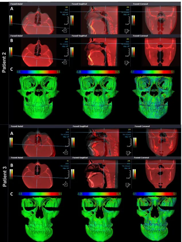
Figure 2 - Patients 2 and 3, (A) multiplanar slices of CBCT volumes before cranial base
superimposition and (B) after cranial base superimposition, in Ondemand3D, showing the
complete correspondence of registration in all areas (C) Skull models after superimposition
showing 3D displacements (registration error) via color maps with scale of 0.5mm, 0.25mm
and 0.01mm. Positive and negative values indicate outward (red) and inward (blue) changes,
Figure 3 - Patients 4 and 5, (A) multiplanar slices of CBCT volumes before cranial base
superimposition and (B) after cranial base superimposition, in Ondemand3D, showing the
complete correspondence of registration in all areas (C) Skull models after superimposition
showing 3D displacements (registration error) via color maps with scale of 0.5mm, 0.25mm
and 0.01mm. Positive and negative values indicate outward (red) and inward (blue) changes,
41
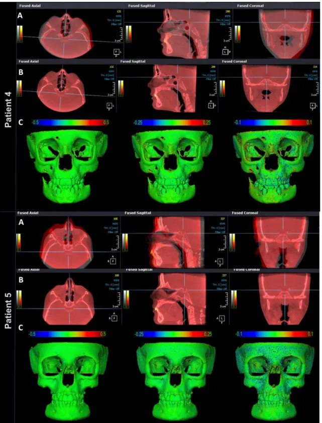
Figure 4 - Patients 6 to 10 - 3D models after superimposition. Each row contains images from
the same patient. Skull models after anterior cranial base superimposition showing 3D
displacements (registration error via color maps with scale of 0.5mm, 0.25mm and 0.01mm.
Positive and negative values indicate outward (red) and inward (blue) changes, respectively.
Figure 5. CBCT superimposition of growing patients subjected to RME with 1-year
follow-up. (A) axial slices view before and after anterior cranial base superimposition in
Ondemand3D. (B) 3D models after superimposition showing 3D displacements via
color maps. In the 0.5mm color map, the black areas represent changes of 0.5 mm,
blue/red areas represent changes less than 0.5mm and green areas represents no
changes. The 5mm color map, the areas in black, red and blue are due to the RME and
normal growth. The changes at the cranial base were minimal as shown in green color
43
Figure 6. CBCT superimposition of non-growing patients subjected to orthognathic surgery
with 1-year follow-up. (A) Sagittal slices view before and after cranial base superimposition in
Ondemand3D. (B) 3D models after superimposition showing 3D displacements via color maps.
In the 0.5mm color map, the black areas represent changes of 0.5 mm, blue/red areas
represent changes less than 0.5mm and green areas represents no changes. The 5mm color
map, the areas in black, red and blue are changes due to the surgical treatment. The changes
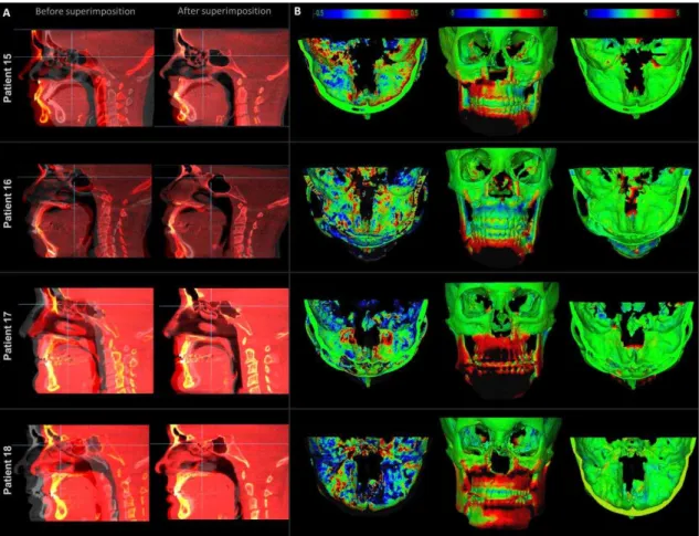
Figure 7. Cranial base superimposition assessment for growing and non-growing patients
subjected to RME and to orthognathic surgery, respectively, with 1-year follow-up. In the
1mm color-coded surface distance maps, the black areas represent changes of 1mm, blue/red
areas represent changes less than 1mm and green areas represents no changes. The
semi-transparent overlays of the pre-treatment (red) and post-treatment (semi-transparent) surface
models show adequate superimposition of the anterior cranial bases.
5. Discussão complementar
O objetivo final do processo de elaboração e execução de uma pesquisa
científica é sua publicação em um periódico de qualidade, contribuindo como
evidência cientifica que suporte a tomada de decisões na prática clínica. Entretanto, as
publicações seguem diretrizes e normatização de formatação, sendo cada vez mais
compacta e priorizando as informações mais relevantes. Com isso, aspectos
45
dos próximos parágrafos foi discutir alguns desses aspectos secundários, porém,
considerados importantes.
Atualmente, a TCCB é uma ferramenta de diagnóstico 3D bem estabelecida e
popularizada na Odontologia. Isso aumentou a demanda por programas para arquivos
DICOM com módulos específicos para mensuração e análise do complexo craniofacial.
Inúmeras empresas iniciaram o desenvolvimento e comercialização de programas para
suprir o emergente mercado das análises 3D. Através de estratégias de marketing,
grandes empresas atraem os profissionais utilizando imagens tridimensionais
sofisticadas, porém, com valor diagnóstico limitado e sem validação científica. Por isso,
pesquisas bem conduzida são imprescindíveis para garantir a qualidade do diagnóstico
3D.
Em uma recente revisão sistemática da literatura, foram encontrados 18
programas para visualização, mensuração e análise das vias aéreas superiores. Apenas
um estudo testou a precisão e acurácia de somente 3 programas, sendo todos para o
sistema operacional Windows (Microsoft). A necessidade de pesquisas de validação de
programas destinados à avaliação das vias aéreas nos levou a desenvolver o estudo
descrito no Artigo 1. Os programas aqui estudados, além de serem populares na
Odontologia, Medicina e Engenharia Biomédica, são compatíveis com os sistemas
operacionais Windows (Microsoft), Macintosh OS X (Apple) e Linux. Esses programas
foram testados, comparados e a evidência científica produzida poderá servir como
base para futuros estudos que utilizem segmentação das vias aéreas superiores.
No artigo 1 (Páginas 14-27), seis diferentes programas foram utilizados para
avaliar o volume da orofaringe por meio de segmentação e construção de modelos
virtuais 3D. Embora os seis programas utilizem segmentação semiautomática, eles
possuem diferentes ferramentas e mecanismos para segmentar as vias aéreas. As
principais vantagens e desvantagens de cada programa são descritas a seguir:
(1) Dolphin3D:
A segmentação das vias aéreas é realizada de forma simples e rápida.
Além disso, permite a identificação da área de menor secção transversal da orofaringe,
informação essencial no diagnóstico das vias aéreas superiores. O processo de
quais se espalham e se difundem através das áreas (
voxels
) pré-estabelecidas pelo
intervalo da escala de cinzas (Figura 1A). A ferramenta que controla o intervalo da
escala de cinza permite um ajuste preciso das áreas a serem segmentadas. Entretanto,
uma vez determinado o intervalo da escala de cinzas, ele se aplica a todos os cortes
axiais, sagitais e coronais das vias aéreas. Com isso, a segmentação preenche espaços
vazios em certas áreas e ultrapassa os limites das vias aéreas em outras regiões,
especialmente nos casos em que as vias aéreas possuem morfologia complexa. Além
disso, o intervalo da escala de cinzas é exibido em unidades próprias do Dolphin3D
sendo incompatível com os programas que utilizam a unidade Hounsfield (Figura 1).
Seria recomendada a atualização desse programa incluindo ferramentas para ajuste e
correção da segmentação em cada imagem 2D (axial, sagital e coronal), quando
necessário. Outra melhoria seria a utilização de unidades da escala de cinza
compatíveis com outros programas. Dentre todos os programas avaliados, foi o que
apresentou maior facilidade para avaliação das vias aéreas superiores.
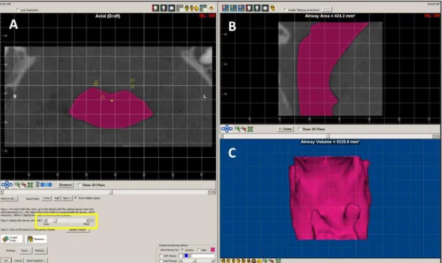
47
Figura 2. Segmentação da orofaringe no programa InVivo Dental. A segmentação é realizada apenas do modo de visualização 3D. Não há parâmetros em imagens axiais, sagitais e coronais para conferir a acurácia da segmentação previamente a construção do modelo 3D a análise do volume.
(2) InVivo Dental:
a avaliação das vias aéreas superiores pode ser realizada de forma
rápida e fácil. Após a renderizacão das vias aéreas, a delimitação da região de interesse
(orofaringe) é realizada através da seleção do intervalo de escala de cinzas em um
modo de visualização exclusivamente em 3D (Figura 2). Isso constitui uma limitação,
pois não permite a inspeção visual dos limites anatômicos das vias aéreas e
impossibilita a conferência da acurácia da segmentação. A impossibilidade de realizar e
visualizar a segmentação nas imagens 2D (axiais, sagitais e coronais) constitui uma
importante desvantagem desta versão do programa InVivo Dental. Por ser um
programa disponível apenas comercialmente, o seu módulo de avaliação das vias
aéreas superiores deixa a desejar e necessita de atualização.
(3) Ondemand3D:
a segmentação das vias aéreas é realizada de forma rápida, através
da inserção de sementes (pontos iniciais de segmentação) diretamente nas imagens
bidimensionais (axial, sagital e coronal). A seguir, a densidade tecidual das vias aéreas
é detectada automaticamente, delineando a área a ser segmentada. Entretanto, existe
refinamento e obtenção de uma segmentação mais acurada. O problema é que o
Ondemand3D não permite um ajuste fino do intervalo de escala de cinza, produzindo
alterações grosseiras das áreas delineadas para segmentação. A ausência de um
controle preciso do processo de segmentação, ou ajuste fino, faz com que mínimas
mudanças no intervalo da escala de cinza selecionado produzam grandes alterações no
volume 3D gerado. Além do mais, o algoritmo do módulo de segmentação do
Ondemand3D é deficiente em detectar precisamente os limites anatômicos das vias
aéreas. O resultado é uma segmentação com falhas tanto na região interna como nos
limites anatômicos das vias aéreas superiores (Figura 3).
49
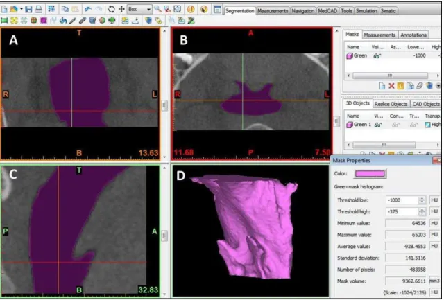
Figura 4. Segmentação da orofaringe no programa Mimics. Em (A) imagem coronal, (B) axial e (C) sagital mostrando a orofaringe corretamente preenchida pela segmentação. Em (D) modelo virtual 3D da orofaringe a partir qual foi computado o volume.