Imaging from temporomandibular joint
during orthodontic treatment: a systematic
review
Introduction: The evolution of imaging in dentistry has provided several advantages for
the diagnosis and development of treatment plans in various dental specialties. Examina-tions as nuclear magnetic resonance, computed tomography and cone beam volumetric tomography, as well as 3D reconstruction methods, have enabled a precise analysis of oro-facial structures. Allied to this fact, the effects of orthodontic treatment on temporoman-dibular joint (TMJ) could be evaluated with the accomplishment of clinical studies with appropriate designs and methodologies. Objective: This study, a systematic literature re-view, had the objective of analyzing the interrelation between orthodontic treatment and TMJ, verifying if orthodontic treatment causes changes in the internal structures of TMJ.
Methods: Survey in research bases MEDLINE, Cochrane, EMBASE, Pubmed, Lilacs and
BBO, between the years of 1966 and 2009, with focus in randomized clinical trials,
lon-gitudinal prospective nonrandomized studies, systematic reviews and meta-analysis.
Re-sults: After application of the inclusion criteria 14 articles were selected, 2 were
random-ized clinical trials and 12 longitudinal nonrandomrandom-ized studies. Conclusions: According to the literature analysis, the data concludes that orthodontic treatment does not occur at the expense of unphysiological disc-condyle position. Some orthodontic mechanics may cause remodeling of articular bone components.
Abstract
Keywords: Temporomandibular joint. Temporomandibular joint dysfunction syndrome. Temporoman-dibular joint disorders. Orthodontics. Magnetic resonance imaging. Tomography.
Eduardo Machado*, Renésio Armindo Grehs**, Paulo Afonso Cunali***
* Specialist in TMD and Orofacial Pain, UFPR. Graduate in Dentistry, UFSM.
** PhD in Orthodontics and Dentofacial Orthopedics, UNESP/Araraquara – SP. Professor of Graduate and Post-graduate Dentistry course, UFSM. *** PhD in Sciences, UNIFESP. Professor of Graduate and Post-graduate Dentistry course, UFPR. Head of the Specialization Course in TMD and
Orofacial Pain, UFPR.
How to cite this article: Machado E, Grehs RA, Cunali PA. Imaging from temporomandibular joint during orthodontic treatment: a systematic
IntROduCtIOn
The effects of orthodontic treatment on Tem-poromandibular Joint (TMJ) are still subject to doubts and discussions. The use of complementary exams has always been a constant in the evalua-tion of this interrelaevalua-tion and can be exemplified by conventional radiographic examinations that were widely used to assess the implications of orthodon-tic treatment on the TMJ. However, this modality of imaging examination has limitations, because the TMJ is one of the structures of the human body more difficult to be well visualized radiographi-cally due to overlapping of several adjacent bony structures. Thus, the effects of orthodontics on TMJ structures are still controversial.
With the advent of imaging examinations with specificity, sensitivity and greater accuracy in the reproduction of articular anatomic structures, such as magnetic resonance imaging (MRI), com-puted tomography and cone-beam volumetric computed tomography as well as 3D reconstruc-tion methods, this interrelareconstruc-tionship can be evalu-ated with greater exactness. Added to this fact, there was accomplishment of clinical studies with designs and more rigorous methodological crite-ria, generating higher levels of evidence.
Thus, the general aim of this study, through a systematic literature review was to analyze within a context of a scientific evidence based dentistry, the implications of orthodontics to the TMJ and check specifically what changes in condylar and articular disc position and joint morphological changes that occur due to orth-odontic treatment.
MAtERIAL And MEtHOdS
We performed a computerized search in MED-LINE, Cochrane, EMBASE, PubMed, Lilacs and BBO in the period from 1966 through February 2009. The research descriptors used were “ortho-dontics”, “orthodontic treatment”, “temporoman-dibular disorder,” “temporoman“temporoman-dibular joint”, “cra-niomandibular disorder”, “TMD”, “TMJ”, “magnetic
resonance imaging” and “tomography”, which were crossed in search engines. The initial list of articles was submitted to review by two reviewers, who ap-plied inclusion criteria to determine the final sample of articles, which were assessed by their title and ab-stract. If there was any disagreement between the results of the reviewers, a third reviewer would be consulted by reading the full version of the article.
Inclusion criteria for selecting articles were: » Studies based on magnetic resonance imag-ing (MRI), computed tomography (CT) and/ or volumetric cone-beam tomography, which assessed the effects of orthodontic treatment in TMJ. Studies based only on electromyogra-phy, cephalometric radiographs and conven-tional radiographs were excluded, as well as studies involving orthognathic surgery.
» Randomized clinical trials (RCT), non-ran-domized prospective longitudinal studies, sys-tematic reviews and meta-analysis.
» Studies in which orthodontic treatment is al-ready completed in the assessed samples. » Studies written in English and Spanish, and published between 1966 and February 2009. Thus, we excluded cross-sectional studies, clini-cal case reports, case series, simple reviews and opin-ions papers, as well as studies in which orthodontic treatment had not yet been completed.
RESuLtS
After applying the inclusion criteria 14 studies were selected and the Kappa index of agreement be-tween reviewers was 1.00. Among these studies, two were randomized clinical trials and 12 were longitu-dinal studies without randomization criteria (Fig 1). Among the selected studies, 11 were based on magnetic resonance imaging and 3 in computed to-mography images, as shown in Figure 2. None of the selected studies used cone-beam computed tomog-raphy for evaluation of the TMJ.
3 2
11 12
FIGURE 1 - Design of included studies. FIGURE 2 - Studies characteristics.
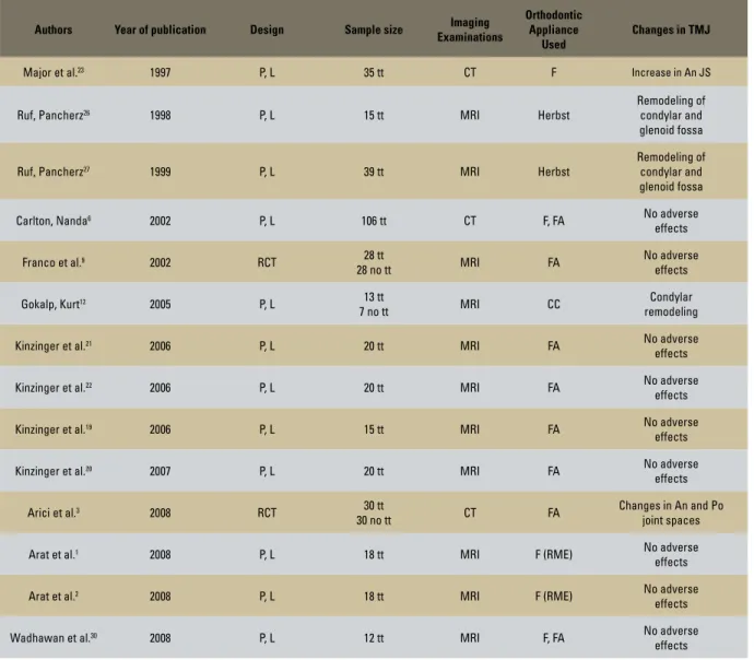
TABLE 1 - Studies based on imaging examination of magnetic resonance imaging, computed tomography and/or cone-beam computed tomography.
P= prospective; L= longitudinal; RCT= randomized clinical trial; tt= treatment; MRI= magnetic resonance imaging; CT= computed tomography; F= fixed appliances; FA= functional appliances; CC= chincup; JS= joint space; An= anterior; Po= posterior; RME= rapid maxillary expansion.
Authors Year of publication Design Sample size Imaging Examinations
Orthodontic Appliance
Used
Changes in TMJ
Major et al.23 1997 P, L 35 tt CT F Increase in An JS
Ruf, Pancherz26 1998 P, L 15 tt MRI Herbst
Remodeling of condylar and glenoid fossa
Ruf, Pancherz27 1999 P, L 39 tt MRI Herbst
Remodeling of condylar and glenoid fossa
Carlton, Nanda6 2002 P, L 106 tt CT F, FA No adverse
effects
Franco et al.9 2002 RCT 28 tt
28 no tt MRI FA
No adverse effects
Gokalp, Kurt12 2005 P, L 13 tt
7 no tt MRI CC
Condylar remodeling
Kinzinger et al.21 2006 P, L 20 tt MRI FA No adverse
effects
Kinzinger et al.22 2006 P, L 20 tt MRI FA No adverse
effects
Kinzinger et al.19 2006 P, L 15 tt MRI FA No adverse
effects
Kinzinger et al.20 2007 P, L 20 tt MRI FA No adverse
effects
Arici et al.3 2008 RCT 30 tt
30 no tt CT FA
Changes in An and Po joint spaces
Arat et al.1 2008 P, L 18 tt MRI F (RME) No adverse
effects
Arat et al.2 2008 P, L 18 tt MRI F (RME) No adverse
effects
Wadhawan et al.30 2008 P, L 12 tt MRI F, FA No adverse
effects
Magnetic resonance imaging Randomized clinical trials
dISCuSSIOn
It becomes increasingly important to analyze the current literature in a critical and rigorous way to verify what level of scientific evidence that the information generates. The application of methodological considerations for research — such as sample size calculation, randomiza-tion, calibrarandomiza-tion, blinding and control of in-volved factors —- are extremely important to qualify the level of evidence generated. And this information must be available for examination and discussion for the reader.28
Currently, the access to scientific evidences is available through many different ways. Because of this facility, the knowledge about the hierarchy of the scientific evidence levels is essential for assess-ing the quality of the study. Thus, meta-analysis, systematic reviews and randomized clinical trials receive the best concepts. Being aware of this fact is important, since the vast majority of articles published in Brazilian journals correspond to stud-ies of low potential for direct clinical application. Magnetic resonance imaging and computed tomography are methods with higher diagnostic accuracy compared with conventional radiology, because of greater anatomic resolution they pro-vide. CT is the ideal method for assessment of bone structures, whereas MRI allows the study of soft tissues, including intra-articular disc. Both methods often complement the study of abnor-malities of the temporomandibular joint (TMJ), thus becoming important tools in the differential diagnosis of various diseases in this region.11
Computed tomography is the examination of choice to evaluate TMJ bony structures, especially for the diagnosis of fractures, joint deformities, an-kylosis and tumors. There is no overlapping of any other structure, enabling assessment of the quality and bone density.5 Similarly, MRI is the gold
stan-dard for the representation of soft tissue and posi-tioning of the TMJ articular disc,17 allowing
infor-mation about the position, function and form of the articular disc and conditions of muscle tissues
and ligaments, as well as assessment of severity of various disorders: trauma, arthritis, arthrosis and neoplastic degeneration.10
Also, the cone-beam computed tomography allows visualization of structures of reduced di-mensions with minimal radiation exposure for pa-tients and less operating time than conventional CT. This imaging modality has several applica-tions, assisting in the diagnosis and in the treat-ment plan in different dental specialties.29 The
cone-beam tomography has a relevant importance in diagnosis, localization and reconstruction of to-mographic images with excellent precision, aiding in therapeutic decisions.4
Clinically, the scientific evidences indicate for a tendency of no association between orth-odontic treatment and temporomandibular disorders (TMD), in other words, orthodontics does not increase the prevalence of signs and symptoms of TMD, with longitudinal and ex-perimental-interventionist studies,7,8,13-16,25
sys-tematic review24 and meta-analysis18
corrobo-rating that. Also, with the analysis of imaging studies, according to the methodological crite-ria adopted by this systematic review, it appears that orthodontic movement does not cause ad-verse effects to the TMJ.6,9,19-22
The systematic literature review shows that the correct occlusal relationship between the teeth did not cause a change in the physiologi-cal position of the condyles and articular discs
in TMJ when MRI and CT were examined,19,21,22
whereas in some cases of TMD an improvement can be obtained as a result of orthodontic treat-ment.9,19,22 Some studies found changes in
con-dylar position3 and in the volumes of the
ante-rior and posteante-rior joint spaces3,23 due to applied
orthodontic mechanics. Furthermore, the use of the chincup caused a morphological change in condylar growth, which may be associated with correction of skeletal malocclusion in conjunc-tion with remodeling in the jaw,12 as well as the
The application of different orthodontic mechanics did not cause incorrect position-ing on the articular disc-condyle relationship. Elastics mechanics,6,23 headgear,6 rapid
max-illary expansion,1,2 Frankel functional
appli-ance,9 Bionator,30 fixed functional orthopedic
appliances,20,21,22 Twin Block30 and functional
mandibular advancement appliance19 did not
cause physiological changes in the positioning of the condyle and articular disc, whereas the implementation or not of extraction protocols did not change this situation.6,23
Great provider of scientific evidence, ran-domized clinical trials were found in low num-ber in this systematic review: only two stud-ies.3,9 This fact is associated with difficulties in
accomplishment of this type of study in patients undergoing orthodontic treatment due to ethi-cal and practiethi-cal questions.18 Likewise, there
were no selected meta-analysis and systematic reviews after application of the inclusion cri-teria. It is important to be noted that all the selected studies presented longitudinal assess-ments, which is the ideal study design to check for risk factors, due to its temporal component.28
The use of imaging examinations — CT, cone-beam CT and MRI — in orthodontic practice, not only for evaluating the occlusal criteria, but also for adjacent structures, tends to become a useful tool. Through 3D reconstruction of the surfaces of condyle and their overlaps, detailed views of adaptive mechanisms and its non-invasive assess-ment may become possible in routine clinical or-thodontics.20 Through these examinations
1. Arat FE, Arat ZM, Tompson B, Tanju S, Erden I. Muscular and condylar response to rapid maxillary expansion. Part 2: mag-netic resonance imaging study of the temporomandibular joint. Am J Orthod Dentofacial Orthop. 2008;133(6 Pt 2):823-9. 2. Arat FE, Arat ZM, Tompson B, Tanju S. Muscular and condylar
response to rapid maxillary expansion. Part 3: magnetic reso-nance assessment of condyle-disc relationship. Am J Orthod Dentofacial Orthop. 2008;133(6 Pt 3):830-6.
3. Arici S, Akan H, Yakubov K, Arici N. Effects of ixed functional
appliance treatment on the temporomandibular joint. Am J Orthod Dentofacial Orthop. 2008;133(6):809-14.
4. Bissoli CF, Ágreda CG, Takeshita WM, Castilho JCM, Medici Filho E, Moraes ML. Importancia y aplicaciones del sistema de
tomograia computarizada cone-beam (cbct). Acta Odontol
Venez. 2007;45(4):589-92.
5. Bumann A, Lotzmann U. Disfunção temporomandibular: diagnóstico funcional e princípios terapêuticos. Porto Alegre: Artmed; 2003.
6. Carlton KL, Nanda RS. Prospective study of posttreatment changes in the temporomandibular joint. Am J Orthod Dento-facial Orthop. 2002;122(5):486-90.
REfEREnCES
7. Egermark I, Carlsson GE, Magnusson T. A prospective long-term study of signs and symptoms of temporomandibular disorders in patients who received orthodontic treatment in childhood. Angle Orthod. 2005;75(4):645-50.
8. Egermark I, Magnusson T, Carlsson GE. A 20-year follow-up of signs and symptoms of temporomandibular disorders and mal-occlusions in subjects with and without orthodontic treatment in childhood. Angle Orthod. 2003;73(2):109-15.
9. Franco AA, Yamashita HK, Lederman HM, Cevidanes LH, Profit
WR, Vigorito JW. Fränkel appliance therapy and the temporo-mandibular disc: a prospective magnetic resonance imaging study. Am J Orthod Dentofacial Orthop. 2002;121(5):447-57. 10. Freitas A. Radiologia odontológica. 6ª ed. São Paulo: Artes
Médicas; 2004.
11. Garcia MM, Machado KFS, Mascarenhas MH. Ressonância
magnética e tomograia computadorizada da articula -ção temporomandibular: além da disfun-ção. Radiol Bras. 2008;41(5):337-42.
12. Gokalp H, Kurt G. Magnetic resonance imaging of the condylar growth pattern and disk position after chin cup therapy: a preliminary study. Angle Orthod. 2005;75(4):568-75.
COnCLuSIOnS
» This systematic literature review finds that the correct occlusal relationship as a result of orthodontic treatment is not ob-tained at the expense of non-physiological positioning of both the condyle and the articular disc. Thus, when orthodontics is used correctly does not cause adverse ef-fects in the TMJ.
» The application of forces during certain orthodontic mechanics, especially orthope-dic situations, can cause alterations in condy-lar growth and bone structures of the TMJ. Thus, the mechanics application should be performed properly and the professional
must have knowledge of these impacts. » In some studies by analysis of imaging
examinations, it was observed that there were improvements in situations of pre-existing TMD at the beginning of orth-odontic therapy. However, these data are only suggestive and more randomized clinical trials are necessary to obtain more precise conclusions.
Contact address
Eduardo Machado
Rua Francisco Trevisan 20, Nossa Sra. de Lourdes CEP: 97.050-230 - Santa Maria / RS, Brazil E-mail: [email protected]
23. Major P, Kamelchuk L, Nebbe B, Petrkowski G, Glover K. Condyle displacement associated with premolar extraction and nonextraction orthodontic treatment of Class I malocclusion. Am J Orthod Dentofacial Orthop. 1997;112(4):435-40. 24. Mohlin B, Axelsson S, Paulin G, Pietila T, Bondemark L,
Brat-tstrom V, et al. TMD in relation to malocclusion and orthodontic treatment. Angle Orthod. 2007;77(3):542-8.
25. Mohlin BO, Derweduwen K, Pilley R, Kingdon A, Shaw WC, Kenealy P. Malocclusion and temporomandibular disorder: a comparison of adolescents with moderate to severe dysfunc-tion with those without signs and symptoms of temporoman-dibular disorder and their further development to 30 years of age. Angle Orthod. 2004;74(3):319-27.
26. Ruf S, Pancherz H. Temporomandibular joint growth adaptation in Herbst treatment: a prospective magnetic resonance imag-ing and cephalometric roentgenographic study. Eur J Orthod. 1998;20(4):375-88.
27. Ruf S, Pancherz H. Temporomandibular joint remodeling in adolescents and young adults during Herbst treatment: a prospective longitudinal magnetic resonance imaging and cephalometric radiographic investigation. Am J Orthod Dento-facial Orthop. 1999;115(6):607-18.
28. Susin C, Rosing CK. Praticando odontologia baseada em evidências. 1ª ed. Canoas: ULBRA; 1999.
29. Xaves ACC, Sena LEC, Araújo LF, Nascimento Neto JBS.
Aplicações da tomograia computadorizada de feixe cônico na
odontologia. Int J Dent. 2005;4(3):80-124.
30. Wadhawan N, Kumar S, Kharbanda OP, Duggal R, Sharma R. Temporomandibular joint adaptations following two-phase therapy: an MRI study. Orthod Craniofac Res. 2008;11(4):235-50.
Submitted: February 2009 Revised and accepted: May 2010 13. Henrikson T, Nilner M. Temporomandibular disorders and need
of stomatognathic treatment in orthodontically treated and untreated girls. Eur J Orthod. 2000;22(3):283-92. 14. Henrikson T, Nilner M. Temporomandibular disorders,
occlu-sion and orthodontic treatment. J Orthod. 2003;30(2):129-37. 15. Henrikson T, Nilner M, Kurol J. Symptoms and signs of
tem-poromandibular disorders before, during and after orthodontic treatment. Swed Dent J. 1999;23(5-6):193-207.
16. Imai T, Okamoto T, Kaneko T, Umeda K, Yamamoto T, Na-kamura S. Long-term follow-up of clinical symptoms in TMD patients who underwent occlusal reconstruction by orthodontic treatment. Eur J Orthod. 2000;22(1):61-7.
17. Kamelchuk L, Nebbe B, Baker C, Major P. Adolescent TMJ tomography and magnetic resonance imaging: a comparative analysis. J Orofac Pain. 1997;11(4):321-7.
18. Kim MR, Graber TM, Viana MA. Orthodontics and temporo-mandibular disorder: a meta-analysis. Am J Orthod Dentofacial Orthop. 2002;121(5):438-46.
19. Kinzinger G, Gulden N, Roth A, Diedrich P. Disc-condyle relationships during Class II treatment with the Functional Man-dibular Advancer (FMA). J Orofac Orthop. 2006;67(5):356-75. 20. Kinzinger G, Kober C, Diedrich P. Topography and morphology
of the mandibular condyle during ixed functional orthopedic
treatment: a magnetic resonance imaging study. J Orofac Orthop. 2007;68(2):124-47.
21. Kinzinger G, Roth A, Gulden N, Bucker A, Diedrich P. Effects
of orthodontic treatment with ixed functional orthopaedic
appliances on the condyle-fossa relationship in the temporo-mandibular joint: a magnetic resonance imaging study (Part I). Dentomaxillofac Radiol. 2006;35(5 Pt 1):339-46.
22. Kinzinger G, Roth A, Gulden N, Bucker A, Diedrich, P. Effects
of orthodontic treatment with ixed functional orthopaedic