Alexandre Perez Marques(a) Andréia Perrella(a)
Emiko Saito Arita(a)
Marlene Fenyo Soeiro de Matos Pereira(a)
Marcelo de Gusmão Paraíso Cavalcanti(a)
(a)Departamento de Estomatologia, Faculdade de Odontologia, Universidade de São Paulo, São Paulo, SP, Brasil.
Corresponding author: Alexandre Perez Marques
Rua Vice-governador Rubens Berardo, 175, Bloco 1, apt. 904 - Gávea
Rio de Janeiro - RJ - Brazil CEP: 22451-070
E-mail: [email protected]
Received for publication on May 12, 2010 Accepted for publication on Sep 10, 2010
Assessment of simulated mandibular
condyle bone lesions by cone beam
computed tomography
Abstract: There are many limitations to image acquisition, using con-ventional radiography, of the temporomandibular joint (TMJ) region. The Computed Tomography (CT) scan is a better option, due to its high-er accuracy, for purposes of diagnosis, surgical planning and treatment of bone injuries. The aim of the present study was to analyze two proto-cols of cone beam computed tomography for the evaluation of simulated mandibular condyle bone lesions. Spherical lesions were simulated in 30 dry mandibular condyles, using dentist drills and drill bits sizes 1, 3 and 6. Each of the mandibular condyles was submitted to cone beam comput-ed tomography (CBCT) using two protocols: 1) axial, coronal and sagit-tal multiplanar reconstruction (MPR); and 2) sagitsagit-tal plus coronal slices throughout the longitudinal axis of the mandibular condyles. For these protocols, 2 observers analyzed the CBCT images independently, regard-ing the presence or not of injuries. Only one of the observers, however, performed on 2 different occasions. The results were compared to the gold standard, evaluating the percentage of agreement, degree of accu-racy of CBCT protocols and observers’ examination. The z test was used for the statistical analysis. The results showed there were no statistically signiicant differences between the 2 protocols. There was greater dif-iculty in the assessment of small-size simulated lesions (drill # 1). From the results of this study, it can be concluded that CBCT is an accurate tool for analyzing mandibular condyle bone lesions, with the MPR pro-tocol showing slightly better results than the sagittal plus coronal slices throughout the longitudinal axis.
Descriptors: Cone-Beam Computed Tomography; Protocols; Bone Diseases; Temporomandibular Joint.
Introduction
Many injuries, inlammatory and degenerative diseases, postural dis-orders, traumas and other abnormalities compromise the equilibrium of temporomandibular joint components, resulting in its disorder or dys-function.1 Complementary radiographic exam is an important
instru-ment to diagnose and evaluate the degree of such anomalies, which affect the temporomandibular joint. Radiographic evidence that characterizes the presence of degenerative disease in the joints includes erosions of the cortical boundary and reduction of the joint biological space.2
presents high speciicity and sensitivity, essential to the diagnosis, planning and treatment of TMJ bone lesions.3 Using CT, it is possible to analyze the
boundary of the mandibular condyle, its position in the mandibular fossa and the involvement of the cortical bone in relation to the bone lesions.4,5
Sev-eral bone alterations that occur in the TMJ, such as erosions and osteophytes in the mandibular condyle, are dificult to detect using conventional radiogra-phy and thus necessitate the use of CT.6
CT has been used to diagnose several bone ab-normalities of the TMJ, from osteophytes to tu-mors.6-8 According to Marques and Moraes,9
ero-sion in the mandibular condyle is prevalent in 7.9% of total TMJ alterations.
Cone beam computed tomography (CBCT) was introduced during the last decade. It uses X-ray exposure in cone shapes instead of slices, as in the spiral CT, for image acquisition. This technology presents less X-ray exposure and lower cost to the patient than the spiral CT, in addition to its capacity to capture images in just one rotation of the X-ray source.10,11
This study aims to demonstrate the sensitivity and speciicity of cone beam computed tomography, and to establish a more adequate protocol for the post-processing of images for the interpretation of simulated mandibular condyle bone lesions.
Material and Methods
The present study was submitted to and ap-proved by the Committee of Ethics in Research of the School of Dentistry, University of São Paulo, un-der protocol # 26/2009. A total of 15 macerated hu-man hu-mandibles (with 30 hu-mandibular condyles) were used in this study. Anatomical specimens of adult mandibles whose mandibular condyles presented bi-laterally a satisfactory aspect of preservation were selected. Each mandibular condyle was divided into ive regions: anterior, lateral, posterior, medial and superior, totaling ive speciic areas. Using a KaVo high-speed handpiece (KaVo do Brasil, Joinville, SC, Brazil) and three surgical carbide ball drill bits for dental use, with different head diameters (# 1, 3, 6), trademark Jet-Carbide Burs (Carbide Burs, Mor-row, OH, USA), holes were made randomly in one
or more portions of each mandibular condyle, that either did or did not reach the cortical and the me-dullar bone, to simulate bone lesions. After drilling the holes, all the mandibles were submitted to cone beam computed tomography.
In order to simulate the X-ray attenuation caused by soft tissues in vivo, the mandibles were inserted one by one into a plastic container with water until they were completely submersed.
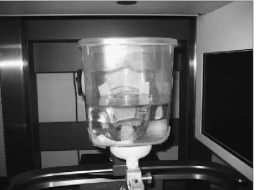
The acquisition of images by CBCT was accom-plished using the i-CAT Classic system (Imaging Sciences International, Hatield, PA, USA) (Figure 1). First, the initial scan was performed with the slicing plane in axial orientation, and leveled with the mandible base up to the region of the mandibu-lar condyle. These images were obtained using the following CBCT acquisition protocol:
• Voxel thickness: 0.25 mm
• Thickness of the sagittal and coronal slice along the longitudinal axis of the mandibular condyle: 1 mm
• Spacing between slices: 1 mm • Acquisition volume: mandible • Time: 40 seconds
• Parameters: 90 kVp and 7 mA
• Field of view (FOV): 13 cm
All the original images from the CBCT were saved on CD-ROM, to be evaluated later at an inde-pendent Dell Precision 390 workstation (Dell Inc., Round Rock, Texas, USA), with a 20” lat screen
monitor - Windows XP (Microsoft, Redmond,
WA, USA), and the iles were also saved together us-ing the i-CAT Vision software (Imaging Sciences International, Hatield, PA, USA).
The single criterion for the analysis of lesions in the tomographic images versus the gold standard was veriication of the presence or absence of the le-sion (hole) in the mandibular condyle.
In relation to the standardization of the analyses, the observers interpreted the tomographic images of the mandibular condyles in the following way:
• Protocol 2 - Sagittal and coronal slices along the longitudinal axis of the mandibular condyle from the CBCT, with 1-mm slice thickness and 1-mm spacing, were also observed at the same independent workstation using the same i-CAT Vision software (Figures 3 and 4).
The systematic evaluation of the tomographic
images was performed by two observers, individu-ally, following the principle of randomization of the images for each observer. Both observers (PhD stu-dents) were calibrated by means of images acquired for this research, in a random form, concerning the agreement of the hypodense images in the dif-ferent regions of the mandibular condyles, on two
Figure 1 - Container with water and the mandible simulating patient position in the i-Cat Visiondevice for CBCT acquisition.
different occasions at an interval of one week. The analyses were performed in the following manner: observer 1 analyzed the images at two distinct times (with an interval of seven days), labeled as observer 1 (irst analysis) and observer 1’ (second analysis). The other observer (observer 2), independently, ana-lyzed the images only once. The second analysis of observer 2 was not carried out because it would not have increased suficiently the number of observers
to a statistically representative sample, and it would have increased costs and time without contributing to the accuracy of the results.
For the analysis of the lesions shown in the CBCT versus the gold standard, the presence or ab-sence of bone lesions (holes) and their locations in all mandibular condyles were evaluated in each pro-tocol of analysis.
For the statistical analysis of this research, the
Figure 3 - Axial slice of the mandibular condyle showing the longitudinal axis of the mandibular condyles and sagittal slices in the medial-lateral direction (i-CAT Vision) showing the hypodense images in the mandibular condyle (arrows).
Figure 4 - Axial slice of the mandibular condyle showing the transversal axis of the mandibular condyles and coronal slices in the antero-posterior direction (i-CAT
percentages of the agreements of each observer were calculated in relation to the gold standard.
For the statistical treatment of the data in this study, the z test was used with a level of signiicance of 5%. Statistically signiicant differences were ob-served when the resultant value of P in the test was less than or equal to 5%.
The information from observer 1 was used to calculate the sensitivity and the speciicity of the protocols of CBCT.
Results
Tables 1 through 6 present the information from the observers in relation to the presence or absence of holes (lesions), and also from the analyses of the two CBCT protocols for assessing simulated bone lesions. The tables also show intra-observer and inter-observer comparisons using the z test, and re-sults of sensitivity and speciicity, aiming at the vali-dation of the methods.
In the statistical analysis, the data recorded by the observers were compared to the gold standard (data shown in Table 1), using the z test.
Table 2 presents the average percentage of agree-ments, in relation to the gold standard, for the
as-sessment of the protocols used, independent of the absence of a hole or the size of the drill bit used to create the hole.
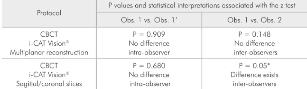
Table 3 presents the z test results for the intra- and inter-observer comparisons, for each protocol used.
As no statistically signiicant differences were found in the intra-observer percentages of agree-ments, and one protocol showed signiicant differ-ence in the inter-observers percentages of agree-ments, the information coming from observer 1 was used to calculate the sensitivity and the speciicity of the protocols utilized, as well as to make the com-parisons of the percentages of agreements between
Table 1 - Gold standard. Number of holes made in the mandibular condyle, by region.
Size of simulated
lesion
Number of lesions made, by region
Anterior Lateral Posterior Medial Superior
Absent 9 8 7 5 7
Drill bit 1 7 8 6 10 7
Drill bit 3 7 7 9 7 8
Drill bit 6 7 7 8 8 8
Protocol
Average comparison of agreements, in relation to the gold standard, taking into consideration all regions
of the mandibular condyles observed
Observer 1 Observer 1’ Observer 2
CBCT i-CAT Vision® Multiplanar reconstruction
0.72 0.70 0.90
CBCT i-CAT Vision® Sagittal/coronal slices
0.70 0.78 0.93
Table 2 - Average comparison of agreements, in relation to the gold standard, taking into consideration all regions of the mandibular condyles observed.
Protocol
P values and statistical interpretations associated with the z test
Obs. 1 vs. Obs. 1’ Obs. 1 vs. Obs. 2
CBCT i-CAT Vision® Multiplanar reconstruction
P = 0.909 No difference intra-observer
P = 0.148 No difference inter-observers
CBCT i-CAT Vision® Sagittal/coronal slices
P = 0.680 No difference intra-observer
P = 0.05* Difference exists
inter-observers
*Statistically significant difference for P ≤ 0.05.
the protocols. Table 4 shows the sensitivity of each protocol used according to the region examined.
Table 5 shows the speciicity of each protocol used according to the region examined by observer 1.
Table 6 presents the percentages of agreements in relation to the gold standard, analyzed using the z test, as a cross-check for the protocols used. In this way, it was possible to establish statistically the existence or not of differences between these proto-cols.
From the results of analyses shown in Table 6, it can be concluded that statistically there are no dif-ferences between the results of the evaluations of multiplanar reconstructions and the evaluations of sagittal/coronal slices in relation to the longitudinal axis of the mandibular condyle.
Discussion
Many studies have been performed about radio-graphic evaluation of the articular degenerative dis-ease located in the temporomandibular joint.12,13
The radiographic patterns, which characterize the presence of degenerative diseases in the TMJ, show erosions of the mandibular condyle and reduc-tions of articular biological space. The transcranial, transfacial, and panoramic radiographs are limited for this type of evaluation, due to the overlaps and distortions peculiar to these methods.2,9
Computed tomography has been used in the last decades, with satisfactory results, for the evaluation of bone alterations in the TMJ. Similar satisfactory results have been observed in the present work.3,14-16
More recently, cone beam computed tomogra-phy has been the object of study for TMJ evaluation by various authors who consider it the best
recom-Protocols Specificity
Anterior Lateral Posterior Medial Superior
CBCT i-CAT Vision® Multiplanar reconstruction
0.67 0.75 0.86 0.80 0.86
CBCT i-CAT Vision® Sagittal/coronal slices
1.00 1.00 1.00 0.80 0.86
Table 5 - Specificity of the protocols utilized for each region examined by observer 1.
Size of simulated lesion
P values for the z test of comparisons of percentages
Anterior Lateral Posterior Medial Superior
Absent 0.211 0.450 0.983 0.999 0.999
Drill bit 1 0.974 0.270 0.991 0.999 0.991
Drill bit 3 0.999 0.604 0.992 0.999 0.207
Drill bit 6 0.999 0.974 0.999 0.966 0.999
Table 6 - Percentages of agreements in relation to the gold standard in the examination of CBCT using the i-CAT Vision software, comparing the evaluation
of multiplanar reconstructions vs. sagittal/coronal slices, using the z test.
Protocol Sensitivity
Anterior Lateral Posterior Medial Superior
CBCT i-CAT Vision® Multiplanar reconstruction
0.47 0.81 0.91 0.60 0.69
CBCT i-CAT Vision® Sagittal/coronal slices
0.42 0.54 0.82 0.56 0.78
mended method for these cases.11,15,17,18
Gaia and Cavalcanti19 performed a study of the
different CT protocols for bone affections of the temporomandibular joint, emphasizing that CT can be considered as the standard image for investiga-tion of TMJ when using axial images and multi-planar reconstruction (MPR). In the present study, the visualization was improved by using sagittal and coronal slices along the longitudinal axis of the mandibular condyle, as they added information re-garding some anatomical regions.
Similarly to the studies of Utumi et al.20 and
Cara et al.,14 the statistical methodology used in this
study considered just one intra-observer analysis in order to avoid increasing costs and time, with-out losing the accuracy of the results, compared to the use of two intra-observer analyses. Addition-ally, a second observer was not considered because it would not increase suficiently the number of ob-servers to characterize a statistically representative sample.
The results of this study corroborate the research of Cara et al.,14 in which they found a high level
of sensitivity and speciicity using multislice CT to identify mandible condyle lesions. Furthermore, our study demonstrated very similar sensitivity and speciicity in both protocols when CBCT images were assessed.
Utumi et al.20 also found a high level of
sensi-tivity and speciicity using multislice CT with para-sagittal reconstruction. The same was found, with sagittal and coronal slices along the axis of the man-dibular condyle, in the present work.
Bone lesions were simulated in dry mandibular condyles, using three sizes of drill bits. This is the same methodology used in the work of Utumi et al.20 This methodology was chosen in order to
ob-tain a Gold Standard, which would permit an easy comparison of the results obtained by the observers, allowing for statistical treatment.
For the two protocols tested, the percentages of agreements (concordance) of hole presence identiied by the observers were calculated in all the regions of the mandibular condyle, and in relation to the size of the hole. It was observed that the larger the size
of the hole, the higher the percentages of agreements by the observers. The results are similar regarding the average size of holes, as well as the absence of any holes, irrespective of the protocol used. Con-cerning the smaller-size holes, the percentage of agreements in relation to the gold standard was low-er. All observers concurred with this fact. Analyz-ing these results, it was noted that even in accurate exams with high resolution like computed tomogra-phy, the smaller the bone lesion, the more dificult its identiication will be, according to Tsiklakis et al.10 and Hussain et al.6
Applying the z test, the results showed that the percentage of agreements between the observers did not differ very much, according to the protocols used. It was observed that no statistically signiicant differences were found (P ≤ 0.05) in the intra-ob-server comparisons, demonstrating the reproducible feature of the technical protocols. Only the protocol of sagittal/coronal slices throughout the longitudi-nal axis of the mandibular condyle showed statis-tically signiicant differences in the inter-observer comparisons. In the other cases, no statistically sig-niicant differences were noted.
Ludlow et al.8 performed a comparative study in vitro of the degree of accuracy for the detection of bone alterations in the TMJ region with the CT multislice, using sagittal and coronal images (mul-tiplanar reconstructions) and panoramic radio-graphs. The multiplanar reconstructions showed greater accuracy for the evaluation of bone lesions in the mandibular condyle than the panoramic im-ages. In comparison to their study, the present work has demonstrated differences in the percentage of agreements concerning the position of the simulat-ed lesions in the mandibular condyle, relatsimulat-ed to the diameter of the drill bits. Concerning the concor-dance, in each analyzed region, it was observed that the images did not present signiicant differences.
Conclusions
• Cone beam computed tomography was validated
litera-ture.
• Both post-processing protocols of CBCT were considered to be adequate in the assessment of bone lesions in the mandibular condyle, with the multiplanar reconstruction protocol showing slightly better results.
Acknowledgements
To CNPq, Brasilia, Brazil, Dr. Marcelo Caval-canti (Universal Project - grants #484848/2006-2 and Research Productivity Scholarship - grants #306509/2006-7).
References
1. Saeed NR, McLeod NM, Hensher R. Temporomandibular joint replacement in rheumatoid - induced disease. Br J Oral Maxillofac Surg. 2001 Feb;39(1):71-5.
2. Akerman S, Jonsson K, Kopp S, Petersson A, Rohlin M. Ra-diologic changes in temporomandibular, hand, and foot joints of patients with rheumatoid arthritis. Oral Surg Oral Med Oral Pathol. 1991 Aug; 72(2):245-50.
3. Perrella A, Borsatti MA, Tortamano IP, Rocha RG, Caval-canti MG. Validation of computed tomography protocols for simulated mandibular lesions: a comparison study. Braz Oral Res. 2007 Apr-Jun;21(2):165-9.
4. Cavalcanti MG, Ruprecht A, Bonomie JM, Vannier MW. Accuracy and precision of spiral CT in the assessment of neo-plastic lesions associated with the mandible. Acad Radiol. 2000 Feb;7(2):94-9.
5. Sales MA, Oliveira JX, Cavalcanti MG. Computed tomogra-phy imaging findings of simultaneous bifid mandibular con-dyle and temporomandibular joint ankylosis: case report. Braz Dent J. 2007;18(1):74-7.
6. Hussain AM, Packota G, Major PW, Flores-Mir C. Role of different imaging modalities in assessment of temporoman-dibular joint erosions and osteophytes: a systematic review. Dentomaxillofac Radiol. 2008 Feb;37(2):63-71.
7. Larheim TA, Bjornland T, Smith HJ, Aspestrand F, Kolben-stvedt A. Imaging temporomandibular joint abnormalities in patients with rheumatic disease. Comparison with sur-gical observation. Oral Surg Oral Med Oral Pathol. 1992 Apr;73(4):494-501.
8. Ludlow JB, Davies KL, Tyndall DA. Temporomandibular joint imaging: a comparative study of diagnostic accuracy for the detection of bone change with biplanar multidirectional tomography and panoramic images. Oral Surg Oral Med Oral Pathol Oral Radiol Endod. 1995 Dec;80(6):735-43. 9. Marques AP, Moraes LC. Prevalência de alterações da
articu-lação temporomandibular por meio de exames de tomografia computadorizada. Rev Bras Odont. 2006;63(3,4):198-201. 10. Tsiklakis K, Syriopoulos K, Stamatakis HC. Radiographic
examination of the temporomandibular joint using cone
beam computed tomography. Dentomaxillofac Radiol. 2004 May;33(3):196-201.
11. Winter AA, Pollack AS, Frommer HH, Koenig L. Cone beam volumetric tomography vs. medical CT scanners. N Y State Dent J. 2005 Jun-Jul; 71(4):28-33.
12. Tsuruta A, Yamada K, Hanada K, Hosogai A, Tanaka R, Koyama J, et al. Thickness of the roof of the glenoid fossa and condylar bone change: a CT study. Dentomaxillofac Radiol. 2003 Jul;32(4):217-21.
13. Yamada K, Tsuruta,A, Hanada K, Hayashi,T. Morphology of the articular eminence in temporomandibular joints and condylar bone change. J Oral Rehabil. 2004 May;31(5):438-44.
14. Cara AC, Gaia BF, Perrella A, Oliveira JX, Lopes PM, Cav-alcanti MG. Validity of single- and multislice CT for assess-ment of mandibular condyle lesions. Dentomaxillofac Radiol. 2007 Jan;36(1):24-7.
15. Cavalcanti MGP. Tomografia computadorizada por feixe cô-nico. Interpretação e diagnóstico para o cirurgião-dentista. 1a ed. São Paulo: Ed. Santos; 2009. 244 p.
16. Hintze H, Wiese M, Wenzel A. Cone beam CT and conven-tional tomography for the detection of morphological tem-poromandibular joint changes. Dentomaxillofac Radiol. 2007 May;36(4):192-7.
17. Mozzo P, Procacci C, Tacconi A, Martini PT, Andreis IA. A new volumetric CT machine for dental imaging based on the cone-beam technique: preliminary results. Eur Radiol. 1998;8(9):1558-64.
18. Pinsky HM, Dyda S, Pinsky RW, Misch KA, Sarment DP. Ac-curacy of three dimensional measurements using cone-beam CT. Dentomaxillofac Radiol. 2006 Nov;35(6):410-6. 19. Gaia BF, Cavalcanti MGP. Afecções ósseas da articulação
tem-poromandibular: protocolos em tomografia computadorizada. Rev Assoc Paul Cir Dent. 2005 Jul-Aug;59(4):297-302. 20. Utumi ER, Perrella A, Albuquerque MA, Adde CA, Rocha