25
LOW-LEVEL LASER THERAPY: EFFECTS ON HUMAN FACE AGED SKIN AND CELL VIABILITY OF HELA CELLS EXPOSED TO UV RADIATION
Sana Mezghani*, Amira Hammami and Mohamed Amri
Laboratory of Functional Neurophysiology and Pathology, Research unit, UR/11ES09, Department of Biological Sciences, Faculty of Science of Tunis, University Tunis El Manar, Tunis, Tunisia
*Corresponding author: [email protected]
Abstract – Chronic and excessive exposure to UV radiation leads to photoaging and photocarcinogenesis. Adequate pro-tection of the skin against the deleterious effects of UV irradiation is essential. Low-level laser therapy (LLLT) is a light source in the red to near-infrared range that has been accepted in a variety of medical applications. In this study, we ex-plored the effect of LLLT in human face aged skin and the cell viability of HeLa cells exposed to UV radiation. We found that LLLT significantly reduced visible wrinkles and the loss of firmness of facial skin in aging subjects. Additionally, treatment of cultured HeLa cells with LLLT prior to or post UVA or UVB exposure significantly protected cells from UV-mediated cell death. All results showed the beneficial effects of LLLT on relieving signs of skin aging and its prevention and protection of the cell viability against UV-induced damage.
Key words:Low-level laser therapy, face skin, signs of aging, UV radiation, cell viability
Received January 15, 2014; Accepted February 24, 2014
INTRODUCTION
Skin aging is the result of the combination of intrin-sic aging (chronological aging) and extrinintrin-sic aging induced by environmental factors, particularly so-lar ultraviolet radiation (actinic aging). Chronic and excessive ultraviolet (UV) radiation correlates with the development of skin photoaging, skin carcinoma and the suppression of skin immunity (Armstrong and Kricher, 2001; Godar, 2005; Yaar and Gilchrest, 2007; Karol, 2009; Fragkiski et al., 2012). Extrinsic skin aging is characterized by elas-tosis in the upper dermis, destruction of its fibrillar structure and moderation of inflammatory infil-tration (Yaar and Gilchrest, 2007; Fragkiski et al.,
source that is usually red to near-infrared (wave-lengths in the range of 630-904 nm). LLLT shows high penetration into tissues and has been used as a light source for multiple health treatments. The wavelengths used in low-level lasers have been demonstrated to enhance the remodeling and repair of bone, to improve the treatment of soft-tissue in-juries, to stimulate wound healing, to restore nor-mal neuronal function following injury and to treat tendinopathy and rheumatoid arthritis (Ty Hopkin et al., 2004; Corazza et al., 2007; Brosseau et al., 2010; Tumilty et al., 2010; Akgul et al., 2014).
In this study, we assessed the putative effects of LLLT on facial skin wrinkles using a human model of aging persons. We also investigated the protective effects of LLLT against UVA- or UVB-induced cy-totoxicity and cell death of human epithelioid HeLa cells.
MATERIALS AND METHODS
Cell culture and treatment
HeLa cells (human epithelial carcinoma cells) were cultured in 35 mm culture dishes and maintained in RPMI medium containing 2 mM L-glutamine (Gib-co-BRL, France) supplemented with 10% fetal bovine serum (FBS) and 100 U/ml of penicillin-streptomy-cin (Gibco-BRL, France), in a humidified 5% CO2 incubator at 37°C. After cell confluence, the culture medium was removed and replaced with fresh culture medium without serum. Then, one set of cells was ex-posed to UVB (0.001 W/cm2, LaboModerne) or to UVA (0.003 W/cm2, LaboModerne) and the other sets of cells were treated for 2 h with LLLT prior to or post UV radiation. Control cells were subjected to the identical procedure without exposure to UV or LLLT. Finally, one set of cells was irradiated only by LLLT to evaluate the cytotoxicity of LLLT radiation.
LLLT exposure system
Laser irradiation was performed with an ABYONIKR 500 diode laser (on a probe designed and built by
ing technical characteristics: the laser contained two wavelengths, working simultaneously, wavelength (λ) = 655 nm (red) and 785 nm (near-infrared), pow-er = 5 mW. For studies conducted on cells, the probe was placed perpendicular to the dish, above the cell monolayer, and a homogeneous fluence was applied to the full extension of the dish.
Clinical study
Ten female subjects aged 40-65 years received LLLT. Irradiation was delivered to the face from a di-ode cluster head. The average energy density of the treatment was 27.77 J/cm2 (treatment time 30 min; number of sessions depended on the patient’s age). During treatment, the cluster head was centered over the wrinkles and held stationary while maintaining a 2-4 cm distance between the treatment head and the skin. Patients and personnel wore laser safety protec-tion glasses during the treatment.
Cell viability assays
At the end of the treatment, the cell number was determined by counting the viable cells in a hemo-cytometer using the Trypan blue (Sigma-Aldrich, France) dye exclusion assay. Viability was expressed as a percentage of the control (not exposed). Four dishes of each group were counted. Cell cytotoxici-ty was quantified by measuring the lactate dehydro-genase (LDH) released in the culture supernatants from damaged cells using a commercially available kit (Biomaghreb, Tunisia).
Statistical analysis
The data are expressed as the means ± S.E.M and were evaluated using the Student’s t-test. The differ-ences were considered to be statistically significant when P = 0.05.
RESULTS
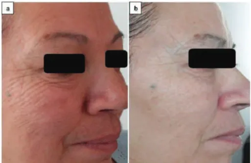
LLLT treatments were performed on 10 subjects aged from 40 to 65 years, exhibiting wrinkles and the loss of firmness facial skin. As shown in Fig. 1, a women of 63 years, treated with LLLT for 30 min/ session (number of sessions 7) exhibited a significant attenuation of wrinkles and crow’s feet, lines were visibly smoothed and the skin’s volume and elasticity was largely restored. The success of LLLT radiation in reducing the signs of facial skin aging was detected in all subjects. The treatment is painless and risk-free.
LLLT exposure reduces UVA or UVB-induced cytotoxicity and cell death in HeLa cells
The results showed that cell viability significantly decreased after UVA or UVB irradiation by 28.2% and 20.48%, respectively, when compared with con-trol cells (not exposed). However, the treatment of cells with LLLT prior to or post UVA or UVB ir-radiation increased cell viability. Pretreatment by laser radiation increased the viability of HeLa cells irradiated by UVA or UVB by 17.02% and 14.43%, respectively. Post-treatment by laser radiation in-creased the cell viability of cells exposed to UVA or UVB by 15.21% and 15.3%, respectively. The cell
viability was 95% when cells were treated only with LLLT (Fig. 2A).
Furthermore, cytotoxicity was quantified by the measurement of LDH released to the culture media from injured cells. According to Fig. 2B, there was a significant increase in LDH leakage after UV expo-sure. This response was significantly reduced by the treatment of cells with LLLT prior to or post UV radi-ation. Pretreatment by laser radiation decreased the release of LDH from HeLa cells irradiated by UVA or UVB by 33.49% and 30.39%, respectively. Post-treat-ment by laser radiation decreased the LDH leakage from cells exposed to UVA or UVB by 29.51% and 29.38%, respectively. Irradiation with LLLT alone did not result in LDH release.
DISCUSSION
We initially investigated the effect of LLLT on the clinical signs associated with aged skin, including wrinkling, lines and the plumpness of the skin. These changes are considered to result from intrinsic ag-ing associated with reduced cellular proliferative capacity and are accelerated by extrinsic factors,
Fig. 1. Face photos of the same patient (63 years old), showing (a) face before laser irradiation and (b) after laser irradiation (7 sessions
particularly sun exposure (Yaar and Gilchrest, 2007; Fragkiski et al., 2012). Our results demonstrated that after treatment with LLLT radiation, persons aged from 40 to 65 years showed a significant attenuation of their wrinkles. and the skin’s volume and elasticity was largely restored. Additionally, our investigation to assess the photoprotective effects of LLLT against UV-cell damage showed that the treatment of cells with LLLT prior to or post UVA or UVB irradiation increased cell viability. These results suggested that the pretreatment by LLLT radiation prevented cells from death and the post-treatment rescued cells al-tered by UV exposure from death and thus altering cell fate. Our results also indicated that LLLT source
light is non-toxic to HeLa cells. Taken together, the results demonstrated the beneficial effects of LLLT in relieving the signs of skin aging, and on the cellu-lar level, LLLT has double effects; it prevented and also protected HeLa cells against UV-mediated cell death. Our results are consistent with other findings that demonstrated that LLLT facilitates collagen syn-thesis, keratinocyte cell motility and growth factor release, increases the regenerative potential of bio-logical tissues and increases the neovascularization and formation of regenerative tissue (Yu et al., 1994; Kreisler et al., 2002; Basso et al., 2013). Others au-thors suggest that the mechanism of low-level lasers is based on the absorption of laser-emitted photons
Fig. 2. Effects of LLLT pretreatment or post-treatment on the viability of HeLa cells irradiated by UVA or UVB. (A) LLLT pretreatment
(2 h) or post-treatment (2 h) increased the viability of cells irradiated with UVA (n) or UVB (n). (B) LLLT pretreatment (2 h) or post-treatment (2 h) decreased the LDH release by cells irradiated with UVA (n) or UVB (n). Cells exposed only to laser radiation served as the positive control. Values are presented as the mean ± SEM. ** P<0.01 and *** P<0.001 compared with the UV group.
% o
f c
el
l v
ia
b
ili
ty
LD
H (U/L)
by the flavin mononucleotide enzyme (FMN) and cytochrome c oxidase, which are activators of cel-lular respiration. Through this absorption, the syn-thesis of ATP from ADP is accelerated, and redox mechanisms are excited, which in turn activates cel-lular metabolism and celcel-lular physiology (Lubart et al., 2005; Karu, 2008; Tafur and Mills, 2008). In this manner, LLLT may act to promote proliferation and/ or cellular homeostasis.
In summary, our results suggest that LLLT signif-icantly reduced the signs of facial skin aging, includ-ing wrinkles and the loss of firmness, prevented and protected human epithelioid cells from cell death in-duced by UV irradiation. We report here for the first time the benefits of LLLT on relieving facial signs of aging, and we believe that these results will be of clinical relevance in helping clinicians to prevent and to attenuate photoaging and the development of skin cancer.
Acknowledgments - We are grateful to Amira Hammami from “La Princesse”, Institut Medico-Esthetic, LASER ESTHETIC, for her investigation on clinical studies.
REFERENCES
Akgul, T., Gulsoy, M. and H.O. Gulcur (2014). Effects of early and delayed laser application on nerve regeneration. Lasers Med. Sci.29, 351–357
Armstrong, B.K. and A. Kricher (2001). The epidemiology of UV induced skin cancer. J. Photochem. Photobiol. B63, 8-18.
Basso, F.G., Oliveira, C. F., Kurachi, C., Hebling, J. and C.A. de Souza Costa (2013). Biostimulatory effect of low-level la-ser therapy on keratinocytes in vitro, Lasers Med. Sci.28, 367–374
Brosseau, L., Robinson, V., Wells, G., Debie, R., Gam, A., Harman, K., Morin, M., Shea, B. and P. Tugwell (2005). Low level laser therapy (Classes I, II and III) for treating rheumatoid arthritis. Cochrane Database Syst. Rev. 4 :CD002049.
Corazza, A.V., Jorge, J., Kurachi, C. and V. S. Bagnato (2007). Photobiomodulation on the angiogenesis of skin wounds in rats using different light sources. Photomed. Laser Surg. 25, 102-106.
Fragkiski, T., Myrto, T., Aikaterini, P., Konstantinos, K. and S. Dimitrios (2012). Extrinsic aging UV-mediated skin carci-nogenesis. Dermato. Endocrinolo. 4, 285-297.
Garcia, R.R. (2011). Atmospheric science: An Arctic ozone hole?
Nature478, 462-463.
Godar, D.E (2005). UV doses worldwide. Photochem. Photobiol. 81, 736-749.
Karol, M.H. (2009). How environmental agents influence the ag-ing process. Biomol. Ther.17 (2), 113-124.
Karu, T.I. (2008). Mitochondrial signaling in mammalian cells activated by red and near-IR radiation. Photochem.
Photo-biol. 84 (5), 1091-1099.
Kligman, L.H., Akin, F. J. and A. M. Kligman (1985). The contri-bution of UVA and UVB to connective tissue damage in hairless mice. J. Invest. Dermatol. 84, 272-284.
Kreisler, M., Christoffers, A. B., Al Haj, H., Willershausen, B. and
B. D’hoedt (2002). Low level 809-nm diode laser-induced in vitro stimulation of the proliferation of human gingival fibroblasts. Lasers Surg. Med.30(5), 365-369.
Lubart, R., Eichler, M. Lavi, R., Friedman, H. and A. Shainberg
(2005). Low-energy laser irradiation promotes cellular re-dox activity. Photomed. Laser Surg. 23, 3-9
Polefka, T.G., Meyer, T. A., Agin, P. P. and R. J. Bianchini (2012). Effects of solar radiation of the skin. J. Cosmet. Dermatol.
11(4), 329.
Schober-Flores, C. (2001). The sun’s damaging effects. Dermatol. Nurs.13(4), 279-286.
Tafur, J. and P. Mills (2008). Low-Intensity Light Therapy: Ex-ploring the Role of Redox Mechanisms. Photomed. Laser Surg. 26,323-328.
Tumilty, S., Munn, J., McDonough, S., Hurley, D. A., Basford, J. R.
and G. D. Baxter (2010). “Low Level Laser Treatment of Tendinopathy: A Systematic Review with Meta-analysis”.
Photomed. Laser Surg. 28 (1), 3.
Ty Hopkin, J., McLoda, T. A., Seegmiller, J. G. and D. Baxter (2004). Low-level laser therapy facilitates superficial wound heal-ing in humans: A triple-blind, sham-controlled study. J. Athl. Train. 39(3), 223-229.
Yaar, M. and B. A. Gilchrest (2007). Photoageing: mechanism, prevention and therapy. Br. J. Dermatol. 157, 874-887.