Recebido em 25.05.2001. / Received in May, 25thof 2001.
Aprovado pelo Conselho Consultivo e aceito para publicação em 22.05.2001. / Approved by the Consultive Council and accepted for publication in May, 22n dof 2002. * Trabalho realizado no Serviço de Dermatologia do Hospital Universitário Walter Cantídio (HUWC) - Universidade Federal do Ceará (UFC). / Work done at the Dermatology Service of the ”Hospital Universitário Walter Cantídio (HUWC) - Universidade Federal do Ceará (UFC)”.
1Residente em Dermatologia do HUWC. / Resident in Dermatology at the HUWC.
2Professor Assistente - Disciplina de Dermatologia- Faculdade de Medicina (UFC). / Assistant Professor - Dermatology Department, Faculty of Medicine (UFC).
3Professor-adjunto - Disciplina de Dermatologia - Faculdade de Medicina (UFC). Chefe do Serviço de Dermatologia do Hospital Universitário Walter Cantídio - UFC/ Adjunct Professor -Dermatology Department - Faculty of Medicine (UFC). Head of -Dermatology Service at the Hospital Universitário Walter Cantídio - UFC
©2003by Anais Brasileiros de Dermatologia
Ceratodermia mutilante de Vohwinkel: relato de três
casos em uma família
*
Vohwinkel´s mutilating keratoderma: report
of three familial cases
*
Lúcia Isabel de Sá Cavalcante
1Érica de Magalhães Holanda
1Thereza Lúcia Prata de Almeida
2José Wilson Accioly-Filho
3Resumo:A ceratodermia hereditária mutilante ou síndrome de Vohwinkel é afecção dermatológica rara caracterizada pelo espessamento cutâneo das palmas, plantas e dorso de mãos e pés, e por constrições em faixa dos dedos (pseudoainhum). São relatados três casos em uma família, envolvendo duas gerações. Em todos havia, adicionalmente, perda auditiva neurossensorial. Discutem-se o diagnóstico diferencial com ou-tras ceratodermias palmoplantares e o tratamento com retinóides. Trata-se da primeira descrição dessa enti-dade no Brasil.
Palavras-chave: ainhum; ceratodermia palmar e plantar
Summary:Keratoderma hereditarium mutilans, or Vohwinkel´s syndrome, is a rare cutaneous di
-sease characterized by thickening of the palms, soles and dorsa of the hands and feet, and by ainhum-like constriction of the fingers. This paper reports a case of three family members, over a span of two generations, who were affected by this disorder. Sensorineural deafness was also observed in each patient. We discuss differential diagnosis and retinoid treatment of this condition. This is the first case report on this disease to come from Brazil.
Key words: ainhum; keratoderma, palmoplantar.
INTRODUÇÃO
As ceratodermias palmoplantares hereditárias repre-sentam um grupo heterogêneo de doenças que se caracteri-zam pelo espessamento cutâneo difuso ou focal das palmas e plantas. Podem ser diferenciadas pelo modo de herança, idade de aparecimento, área afetada, anormalidades associa-das, achados histológicos, prognóstico e resposta terapêutica.1 A ceratodermia hereditária mutilante ou síndrome de Vohwinkel (SV) é doença de herança preferencialmente autossômica dominante (um tipo recessivo já foi relatado), descrita originalmente por Vohwinkel em 1929.2Em mea-dos da década de 1980, apenas 30 casos haviam sido rela-tados na literatura.3É mais freqüentemente observada em mulheres e em caucasianos, e em geral inicia-se na infân-cia. Caracteriza-se por: 1) hiperceratose palmoplantar difusa, com aparência de favo de mel; 2) faixas constritivas digitais
INTRODUCTION
Heriditary palmoplantar keratodermas represent a heterogeneous group of diseases characterized by diffuse or focused cutaneous thickening of the palms and soles. They can be differentiated by hereditary means, age of appearance, area affected, associated abnormalities, histo-logical findings, prognosis and therapeutic response.1
conhecidas como pseudoainhum;*e 3) placas ceratósicas em forma de estrela-do-mar no dorso de mãos e pés, também envolvendo os punhos, antebraços, cotovelos e joelhos. Esporadicamente, têm-se observado associações com alope-cia universal congênita ou cicatrialope-cial,4,5dermatoses ictiosifor-mes,3,6-8 paraplegia espástica e miopatia,9 perdas auditivas neurossensoriais e surdez,5,8-14 retardo mental,15,16 anormalida-des craniofaciais,11 acantose nigricante,17 bolhas plantares, coxins falangianos3,9,18,19e anormalidades ungueais.20,21
Os autores relatam três casos de SVem uma família (envolvendo duas gerações), associados com perda auditiva neurossensorial. São discutidos o diagnóstico diferencial e a terapêutica. Trata-se do primeiro relato dessa entidade neste país.
RELATO DOS CASOS Caso 1
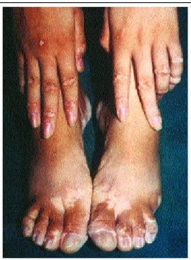
Paciente do sexo feminino, 17 anos de idade, bran-ca, estudante, nascida de parto normal sem complicações, sétima filha de uma família de 10 irmãos. Pais não consan-güíneos. Sua doença teve início aos dois anos de idade com o aparecimento de "calosidades" nas palmas e calcanhares. Segundo referia a paciente, as lesões progrediram continua-mente em forma de "verrugas", coalescendo até formar pla-cas espessas e difusas, pruriginosas, nas palmas e plantas. Concomitantemente houve aparecimento progressivo de constrições envolvendo os quiro e pododáctilos, tendo dois dígitos sido amputados cirurgicamente aos 11 anos de idade. Negava dificuldades auditivas. Pai e irmão têm doen-ça semelhante. Ao exame dermatológico, observavam-se lesões em placa, hiperceratóticas, eritematosas, nas palmas, plantas e faces dorsais das mãos e pés. As áreas de hiperce-ratose palmoplantar apresentavam aspecto de favo de mel (Figuras 1 e 2), enquanto as faces dorsais das mãos e torno-zelo exibiam placas lineares em forma de estrela-do-mar (Figura 3). No dorso dos dedos, cotovelos e joelhos eram observadas lesões papulosas, ceratóticas, semelhantes a verrugas (Figura 4), assim como amputação dos quintos pododáctilos direito e esquerdo, e constrição importante entre as falanges distais e proximais dos quartos quirodácti-los direito e esquerdo (Figura 4). O exame físico não reve-lou nenhuma outra anormalidade. Exames laboratoriais de rotina (hemograma completo, bioquímica do sangue, sumá-rio de urina, radiografia de tórax) foram normais. A audio-metria detectou perdas auditivas neurossensoriais bilaterais
known as pseudoainhum;* and 3) keratotic plaques in starfish-shapes on the dorsa of the hands and feet, which may also involve wrists, forearms, elbows and knees. There have been sporadic associations observed with congenital or cicatricial alopecia universalis,4,5 ichtyosiform der-matoses,3,6-8 spastic paraplegia and myopathy,9loss of neu-rosensorial hearing and deafness,5,8-14mental retardation,15,16 craniofacial abnormalities,11acanthosis nigricans,17plantar blisters, digital cushions3,9,18,19 and ungual abnormalities.20,21 The authors report three cases of SV in a family (spanning two generations), associated with loss of neu-rosensorial hearing. The diagnostic and therapeutic differential is discussed. This is the first case report on the disease from Brazil.
CASE REPORT Case 1
A 17-year-old Caucasian female patient, a student who was born from normal cesarean section without com-plications, the seventh daughter of a ten-child family. Parents are not consanguine. Her disease began at the age of two with the appearance of "callosities" in the palms and heels. According to the patient's account, the lesions pro-gressed continually in the form of "verrucas", until coalesc-ing into thick, diffuse and pruriginous plaques on the palms and soles. There was a concomitantly progressive appear-ance of constrictions involving the fingers and toes, with two digits being surgically amputated at age eleven. The patient denies having experienced hearing problems. The father and brother have a similar disease. In the dermatologic exam, hyperkeratotic, erythematous lesions in plaques were observed in the palms, soles and dorsa of the hands and feet. The hyperkeratotic palmoplantar areas demonstrated a hon-eycomb-like aspect (Figures 1 and 2), while the dorsa of the hands, and ankle showed linear plaques in a starfish shape (Figure 3). On the dorsa of the fingers, elbows and knees, papulous, keratotic and verruca-like lesions were observed (Figure 4), as was the amputation of the right and left fifth toe, and a major constriction between the distal and proxi-mal phalanges of the right and left fourth finger (Figure 4). The physical examination did not reveal any abnormality. Routine laboratory tests (complete biochemical blood hemo-gram, urine summary, thorax radiography) were normal. The audiometer detected a loss in bilateral neurosensorial hearing for acute high-frequency sounds. The radiological
*A palavra ainhum na língua falada pelos negros patuás
do Brasil significa "fissura". A nomenclatura da condição, contu-do, permanece confusa. Uma revisão literária demonstra que os termos "ainhum" e "pseudoainhum" são quase sempre usados
indistintamente. No entanto, Rook et al.30definem ainhum como
"o desenvolvimento de uma faixa constritiva ao redor de um dedo, ocorrendo espontaneamente no negro", enquanto pseudoainhum é "uma seqüência similar de eventos ocorrendo em uma variedade de condições ou como uma anomalia isolada do desenvolvimen-to". Por esse motivo, emprega-se neste texto o segundo termo.
* The word "ainhum" in the patois spoken by
Afro-Brazilians means "fissure". The terminology of the condition remains confused, however. A literature review shows that the term "ainhum" and "pseudoainhum" are almost always used
indistinctly. On the other hand, Rook et al.30define ainhum as "the
Figure 3: Linear plaques with starfish aspect (Case 1).
examination of the hands and feet demonstrated discrete bone rarefaction and osteoporosis distal from the constrict-ing band, and absence of distal phalanges in the right and left fifth toes. The histopathologic exam of the palmar region and keratotic papule of the knee revealed marked hyperker -atosis, hypergranulosis, moderate acanthosis and scarce superficial perivascular mononuclear cellular infiltrate. Based on clinical and laboratory findings, the diagnosis of Vohwinkel's syndrome associated with neurosensorial hear-ing loss was established. The patient was treated with etreti-nate, initially 1 mg/kg daily dose. There was progressive reduction of hyperkeratosis and partial regression of the band constrictions, especially observable after the three first months of treatment. Later, the dose was tapered to 0.5 mg/kg daily, with maintenance of therapeutic results. Six months after initiation, the treatment had to be interrupted because of a lack of medication at the policlinic. However a few months later, the treatment was reinitiated, this time with acritretin, 0.4 mg/kg daily. Significant improvement was obtained again of the keratoderma, with reduction of the band constric-tions. During the entire treatment, the patient tolerated the use of these aromatic retinoids well, with mod-erate cheilitis occurring only. The laboratory exams continued until the normal limits. Contraceptive measures were adopted.
para sons agudos de elevada freqüência. O exame radiológi-co das mãos e dos pés demonstrou discretas rarefações ósseas e osteoporose distal às bandas constritivas, e ausência das falanges distais dos quintos pododáctilos direito e esquerdo. O exame histopatológico da região palmar e pápu-la ceratótica do joelho revelou marcada hiperceratose, hiper-granulose, acantose moderada e escasso infiltrado celular mononuclear perivascular superficial. Baseado nos achados clínicos e laboratoriais, o diagnóstico de síndrome de Vohwinkel associada a perdas auditivas neurossensoriais foi estabelecido. A paciente foi tratada com etretinato, inicial-mente na dose de 1mg/kg/dia. Houve redução progressiva da hiperceratose e regressão parcial das constrições em banda, especialmente observáveis após os três primeiros meses de tratamento. Posteriormente, a dose
foi reduzida para 0,5mg/kg/dia, com manutenção dos resultados terapêuti-cos. Seis meses após o início do tra-tamento foi obrigada a interrompê-lo por falta do medicamento no posto de saúde. Entretanto, alguns meses após, reiniciou-o, dessa vez com aci-tretina 0,4mg/kg/dia. Obteve nova-mente melhora importante da cerato-dermia e redução das constrições em faixa. Durante todo o tratamento, tolerou bem o uso desses retinóides aromáticos, ocorrendo apenas queili-te moderada. Os exames laborato-riais permaneceram nos limites da normalidade. Medidas contracepti-vas foram adotadas.
Figura 1: Hiperceratose palmar difusa em favo de mel e constrição anular (pseudoainhum) no quinto quirodáctilo esquerdo (Caso 1). / Figure 1: Diffuse palmer honeycomb hyperkeratosis and ring-shaped constriction (pseudoainhum)
in the fifth left finger (Case 1).
Figura 2: Hiperceratose plantar difusa, padrão em favo de mel (Caso 1). / Figure 2: Diffuse plantar hyperkeratosis, honeycomb
pattern (Case 1).
Figura 5: Hiperceratose difusa em favo de mel com numerosas fissuras na região plantar
(Caso 2).
Figure 4: Keratotic papules in the dorsa of the toes and absence of the fifth right and left toes and ring-shaped constriction (pseudoainhum) in the fourth right and left toes (Case 1).
Figura 4: Pápulas ceratóticas no dorso dos dedos dos pés e ausência dos quintos pododáctilos direito e esquerdo e
constrições anulares pseudoainhum) nos quartos pododáctilos direito e esquerdo (Caso1).
Caso 2
Paciente do sexo masculino, 13 anos de idade, estudante e agricul-tor, nascido de parto normal sem complicações. É o nono filho de uma família de 10 irmãos. Referia que desde os dois anos de idade apresen-tava espessamento da pele nas regiões plantares. Aos cinco anos surgiram pápulas ceratóticas no dorso dos dedos das mãos e pés, joe-lhos e cotovelos. Aos 10 anos, tor-nou-se evidente o envolvimento pal-mar. Queixava-se do surgimento de
fissuras dolorosas nas regiões palmoplantares. A dor melho-rava com o uso de "sebo-gordura animal". Negava prurido nas lesões e deficiência auditiva. Ao exame dermatológico, observaram-se hiperceratose difusa de média intensidade nas regiões palmoplantares com aspecto em favo de mel (Figura 5); placas ceratóticas na face dorsal das regiões do metacar-po e metatarsofalangianas, cotovelos, joelhos e maléolos; lesões ceratóticas, de aspecto linear, tipo estrela-do-mar, nos calcâneos e tornozelos. Exames laboratoriais: hemograma completo, bioquímica do sangue, sumário de urina - sem alte-rações. Histopatológico: pele do tipo acral, exibindo hiperce-ratose compacta, acantose irregular, hipergranulose e papilo-matose, compatível com ceratodermia. Audiome tria: perda auditiva de leve a moderada à esquerda e de moderada a grave à direita para sons agudos de alta freqüência (padrão neurossensorial). Foi prescrito o uso de tópicos à base de uréia e ácido salicílico. Não foi possível o emprego do etreti-nato porque o paciente residia a longa distância e tinha difi-culdades para retornar ao
ambulatório.
Caso 3
Paciente do sexo mas-culino, 57 anos, agricultor, pri-meiro filho de uma família de 10 irmãos. Informa início da doença aos seis anos de idade
Case 2
A 13-year-old male student and farmer, born of normal cesarean section without complications. He is the ninth son of a ten-sibling family. He reported experiencing thickening of the skin in the sole regions ever since the age of two. At the age of five keratotic papules arose on the dorsa of the hands and feet, knees and elbows. At the age of ten, involvement of the sole became evident. He com-plained of the emergence of painful fissures in the palmoplantar regions. Pain subsided with the use of "animal sebum-fat". He denied having pruritus in the lesions and experiencing hearing defi-ciency. In the dermatologic exam, medium-intensity diffuse hyperkeratose in the palmoplantar regions with a honeycomb aspect was observed (Figure 5); keratotic plaques in the dorsa of the metacarpal and metatarsophalangeal regions, elbows, knees and malleolus; keratotic lesions of a linear aspect, starfish-type, in the calcaneum and ankle. Laboratory exams: complete hemogram, biochemical assay for the blood, urine test summary - without alterations. Histopathologic exam: acral skin type, exhibiting compact hyperkeratosis, irregular acanthosis, hypergranulosis and papilomatosis, compatible with keratoderma. Audiometer: mild-to-moderate loss of hear-ing on the left side and moderate-to-severe on the right for acute high-frequency sounds (neurosensorial standard). Use of urea- and salicylic-acid-based topics were prescribed. It was not possible to use etretinate because the patient lives far from the outpatient's clinic and has difficulty commuting there.
Case 3
A 57-year-old patient, a farmer, the first-born child to a ten-children family. He reported the onset of the dis-ease at the age of six, with emergence of keratotic papules in the dorsa of the
hands and feet. At age 10 he referred to thickening of the skin in the palms and soles. At age 20 he observed the devel-opment of band constrictions in the fifth toe, incapacitating him to work. Some time later, he submitted to amputation of both digits. He referred to a reduction of bilateral acute hearing 15 years ago (more accentuated on the right side). Upon the physical examination, standard honeycomb-shaped palmoplantar hyperkeratosis with painful fissures, paupulous lesions, of linear distribution (in starfish) on the dorsa of the hands and calcaneous, keratotic papules over the bone eminence of the metacarpal and metatarsophal-nageal articulations, elbows, knees and malleolous regions. Bilateral absence of the fifth toes. Laboratory exams: com-plete hemogram, biochemical assay of the blood - without alterations. Radiological exam of the hands and feet reveal loss of the fifth right and left toes. The histologic exam demonstrated acanthotic epidermis with hypergranulosis and papilomatosis, thick corneal layer with compact ortho-keratosis -- a diagnosis compatible with keratoderma. The audiometer showed moderate to severe loss of hearing on the right side and mild deafness on the left, for acute high-frequency (neurosensorial) sounds. As with the previously described patient, use of keratolitic topics was prescribed (urea cream 15%, salicylated vaseline 5%). It was not pos-sible to use retinoids, because the patient did not dispose of the financial conditions to commute to the capital, Fortaleza (in Ceara State (CE)) so as to undergo adequate clinical and laboratory follow-up.
DISCUSSION
Clinical findings in the three cases correspond to those described in mutilating hereditary keratoderma or Vohwinkel's syndrome. It shows soft keratoderma of the palms and soles (honeycomb-like), with other areas of involvement, including the extensor side of the knees, elbows, areas over the metacarpalphalangian articulations and Achilles tendon (i.e. keratotic papules occasionally dis -posed in linear plaques producing starfish images). Two of the patients (father and daughter) showed constricting bands around some digits of the hands and/or feet, both having undergone surgical amputation of a few digits. In all patients, manifestation began at pre-school age (at about two years of age) and became increasingly severe until ado-lescence. Individual differences were nonetheless made present, like the development of these constricting bands in the father at around age 20, while his daughter developed them at about age 10. Such findings correspond to those described in the literature.1-21
Patient heredograms confirmed the dominant pat-tern of transmission such as described in the literature.1
The large variability in phenotypic expression of the disease and associated clinical findings has been described in patients with Vohwinkel's syndrome.1-21At least two vari-ants are currently accepted. The first is associated with gen-eralized ichthyosis, with no deafness. Linear keratotic
com surgimento de pápulas ceratóticas no dorso das mãos e pés. Aos 10 anos referia espessamento da pele das palmas e plantas. Aos 20 anos observou desenvolvimento de constri-ções anulares nos quintos pododáctilos, que o incapacitavam para o trabalho. Algum tempo após, submeteu-se a amputação de ambos os dígitos. Referia diminuição da acuidade auditiva bilateralmente há 15 anos (mais acentuada no lado direito). Ao exame físico, apresentava hiperceratose palmoplantar padrão em favo de mel com fissuras dolorosas, lesões papulo-sas, de distribuição linear (em estrela-do-mar) no dorso das mãos e calcâneos, pápulas ceratóticas sobre eminências ósseas das articulações metacarpo e metatarsofalangianas, cotovelos, joelhos e regiões maleolares. Ausência bilateral dos quintos pododáctilos. Exames laboratoriais: hemograma completo, bioquímica do sangue - sem alterações. Exame radiológico de mãos e pés revela perda dos quintos pododác-tilos direito e esquerdo. O exame histopatológico demonstrou epiderme acantótica, com hipergranulose e papilomatose, espessa camada córnea com ortoceratose compacta, sendo o diagnóstico compatível com ceratodermia. Na audiometria foi verificado perda auditiva de moderada a grave à direita e leve à esquerda, para sons agudos, de alta freqüência (neurossen-sorial). Tal como para o paciente previamente descrito, foi prescrito o uso de tópicos ceratolíticos (creme de uréia a 15% , vaselina salicilada a 5%). Não foi possível o uso de retinói-des, pois o paciente não dispunha de condições financeiras para deslocar-se à capital (Fortaleza, CE) e, assim, fazer seguimento clínico-laboratorial adequado.
DISCUSSÃO
Os achados clínicos nos três casos correspondem aos descritos na ceratodermia hereditária mutilante ou síndrome de Vohwinkel. Apresentavam maciça ceratodermia das pal-mas e plantas (em favo de mel), com outras áreas de envolvi-mento, incluindo as faces extensoras dos joelhos, cotovelos, áreas sobre as articulações metacarpofalangianas e tendão de Aquiles (pápulas ceratóticas ocasionalmente dispostas em placas lineares produzindo imagens em estrela-do-mar). Dois dos pacientes (pai e filha) apresentavam faixas constritivas ao redor de alguns dígitos das mãos e/ou pés, tendo sido realiza-da, em ambos, amputação cirúrgica de alguns deles. Em todos os pacientes, o quadro teve início na idade pré-escolar (por volta dos dois anos de idade) e progrediu em gravidade até a adolescência. Algumas diferenças individuais, no entanto, mostravam-se presentes, como o desenvolvimento dessas fai-xas constritivas no pai por volta dos 20 anos de idade, enquan-to sua filha as desenvolveu em enquan-torno dos 10 anos de idade. Tais achados correspondem aos descritos na literatura.1-21
O heredograma dos pacientes confirma o padrão dominante de transmissão tal como descrito na literatura.1
linea-Heredograma da família
Family Heredogram
res configuradas com forma de estrela-do-mar não são observá-veis. Define-se como variante ictiótica ou de Camisa.3 Mutações nos genes que codificam a loricrina (cromossoma 1q21), uma proteína rica em glicina envolvida na formação do envelope córneo celular (estrutura rígida e insolúvel formada abaixo da membrana plasmática dos ceratinócitos durante a diferenciação terminal), foram descritas.22-25A segunda, na qual se enquadram os três pacientes descritos neste artigo, está asso-ciada com perda auditiva neurossensorial. Acredita-se que seja conseqüente à mutação no gene GJB2, que codifica a proteína de fenda de junção, conexina 26 (CX26).26 Mutações nesse gene também podem determinar surdez não sindrômica. Não está esclarecida a forma como duas diferentes doenças podem originar-se de diferentes mutações dentro do mesmo gene.27A síndrome de Vohwinkel é o resultado direto de defeito no pro-cesso de diferenciação terminal (ceratinização). Alguns traba-lhos têm observado aumento nos níveis de betaglucoronidase (sangue, urina, pele), que costuma estar elevada em condições em que haja aumento da proliferação celular.28 Estudos ultra-estruturais efetuados por Palungwachira et al., em 1992,
revela-ram que as células espinhosas e granulosas da epiderme das regiões afetadas continham mitocôndrias tumefeitas e muitos desmossomas, e que os corneócitos continham numerosos grâ-nulos aderidos à membrana e vacúolos lipídeos, o que resulta-ria em ceratinização patológica.29
Os achados histopatológicos não são específicos.2-7,8-21 Costuma-se observar hiperceratose ortoceratótica, hipergra-nulose, acantose com papilomatose e, ocasionalmente, um discreto infiltrado crônico na derme superficial. Estes, cor-respondem aos observados nos pacientes aqui relatados.
O diagnóstico diferencial da síndrome de Vohwinkel inclui outros tipos de ceratodermia que podem estar asso-ciados com auto-amputação dos dígitos: síndrome de Olmsted, ceratodermia acral, paquioníquia congênita, cera-todermia palmoplantar de Sybert, mal de Meleda e cerato-dermia palmoplantar de Gamborg-Nielsen.1
Outras doenças não hereditárias que podem causar faixas constritivas com ou sem hiperceratose incluem: han-seníase, sífilis terciária, framboesia, ainhum, esclerodermia, faixas amnióticas, envenenamento por ergotamina, tumores da medula espinhal,
síndro-me de Reynold e câncer da mama, siringomielia, entre outras.30,31
O tratamento da sín-drome de Vohwinkel visa ao alívio paliativo da ceratoder-mia e prevenção da auto-amputação dos dígitos. A
papules configured in starfish form are not observable. It was defined as an ichthyotic or Camisa variant.3Mutations were described in the genes codifying loricrin (chromosome 1q21): a protein rich in glycine involved in the formation of the cellular cornea envelop (i.e. the rigid and insoluble struc-ture formed below the plasmatic membrane of the ker-atinocytes during terminal differentiation). 22-25 The second, which the three patients described in this article manifested, is associated with loss of sensorineural hearing. It is believed to be a consequence of the mutation in gene GJB2 that codi-fies the joint cleft protein, connexin-26 (CX26).26Gene muta-tions might also determine non-syndrome deafness. It is unclear how these two different diseases could originate in different mutations within the same gene.27Vohwinkel's syn-drome is the direct result of a processing defect in terminal differentiation (keratinization). Some papers have observed an increase in betaglucoronidasis levels (blood, urine, skin), which is usually high in conditions where cellular prolifera-tion increases.28 Ultrastructural studies carried out by Palungwachira et al. in 1992 revealed that thorny-headed and granulous cells of the epidermis in affected regions con-tained tumefied mitochondria and many desmosomes, and that corneocytes contained numerous granules adhered to the membrane and lipid vacuoles, which results in patholog-ical keratinization.29
Histopathologic findings are not specific.2-7,8-21It is cus-tomary to observe orthokeratotic hyperkeratosis, hypergranu-lose, acanthosis with papillomatosis and, occasionally, a dis-crete chronic infiltrate in the superficial dermis. These corre-spond to those observed in the patients discussed in this case report.
The differential diagnosis of Vohwinkel's syndrome includes other types of keratoderma that can be associated with auto-amputation of the digits: Olmsted syndrome, acral keratoderma, congenital pachyonychia, Sybert's pal-moplantar keratoderma, mal de Meleda and Gamborg-Nielsen palmar and plantar keratoderma.1
Other non-hereditary diseases that may cause cons-tricting bands with or without hyperkeratosis include: leprosy, terciary syphilis, frambesia, ainhum, scleroderma, amniotic bands, ergotamine poisoning, spinal medulla tumors, Reynold's syndrome and breast cancer, syringo-myelia among others.30,31
against auto-amputation of the digits. Surgical release of the constricting bands may be used to prevent auto-ampu-tation, which is without a doubt the most incapacitating complication of this disease.32 Up to the retinoid era, the treatment of this condition was limited to the use of local keratolitic (urea and salicylic acid) and surgical interven-tion.5,19
Founded in reports on the beneficial use of aro -matic retinoids in mal de Meleda and non-bullous ichthyosiform keratoderma with ring-shaped digital con-strictions, Chang Sing Pang et al. in 19819 successfully treated a patient with mutilating hereditary keratoderma with etretinate. They observed that there was resolution of the constricting bands (pseudoainhum) in the course of treatment. A series of citations on the use of etritinate and isotretinate follows, with reversion of the keratoderma and pseudoainhum, though relapse did occur once medication was suspended.3,8,33 The first patient focused on in this paper made use of etretinate and, later, acitretine, and obtained results similar to those cited in the literature. In spite of the palliative action of the retinoids in relation to keratoderma and the fact that the patient was at fertile age, which obligated the authors to carefully instruct her to use contraceptive medication, use of the drug was chosen so as to reduce the incapacity provoked by this dis -ease. Above all, surgery was avoided with all its associat-ed risks as a way to correct the constricting bands. The other two family members (father and brother) were not given follow-up treatment due to the distance at which they lived from the outpatient's clinic and the difficulties in commuting there.
The therapeutic effect of retinoids are believed to lie in the reduction of the epithelial cell cohesion and inhibi-tion of pathologic keratinizainhibi-tion.35Cole19 believes that the primary determining factor of pseudoainhum would be excessive and abnormal keratinization.36Therefore, it seems that the pathological cornification in Vohwinkel's syndrome may lead to digital constrictions and that, as observed in the cases reported in this paper and by other authors, this process may be reverted by aromatic retinoids. Even so, it has been demonstrated that the constricting bands consist of fibrous conjunctive tissue, resembling cicatricial tissue, which raises doubts as to the way the aromatic retinoids may revert this process.37
To conclude, three case reports in one family were presented, involving two generations affected by mutilating hereditary keratoderma or Vohwinkel's syndrome. All showed neurosensory deafness. The use of aromatic reti-noids, in spite of the palliative care, led to improving the quality of life of one of these patients. q liberação cirúrgica das faixas de constrição pode ser utilizada
para prevenir a auto-amputação, o que é, sem dúvida, a com-plicação mais incapacitante dessa doença.32Até a era dos reti-nóides, o tratamento dessa condição limitava-se ao uso de agentes ceratolíticos locais (uréia e ácido salicílico) e inter-venção cirúrgica.5,19
Fundamentados em relatos do uso benéfico de reti-nóides aromáticos no mal de Meleda e na ceratodermia ictiosiforme não bolhosa com constrições digitais anula-res, Chang Sing Pang et al., em 1981,9trataram com êxito
um paciente com ceratodermia hereditária mutilante com etretinato. E observaram que houve resolução das faixas constrictivas (pseudoainhum) ao longo desse tratamento. Seguiu-se uma série de citações do uso de etretinato e iso-tretinoína, com reversão da ceratodermia e pseudoainhum, embora ocorresse recaída uma vez suspensa a medica-ção.3,8,33 A primeira paciente aqui focalizada, tendo feito uso de etretinato e, posteriormente, de acitretina, obteve resultados semelhantes aos citados na literatura. Apesar da ação paliativa dos retinóides em relação à ceratodermia e do fato de a paciente estar em idade fértil, o que obrigou os autores a orientá-la cuidadosamente em relação a medi-das contraceptivas, optou-se por seu uso para reduzir a incapacitação provocada pela doença. Além do mais, evi-tar-se-ia uma intervenção cirúrgica, com riscos associa-dos, para a correção das faixas constritivas. Os outros dois membros da família (pai e irmão) não foram seguidos por residirem a longa distância e ter dificuldades para retornar ao ambulatório.
Acredita-se que o efeito terapêutico dos retinóides resida na diminuição da coesão celular epitelial e inibição da ceratinização patológica.35 Cole19 acredita que o fator primário determinante do pseudoainhum seja uma ceratini-zação excessiva e anormal.36Assim, parece que a cornifica-ção patológica na síndrome de Vohwinkel pode levar a constrições digitais e que, como observado nos casos apre-sentados e por outros autores, esse processo pode ser rever-tido por retinóides aromáticos. Mesmo assim, tem-se demonstrado que as faixas constritivas consistem de tecido conjuntivo fibroso, assemelhando-se a tecido cicatricial, o que levanta dúvidas quanto à maneira como os retinóides aromáticos podem reverter esse processo.37
Em suma, apresenta-se relato de três casos em uma família, envolvendo duas gerações, afetados pela ceratodermia hereditária mutilante ou síndrome de Vohwinkel. Todos apresentavam perda auditiva neuros-sensorial. O uso de retinóides aromáticos, apesar de paliativo, permitiu melhorar a qualidade de vida de um
Nat Genet 1996; 13:70-7.
23. Korge BP, Ishida - Yamamoto A, Pünter C, Dopping-Hepenstal PJC, Iizuka H, Stepheson A, Eady RAJ, Munro CS. Loricrin muta-tion in Vohwinkel´s keratoderma is unique to the variant with ichthyosis. J Invest Dermatol 1997; 109:604-10.
24. Armstrog DKB, Mckenna KE, Hughes AE. A novel inser-tional mutation in loricrin in Vohwinkel´s keratoderma. J Invest Dermatol 1998; 111:702-4.
25. Takahashi H, Ishida-Yamamoto A, Kishi A, Ohara K, Iisuka H. Loricrin gene mutation in a japanese patient of Vohwinkel´s syn-drome. J.Dermatol Sci 1999; 19: 44-7.
26. Maestrini E, Korge B, Ocana-Sierra J, Calzolari E, Cambiaghi S, Scudder PM, Hovnanian A, Monaco AP, Munro CS. A missense mutation in connexin 26, D66H, causes mutilating Keratoderma with sensorineural deafness (Vohwinkel´s syndrome) in the three unrelated families. Hum Mol Genet 1999; 8:1237-43.
27. White TW. Functional analysis of human Cx 26 mutations asso-ciated with deafness. Brain Res Brain Res Rev 2000; 32:181-3. 28. Hughes TJ, Hughes ML, Caldwell BD. Molecular quantification of human beta-glucuronidase levels in patient with Vohwinkel´s syndrome. J Am Podriatr Med Assoc. 2001; 91:114-20.
29. Palungwachira P, Iwahara K, Ogawa H, Keratoderma heredi-taria mutilans. Etretinate treatment and electron microscopic stud-ies. Australas J Dermatol 1992; 33:19-30.
30. Rook A, Wilkinson DA, Ebling FJG. Textbook of Dermatology. 3rd. ed. Oxford: Blackwell Scientific Publications, 1979: 1638-9
31. Wollina U, Graefe T, Oelzner P, Hein G, Schreiber G. Pseudoainhum of all fingers associated with Reynold´s syndrome and breast cancer: report of a case and review of the literature. J Am Acad Dermatol 2001; 44:381-4.
32. Pisoh T, Bhatia A, Oberlin C. Surgical conection of pseudo-ain-hum in Vohwinkel´s syndrome. J Hand Surg 1995; 20 B: 338-41. 33. Rivers J, Duke E, Justus D. Etretinate: mangement of keratoma hereditaria mutilans in four family members. J Am Acad Dermatol 1985; 13:43-9.
34. Goldfarb MT, Woo TY, Rasmussen JE. Keratoderma heredi-taria mutilans (Vohwinkel´s syndrome) : a trial of isotretinoin. Pediatr Dermatol 1985; 2: 216-8.
35. Dicken CH, Connolly SM. Systemic retinoids in dematology. Mayo Clin Proc 1982; 57:51-7.
36. Cole GJ. Ainhum: an account of 54 patients with special refer-ence to etiology and treatment. J Bone Joint Surg 1965; 47:43-51. 37. Burgdorf WHC. Ainhum and pseudoainhum. In: Fitzpatrick TB , Eisen AZ , Wolff K , eds. Dermatology in General Medicine, 2nd .ed. New York: McGraw-Hill, 2000; 1208-9.
REFERÊNCIAS / REFERENCES
1. Lucker GPH, Van De Kerkhof PCM, Steijlen PM. The heredi-tary palmoplantar keratoses: an updated review and classification. Br J Dermatol 1994; 131:1-14.
2. Vohwinkel KH. Keratoma hereditaria mutilans. Arch Dermatol Syphilol 1929; 158:354-64.
3. Camisa C, Rossana C. Variant of keratodermia hereditaria muti-lans (Vohwinkel ´s syndrome). Arch Dermatol 1984; 120:1323-8. 4. Bhatia K, Chaudhary S, Pahwa U, Mehrotra G. Keratoderma hereditaria mutilans with congenital alopecia universalis (atrichia congenita). J Dermatol 1989; 16:231-6.
5. Gibbs RC, Frank SB. Keratoderma hereditaria mutilans (Vohwinkel): differentiating features of conditions with constric-tion of digits. Arch Dermatol 1966; 94:619-25.
6. Wirt F. Keratoma hereditarium mutilans. Arch Dermatol Syphil 1930; 159:311-2.
7. Greschebin S. Observation on ainhum: does it exist as an enti-ty? Urol Cutan Rev 1936; 40:98-102.
8. Wereide K. Mutilating palmoplantar keratoderma sucessfully treated with etretinate. Acta Derm Venereol (Stockh) 1984; 64:566-9.
9. Chang Sing Pang AFI, Oranje AP, Vuzevki VD, Stolz E. Sucessful treatment of keratoderma hereditaria mutilans with an aromatic retinoid. Arch Dermatol 1981; 117:225-8.
10. Mc gibbon DH, Watson R. Vohwinkel´s syndrome and deaf-ness. J Laryngol Otol 1977; 91:853-7.
11. Sensi A, Bettoli V, Zampino M, Gandini E, Calzolari E. Vohwinkel´s syndrome (mutilating keratoderma) associated with craniofacial anomalies. Am J Med Genetics 1994; 50:201-3. 12. Peris K, Salvati E, Torlone G, Chimenth S. Keratoderma hereditarium mutilans (Vohwinkel´s syndrome) associated with congenital deaf-mutism. Br J Dermatol 1995; 132:617-20. 13. Solis RR, Diven DG, Trizna Z. Vohwinkel´s syndrome in three generations. J Am Acad Dermatol 2001; 44: 376-8.
14. Bell M, Hoede N, Schopf RE. [Pseudoainhum in Vohwinkel´s disease. Keratoma palmoplantare mutilans]. Hautarzt 1993; 44:738-41.
15. Krischnaram A, Reddy B, Cary B, Buranarachagan A. Vohwinkel´s disease. Indian J Dermatol Venereol Lepr 1984; 50:115-8.
16. Kikuchi I, Nagata T, Abe S. Keratosis palmoplantaris muti-lans. J. Dermatol 1976; 3:85-8.
17. Chadhuri A, Haldar B. Keratoderma hereditaria mutilans with acanthosis nigricans (Vohwinkel´s disease). Indian J Dermatol Venereol Lepr 1980; 46:229-304.
18. Schamroth JM. Mutilating Keratoderma. Int J Dermatol 1986; 25: 249-51.
19. Cole RD, Mccauley MG, Way BH. Vohwinkel´S keratoma hereditarium mutilans. Int J Dermatol 1984; 23:131-4.
20. Sierra JO, Blesa G, Montero E. Syndrome de Volwinkel (étude de quatre cas). Ann Dermatol Syphil 1975; 102:41-5.
21. Sekkat A, Benhayoune TS. A propos d´un cas de keratodermie ainhumoide et mutilante. Ann Dermatol Venereol 1980; 107:447-9.
22. Maestrini E, Monaco AP, Mcgrath JA, Ishida-Yamamoto A, Camisa C, Hovnanian A, Weeks DE, Lathrop M, Uitto J, Christiano AM. A molecular defect in loricrin, the major compo-nent of the cornified envelope, underlies Vohwinkel´s syndrome.
ENDEREÇO PARA CORRESPONDÊNCIA: / MAILINGADDRESS: José Wilson Accioly-Filho