Review article
A síndrome de Marfan: uma revisão geral
Shi-Min Yuan
I, Hua Jing
IIDepartment of Cardiothoracic Surgery, Jinling Hospital, School of Clinical Medicine, Nanjing University, Nanjing 210002, Jiangsu Province, People’s Republic of China
IPostdoctoral Researcher, Department of Cardiothoracic Surgery, Jinling Hospital, School of Clinical Medicine, Nanjing University, Nanjing 210002, Jiangsu Province, People’s Republic of China. IIProfessor and Head, Department of Cardiothoracic Surgery, Jinling Hospital, School of Clinical Medicine, Nanjing University, Nanjing 210002, Jiangsu Province, People’s Republic of China.
ABSTRACT
Marfan’s syndrome is an autosomal dominant condition with an estimated prevalence of one in 10,000 to 20,000 individuals. This rare hereditary connective tissue disorder affects many parts of the body. The diagnosis of Marfan’s syndrome is established in accordance with a review of the diagnostic criteria, known as the Ghent nosology, through a comprehensive assessment largely based on a combination of major and minor clinical manifestations in various organ systems and the family history. Aortic root dilation and mitral valve prolapse are the main presentations among the cardiovascular malformations of Marfan’s syndrome. The pathogenesis of Marfan’s syndrome has not been fully elucidated. However, ibrillin-1 gene mutations are believed to exert a dominant negative effect. Therefore, Marfan’s syndrome is termed a ibrillinopathy, along with other connective tissue disorders with subtle differences in clinical manifestations. The treatment may include prophylactic β-blockers and angiotensin II-receptor blockers in order to slow down the dilation of the ascending aorta, and prophylactic aortic surgery. Importantly, β-blocker therapy may reduce TGF-β activation, which has been recognized as a contributory factor in Marfan’s syndrome. The present article aims to provide an overview of this rare hereditary disorder.
RESUMO
Síndrome de Marfan é uma condição autossômica dominante com prevalência estimada de 1 em 10.000 a 20.000 indivíduos. É uma rara desordem hereditária do tecido conjuntivo que afeta muitas partes do corpo. O diagnóstico da síndrome de Marfan é feito de acordo com uma revisão dos critérios diagnósticos conhecida como a nosologia Ghent, por meio de uma avaliação abrangente, em grande parte baseada em uma combinação de pequenas e grandes manifestações clínicas em vários sistemas de órgãos e na história familiar. Dilatação da raiz aórtica e prolapso da valva mitral são as principais apresentações entre as malformações cardiovasculares da síndrome de Marfan. A patogênese da síndrome de Marfan não foi totalmente esclarecida, mas acredita-se que mutações genéticas de ibrillina-1 exercem um efeito negativo dominante. Portanto, a síndrome de Marfan é denominada como ibrilinopatia, juntamente com outras desordens do tecido conectivo, com sutis diferenças nas manifestações clínicas. O tratamento pode incluir β-bloqueadores proiláticos e bloqueadores dos receptores da angiotensina II, a im de retardar a dilatação da aorta ascendente e cirurgia proilática da aorta. De importância, a terapia com β-bloqueadores pode reduzir a ativação de TGF-β, que foi reconhecido como um fator contribuinte da síndrome de Marfan. O presente artigo visa proporcionar uma visão global desta rara desordem de hereditariedade.
KEY WORDS: Aortic aneurysm. Arachnodactyly. Connective tissue diseases. Marfan syndrome. Mitral valve prolapse.
PALAVRAS CHAVE: Aneurisma aórtico. Aracnodactilia.
Doenças do tecido conjuntivo. Síndrome de Marfan. Prolapso da valva mitral.
INTRODUCTION
Marfan’s syndrome is an autosomal dominant condition with an es-timated prevalence of one in 10,000 to 20,000 individuals. It is a rare hereditary connective tissue disorder that afects many parts of the body.1 here is no geographic, ethnic or gender predilection. It is also known as arachnodactyly, since this is one of the signs of Marfan’s syndrome, which is characterized by abnormally long, slender or spidery ingers and toes.2 Liu Bei (A.D. 161-223), the founder of the Shu Han dynasty of the hree Kingdoms, and also the protagonist of “he Romance of hree Kingdoms”, one of the famous ancient Chinese works,3 and for-mer Afor-merican President Abraham Lincoln (1809-1865) are thought to have had Marfan’s syndrome, in that they manifested several key clinical features.3,4 In 1896, Marfan’s syndrome was irst described in a 5.5 year-old girl by a French pediatrician named Antonin Marfan.5
Marfan’s syndrome: an overview
Sao Paulo Med J. 2010;128(6):360-6
361
METHODS
A literature search was conducted in the Lilacs, PubMed, Embase and Cochrane Library databases using the search term “Marfan’s syn-drome”. A manual search of abstracts of articles was made to identify those relating to the topic. he results are presented in Table 1.
CLINICAL MANIFESTATIONS
he clinical manifestations of Marfan’s syndrome become more ev-ident with age. he most common symptom of Marfan’s syndrome is myopia, and 60% of the individuals with Marfan’s syndrome have ec-topia lentis. Individuals who have Marfan’s syndrome are also at high-er risk of retinal detachment, glaucoma and early cataract formation. Other common symptoms of Marfan’s syndrome involve the skeleton and connective tissue systems, including joint laxity, dolichostenom-elia, pectus excavatum or pectus carinatum, and scoliosis.12 Cardiovas-cular malformations are the most life-threatening presentations of Mar-fan’s syndrome. Aortic root dilation and mitral valve prolapse are sig-niicant clinical indings in patients with Marfan’s syndrome,9 and have been recognized as being as prevalent as ocular defects in Marfan’s syn-drome.13 Sisk et al.14 found aortic root dilation and mitral valve prolapse in all their 13 cases of Marfan’s syndrome, which presented at ages of less than four years. Other cardiovascular manifestations in infants may include coarctation of the aorta, atrial septal defect, patent ductus arte-riosus, pulmonary artery stenosis, persistent left superior vena cava etc.15 he aorta is the principal site of the lesions; particularly, the aortic root
tends to develop dilation, aneurysm and dissection. In addition, mitral valve prolapse may also be observed.16 Aortic root dilation was found in 60% of a series of patients with Marfan’s syndrome (74% males, 33% females) while mitral valve prolapse was found in 91% (87% males, 100% females).13 he histopathology of the aortic wall is characterized by widespread fragmentation of the elastin component, and the elas-tin ibers are often thin. Aortic dissection, which is commonin Mar-fan’s syndrome, is usually due to an intimal tear in theproximal ascend-ing aorta, with dissection involvascend-ing the sinotubularjunction and aortic sinuses, thus resulting in prolapse of one ormore commissures.16 he principal pathological indings from the mitral valve are annular dila-tion, ibromyxomatous changes to the lealets and chordae, elongation and rupture of chordae and deposition of calcium.17
PATHOGENESIS
he pathogenesis of Marfan’s syndrome has not been fully elucidat-ed. However, ibrillin-1 gene mutations are believed to exert a dominant negative efect.18 Fibrillin is an extracellular matrix protein that forms a major component of microibrils of the extracellular matrix of both elas-tic and non-elaselas-tic connective tissues and which is essential for normal elastic ibrillogenesis.19 he ibrillin-1 gene contains 65 exons, located at chromosome 15q-21.1. Fibrillin-1 mutation disrupts microibril for-mation, thereby resulting in ibrillin protein abnormalities, and subse-quently weakening the connective tissue.20
Changes to the transforming growth factor-beta (TGF-β)-signaling pathway may lead to diverse Marfan phenotypes.21 he gene defect
ul-Database Search strategies Results
PubMed
Marfan’s syndrome 5293 found 5293 related
1800 case reports 44 clinical trials 234 comparative studies 3 meta-analyses
6 randomized controlled trials 633 reviews
2573 others Embase (Excerpta Medica
databases) (1966-2010)
Marfan’s syndrome 3801 found 3801 related
2371 articles 11 articles in press 0 Cochrane reviews 31 conference abstracts 147 conference papers 33 controlled clinical trials 111 editorials
207 letters 5 meta-analyses 69 notes
10 randomized controlled trials 654 reviews
105 short surveys 8 systematic reviews Lilacs (Literatura
Latino-Americana e do Caribe em Ciências da Saúde)
Marfan syndrome 116 found 116 related
6 case cohorts 53 case reports 1 classical article 1 clinical trial
1 comparative review study 7 prospective cohort studies 27 reviews
19 retrospective cohort studies 1 textbook
Cochrane Marfan syndrome 8 found 8 related 8 clinical trials
diagnosing conduit leakage, coronary artery aneurysm, distal aortic dis-sections and prosthetic valve dysfunction.33 he risk of aortic dissec-tion, which is the most serious manifestation of Marfan’s syndrome, in-creases as the aorta enlarges. herefore, elective composite graft surgery is recommended when the aortic root size reaches 60 mm, regardless of symptom status, or 55 mm in the presence of severe aortic regurgitation. Surgical replacement of the aortic root with a composite graft does not end the progress of the disorder.33
MRI delineates the presence and extent ofaortic aneurysms and reveals the relationship between the aneurysm andarch vessels. It also demonstrates intimal laps and individual lumina intypes A and B aor-tic dissections.34 MRI has been found to be equivalent to computed tomography for depictingaortic, dural and hip abnormalities in pa-tients who had not undergonesurgery, but superior to computed to-mography for postoperative evaluations onpatients who received aor-tic valve replacement with Björk-Shiley or St. Jude valves, which pre-clude adequate evaluation of the aortic root on computed tomography scans.35 MRI may also facilitate measurement of the distances between
Chart 1. Major and minor diagnostic indings relating to Marfan’s syndrome26
Major criteria
1. Enlarged aorta 2. Tear in the aorta 3. Dislocation of the lens 4. Family history of the syndrome
5. At least four skeletal problems, such as lat feet or curved spine
6. Dural ectasia (enlargement of the lining that surrounds part of the spinal cord)
Minor criteria
1. Short sightedness (myopia) 2. Unexplained stretch marks 3. Loose joints
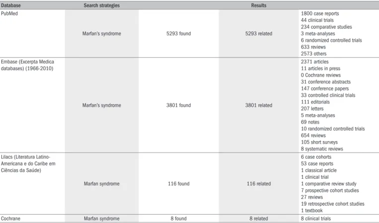
Figure 1. Aortogram showing dilated aortic root (arrow) of 5 cm in diameter, in a 44-year-old male patient with Marfan’s syndrome.
timately causes erratic binding between the ibrillin and connective tissue matrix. Mutations to transforming growth factor-beta recep-tor 2 (TGFβR2) in patients with Marfan’s syndrome type II (MFS2 mapped at 3p24.2-p25) have demonstrated alternative evidence for ab-normal TGF-β signaling in the pathogenesis of Marfan’s syndrome.22
Gene mutation may be absent in Marfan’s syndrome, and patients with the gene mutation do not necessarily develop the clinical manifes-tations of Marfan’s syndrome. he current theories support the notion that ibrillin mutations have an impact on the development of Mar-fan’s syndrome. However, only 28%-66% of the patients with MarMar-fan’s syndrome have been found to have ibrillin-1 mutations.23,24 Moreover, ibrillin-1 mutations have also been found in patients with familial aor-tic aneurysms, mitral valve prolapse or Marfan-like skeletal abnormali-ties, such as scoliosis or pectus excavatum.18 hey have also been found in a wide range of connective tissue disorders collectively known as ibrillinopathies, ranging from mild phenotypes, such as isolated ectopia lentis, to severe disorders including neonatal Marfan’s syndrome, which generally leads to death within the irst two years of life.25
DIAGNOSIS AND DIFFERENTIAL DIAGNOSIS
An early presentation of Marfan’s syndrome includes tall stature, ec-topia lentis, scoliosis, mitral valve prolapse, aortic root dilation and aor-tic dissection. he diagnosis of Marfan’s syndrome should be made in accordance with the revised diagnostic criteria, known as the Ghent no-sology, which involves major and minor diagnostic indings (Chart 1)26 and is largely based on clinical manifestations from various organ sys-tems and on the family history. A tall, thin body habitus, long limbs, arachnodactyly, pectus deformities and sometimes scoliosis with a posi-tive family history in a young individual may be suggesposi-tive of a diagno-sis of Marfan’s syndrome.27
Aortograms may show a dilated aortic root (Figure 1), or difuse an-eurysmal dilation of the ascending aorta in severe cases,26 here may be intimal laps in the presence of aortic dissection.28 In the early years, aor-tography demonstrated aortic dilation and aortic regurgitation, but this may occasionally miss the diagnosis of aortic dissection.29
Marfan’s syndrome: an overview
Sao Paulo Med J. 2010;128(6):360-6
363
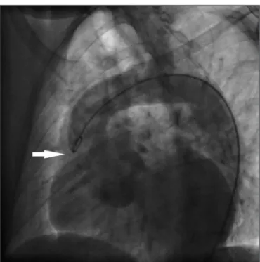
Figure 2. Echocardiography showing aortic root dilation to 67 mm in diameter on a long-axis view, in a 34-year-old male patient with Marfan’s syndrome.
section may occur at any time during pregnancy or even postpartum, but most often in the third trimester.53 Cesarean section is preferred in women with aortic dilation.26 An aortic root diameter of less than 40 mm would result in favorablematernal and fetal outcomes.54 he
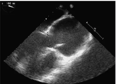
fe-Figure 3. Echocardiography on a 29-year-old male patient with Marfan’s syndrome showing (A) mitral valve prolapse (anterior mitral lealet lail) (arrow) on a four-chamber view, and (B) aortic root dilation and aortic dissection with an intimal lap (arrow) in the aortic cavity on a long-axis view.
Work-up Target
Echocardiogram, aortogram, magnetic resonance imaging and computed tomography
Measurement of the aortic root and detection of valve prolapse
Slit lamp examination Lens abnormalities
X-ray studies on skeletal system Evaluations of hand, spine, pelvis, chest, foot and skull for characteristic abnormalities Magnetic resonance imaging Dural ectasia
Prenatal testing At approximately 10-12 weeks, using chori-onic villus sampling, on a prospective parent who has Marfan syndrome
Genetic testing Genetic testing may be helpful, but is very costly and time-consuming for different gene mutations
Table 2. Main diagnostic work-up for Marfan’s syndrome
the non-coronary, right coronary and left coronary cusps and the aor-tic root area on short-axis views of the aoraor-tic root. An asymmetrical aortic root might be of clinical importance in unexpected aortic root dissection.36
Morphological studies on the aponeurosis have revealed a collagen-type diference in Marfan, compared with the usual Dupuytren disease, with fetal type III in the former, in which the collagen bundles were more dispersed and less compact and resistant, and adult type I in the latter.37 Oxidative research has revealed that aortic 8-isoprostane was 32-50% greater in the Marfan group than in the control, and that SOD-1 and SOD-2 expressions were decreased in Marfan aortas, while xan-thine oxidase, iNOS and the enzymatic subunits of NAD(P)H oxidase were increased.38 Genomic DNA analysis may occasionally miss the FBN1 mutations in Marfan patients.39 Reverse transcription PCR am-pliication of the ibrillin gene mRNA has detected a 123-bp deletion in afected individuals.40 In neonatal Marfan syndrome, FBN1 mutations have been noted in a region encompassing exons 24 to 32.19 Detection of enzymatic mutation is a more reliable and sensitive approach towards FBN1 mutation detection relating to Marfan’s syndrome. In this way, causative mutations, such as R565X and R1523X, and polymorphisms that were missed by heteroduplex analysis have been observed.41 Work-ups through which a diagnosis of Marfan’s syndrome can be made are listed in Table 2.
Among the diferential diagnoses for Marfan’s syndrome are ho-mocystinuria, familial mitral valve prolapse syndrome, familial annu-loaortic ectasia, isolated ectopia lentis, Ehlers-Danlos syndrome types II and III, Stickler syndrome (hereditary arthro-ophthalmopathy) and Klinefelter syndrome (Table 3).42-50
PREGNANCY
dis-Sao P
aulo Med J
. 2010;128(6):360-6
Jing H
Familial thoracic aortic aneurysm syndrome
Aortic root aneurysm and dissection Aneurysms of the thoracic aorta
No eye or musculoskeletal indings. No systemic manifestations of Marfan’s syndrome. Relevant family history in familial dissections.
No differentiating tests Breakdown of the extracellular matrix proteins elastin and collagen by proteases such as collagenase, elastase, various matrix metalloproteinases, and plasmin (formed from plasminogen by urokinase plasminogen activator and tissue-type plasminogen activator)45
Bicuspid aortic valve and ascending aortic aneurysm
Maximum dilation often occurs further up in the ascending aorta, beyond the sinotubular junction
Occasionally occurs with Marfan’s syndrome.
No eye or musculoskeletal indings Echo, thorax computed tomography or MRI can reveal signs of bicuspid aortic valve.
Inadequate production of ibrillin-1
Ehlers-Danlos syndrome An inherited heterogeneous group of connective tissue disorders, characterized by abnormal collagen synthesis, affecting skin, ligaments, joints, blood vessels and other organs. Marked joint hypermobility, papyraceous scars and mitral valve prolapse
Aortic aneurysm or aortic dissection at any age
Type IV variety most commonly affects the aorta, characterized by thin skin and bleeding disorders with increased bruising.
Skin biopsy for abnormal collagen and DNA testing for gene mutation.
EDS IV (mutations in the type III collagen gene), EDS VI (homozygous and compound heterozygous mutations in the lysyl hydroxylase gene), EDS VIIA and VIIB (mutations in the type I collagen genes), EDS VIIC (deiciency of procollagen N-proteinase) and EDS IX (decreased lysyl oxidase activity)46
Erdheim disease Loss of elastic and muscle ibers in the aortic media, with accumulation of mucopolysaccharide, sometimes in cyst-like spaces between the ibers.47
Aortic root dilation or rupture
No eye or musculoskeletal indings or family history.
No differentiating tests
Homocystinuria Marfanoid habitus, ectopia lentis, mental retardation and osteoporosis
Ectopia lentis A deiciency of cystathionine β synthase. Tall stature, long-bone overgrowth and ectopia lentis, but typically without aortic enlargement or dissection.
Raised concentrations of plasma homocystine
Mutations in the CBS, MTHFR, MTR and MTRR genes48
Loeys-Dietz syndrome Loeys-Dietz syndrome is a genetic disorder that affects blood vessels in the body, especially the aorta. It was irst described in the medical literature in January 2005.
Aortic dilation, aneurysm and dissection, and mitral valve prolapse
No associated lens dislocation. Aortic dissection occurs at much smaller diameter. Biid uvula or cleft palate, arterial tortuosity and hypertelorism
Not currently available Mutations in the genes encoding transforming growth factor beta receptor 1 (TGFBR1) or 2 (TGFBR2)
Shprintzen-Goldberg syndrome
Craniosynostosis (involving the coronal, sagittal or lambdoid suture), distinctive craniofacial features, skeletal abnormalities (dolichostenomelia, arachnodactyly, camptodactyly, pes planus, pectus excavatum, pectus carinatum, scoliosis, joint hypermobility or contractures), neurological abnormalities, mild-to-moderate mental retardation and brain anomalies
Mitral valve prolapse, mitral regurgitation and aortic regurgitation
Aortic root dilation is most likely not found. Not available Uncertain, but maybe ibrillin-1 mutations.
MASS phenotype Mitral valve prolapse, mild aortic dilation, striae atrophicae and skeletal involvement
Mitral valve prolapse, aortic root dilation and skin and skeletal conditions
Long limbs, deformity of the thoracic cage, striae atrophicae, mitral valve prolapse and mild and non-progressive dilation of the aortic root
Careful follow-up is needed to distinguish the MASS phenotype from emerging Marfan’s syndrome, especially in children.
Fibrillin-1 mutations
Congenital contractural arachnodactyly (Beals syndrome)49
Multiple lexion contractures, arachnodactyly, severe kyphoscoliosis, abnormal pinnae and muscular hypoplasia
Skeletal features with Marfan’s syndrome such as marfanoid habitus, arachnodactyly, camptodactyly and kyphoscoliosis
Multiple joint contractures (especially elbow, knee and inger joints), and crumpled ears in the absence of signiicant aortic root dilation are characteristic of Beals syndrome and rarely found in Marfan syndrome.
Crumpled appearance of ear helix and congenital contractures, typically without ocular and cardiovascular complications
A mutation in the ibrillin-2 gene on chromosome 5q23
Isolated ectopia lentis A rare syndrome characterized by dislocation of eye lenses, which often occurs at birth.
Ectopia lentis, maybe associated with mild skeletal indings.
Mental retardation, impaired joint mobility and stiff joints, but without cardiovascular abnormalities
No Fibrillin-1 mutations
Stickler syndrome (hereditary arthro-ophthalmopathy)
A relatively common genetic disorder characterized by very lexible (hyperextensible) joints, typical facial characteristics, hearing loss and severe nearsightedness with associated eye problems.
Tall stature, mitral valve prolapse and retinal detachment
Retrognathia, midfacial hypoplasia, without aortic involvement
No Mutation in the COL2A1 gene on chromosome 12 in region 12q13.11-q13.2
Klinefelter syndrome (47, XXY or XXY syndrome)
Men with Klinefelter syndrome present with sequelae of hormonal and spermatogenic testicular failure, such as infertility, low testosterone, erectile dysfunction and low bone mineral density
Marfanoid habitus Small testes and genitalia, and learning dificulty
Serum FSH levels were elevated 50
Marfan’s syndrome: an overview
Sao Paulo Med J. 2010;128(6):360-6
365
tus has a 50% chance of acquiring the disease.55 Cardiopulmonary by-pass during pregnancyis associated with maternal mortality of 3% and fetalmortality of 20%.56
MANAGEMENT
It was recently suggested that the current strategies for Marfan’s management should be blood pressure control and restrictions on phys-ical activities.57 Nevertheless, Marfan’s syndrome can be efectively man-aged if it is under integrated care provided by a team of specialists from all relevant specialties.58
Improvements in life expectancy could be achieved more readily through management of cardiovascular disorders, including mitral valve prolapse, aortic dilation and aortic dissection. he treatment may include prophylactic β-blockers to slow down the dilation of the ascending aorta, and prophylactic aortic surgery. Short to midterm follow-up results have suggested that β-blockers are useful for preventing progressive dilation of the aorta.59 he mean slope of the regression line for the aortic root di-mensions has been found to be signiicantly lower in the treatment group than in the controls.59,60 Recent research on the etiology of Marfan’s syn-drome in mouse models has suggested that aortic root dilation and ibril-lin-1 abnormalities are probably caused by excessive TGF-β signaling, and that TGF-β antagonists, including angiotensin II-receptor blockers, may signiicantly slow down the progression of aortic root dilation61 or pre-vent certain manifestations of Marfan’s syndrome, including aortic aneu-rysm.44 Ahimastos et al.62 observed that perindopril therapy may reduce arterial stifness, central and peripheral pulse wave velocities, aortic root diameters (in both end-systole and end-diastole) and TGF-β, among pa-tients with Marfan’s syndrome in comparison with placebo.
Prophylactic aortic root replacement with a composite graft is rec-ommended when aortic root dilation predisposes towards aortic rup-ture and the potential aortic dissection could theoretically have been prevented, as suggested by current guidelines: (1) aortic root diameter ≥ 55 mm; (2) positive family history of aortic dissections and aortic root diameter ≥ 50 mm; and (3) aortic root growth ≥ 2 mm/year.11
Aortoplasty with aortic valve replacement has been successful in in-fants with Marfan’s syndrome who presented annuloaortic ectasia and aortic regurgitation.63 However, the Bentall operation is a preferred pro-cedure for Marfan patients. From this, low operative mortality and long-term survival can be expected, with a survival rate of 80% after ive years and 60% after 10 years. Clinical observations have shown that compos-ite valve replacement and valve-sparing repair procedures were associat-ed with early postoperative mortality rates of 6.8% and 0%, respective-ly.64 Gillinov et al.65 reported, among patients with Marfan’s syndrome undergoing aortic root replacement, that the mean aortic root diameter was 6.2 ± 0.2 cm. Aortic root replacement was carried out in 96% of the cases, and a mitral valve procedure in 42%, with a 10-year survival rate of 79% ± 10%. Alexiou et al.66 found that the operative mortality rate was 6.1%, while the procedure led to 10-year freedom from throm-boembolism, hemorrhage and endocarditis of 88%, 89.8% and 98.4%, respectively. de Oliveira et al.67 achieved satisfactory freedomfrom reop-eration, which was 75% after 10 years for root replacementand 100%
for the valve-sparing patients, and excellent 10-year survival, which was 87%and 96% for the two procedures, respectively.
CONCLUSIONS
Marfan’s syndrome is a rare hereditary connective tissue disorder af-fecting many parts of the body. Establishment of a diagnosis of Marfan’s syndrome is based on the Ghent nosology, which involves comprehensive evaluation of major and minor systemic manifestations. he pathogenesis of Marfan’s syndrome has not been fully elucidated, but ibrillin-1 gene mutations are believed to exert a dominant negative efect through ex-cessive TGF-β signaling pathways. Cardiovascular malformations, chiely aortic root dilation and mitral valve prolapse, are the most life-threatening symptom of Marfan’s syndrome, since these patients are at risk of acute aortic dissection. Prophylactic aortic root replacement with a composite graft is recommended when the dilated aortic root has a tendency to rup-ture and the potential aortic dissection could theoretically have been pre-vented. Regular cardiovascular, ocular and skeletal surveillance by means of echocardiography, slit lamp examination of the eye, and magnetic reso-nance is recommended upon diagnosis or after the operation.
REFERENCES
1. Haneline M, Lewkovich GN. A narrative review of pathophysiological mechanisms associated with cervical artery dissection. J Can Chiropr Assoc. 2007;51(3):146-57.
2. Gelb BD. Marfan’s syndrome and related disorders--more tightly connected than we thought. N Engl J Med. 2006;355(8):841-4.
3. Department of Cardiac Surgery, Veteran General Hospital. Marfan Syndrome (MFS). Availa-ble from: http://www.marfan.org.tw/index.php?job=art&articleid=a_20061102_005118. Accessed in 2010 (Apr 16).
4. Marfan syndrome. Available from: http://science.jrank.org/pages/4127/Marfan-Syndro-me.html. Accessed in 2010 (Sep 29).
5. Van de Velde S, Fillman R, Yandow S. Protrusio acetabuli in Marfan syndrome. History, diag-nosis, and treatment. J Bone Joint Surg Am. 2006;88(3):639-46.
6. Yetman AT, Bornemeier RA, McCrindle BW. Long-term outcome in patients with Marfan syndrome: is aortic dissection the only cause of sudden death? J Am Coll Cardiol. 2003;41(2):329-32. 7. Pahuja D. Marfan syndrome: diagnosis and workup of cardiac manifestations. The Internet
Journal of Cardiology. 2006;3(1). Available from: http://www.ispub.com/journal/the_in- ternet_journal_of_cardiology/volume_3_number_1_9/article/marfan_syndrome_diagno-sis_and_workup_of_cardiac_manifestations.html. Accessed in 2010 (Sep 29). 8. Geva T, Hegesh J, Frand M. The clinical course and echocardiographic features of Marfan’s
syndrome in childhood. Am J Dis Child. 1987;141(11):1179-82.
9. Morse RP, Rockenmacher S, Pyeritz RE, et al. Diagnosis and management of infantile marfan syndrome. Pediatrics. 1990;86(6):888-95.
10. Silverman DI, Gray J, Roman MJ, et al. Family history of severe cardiovascular disease in Marfan syndrome is associated with increased aortic diameter and decreased survival. J Am Coll Cardiol. 1995;26(4):1062-7.
11. Groenink M, Lohuis TA, Tijssen JG, et al. Survival and complication free survival in Marfan’s syndrome: implications of current guidelines. Heart. 1999;82(4):499-504.
12. Tsipouras P, Silverman DI. The genetic basis of aortic disease. Marfan syndrome and beyond. Cardiol Clin. 1999;17(4):683-96.
13. Brown OR, DeMots H, Kloster FE, et al. Aortic root dilatation and mitral valve prolapse in Marfan’s syndrome: an ECHOCARDIOgraphic study. Circulation. 1975;52(4):651-7. 14. Sisk HE, Zahka KG, Pyeritz RE. The Marfan syndrome in early childhood: analysis of 15
patients diagnosed at less than 4 years of age. Am J Cardiol. 1983;52(3):353-8. 15. Papaioannou AC, Agustsson MH, Gasul BM. Early manifestations of the cardiovascular
disor-ders in the Marfan syndrome. Pediatrics. 1961;27:255-68.
17. Kirali K, Yakut N, Güler M, et al. Surgical treatment of siblings with Marfan syndrome. Asian Cardiovascular & Thoracic Annals. 1999;7(2):138-41. Available from: http://asianan-nals.ctsnetjournals.org/cgi/reprint/7/2/138?maxtoshow=&hits=10&RESULTFORMAT=& fulltext=aortic&searchid=1&FIRSTINDEX=180&resourcetype=HWFIG. Accessed in 2010 (Sep 29).
18. Erentuğ V, Polat A, Kirali K, Akinci E, Yakut C. Marfan sendromunda kardiyovasküler tutulum ve tedavi [Cardiovascular manifestations and treatment in Marfan syndrome]. Anadolu Kar-diyol Derg. 2005;5(1):46-52.
19. Robinson PN, Godfrey M. The molecular genetics of Marfan syndrome and related microi-brillopathies. J Med Genet. 2000;37(1):9-25.
20. Cañadas V, Vilacosta I, Bruna I, Fuster V. Marfan syndrome. Part 1: pathophysiology and diagnosis. Nat Rev Cardiol. 2010;7(5):256-65.
21. Alghamdi AA, Van Arsdell GS. Replacement of aortic root and ascending aorta in adult congenital heart disease. Expert Rev Cardiovasc Ther. 2007;5(6):1087-94.
22. Ramirez F, Dietz HC. Marfan syndrome: from molecular pathogenesis to clinical treatment. Curr Opin Genet Dev. 2007;17(3):252-8.
23. Fibrillin Mutations. Do they really cause Marfan syndrome? Available from: http://www.ctds. info/ibrillin.html. Accessed in 2010 (Set 29).
24. Loeys B, Nuytinck L, Delvaux I, De Bie S, De Paepe A. Genotype and phenotype analysis of 171 patients referred for molecular study of the ibrillin-1 gene FBN1 because of suspected Marfan syndrome. Arch Intern Med. 2001;161(20):2447-54.
25. Robinson PN, Booms P, Katzke S, et al. Mutations of FBN1 and genotype-phenotype corre-lations in Marfan syndrome and related ibrillinopathies. Hum Mutat. 2002;20(3):153-61. 26. Stanley P, Roebuck D, Barboza A. Takayusu’s arteritis in children. Tech Vasc Interv Radiol.
2003;6(4):158-68.
27. Dean JC. Marfan syndrome: clinical diagnosis and management. Eur J Hum Genet. 2007;15(7):724-33.
28. Slavotinek J, Kendall SW, Flower CD, et al. Radiological evaluation of the ascending aorta following repair of type A dissection. Cardiovasc Intervent Radiol. 1993;16(5):293-6. 29. McLeod AA, Monaghan MJ, Richardson PJ, Jackson G, Jewitt DE. Diagnosis of acute aortic
dissection by M-mode and cross-sectional echocardiography: a ive-year experience. Eur Heart J. 1983;4(3):196-202.
30. Come PC, Fortuin NJ, White RI Jr, McKusick VA. Echocardiographic assessment of cardio-vascular abnormalities in the Marfan syndrome. Comparison with clinical indings and with roentgenographic estimation of aortic root size. Am J Med. 1983;74(3):465-74. 31. Elkayam U, Ostrzega E, Shotan A, Mehra A. Cardiovascular problems in pregnant women with
the Marfan syndrome. Ann Intern Med. 1995;123(2):117-22.
32. Kiotsekoglou A, Sutherland GR, Moggridge JC, et al. The unravelling of primary myocardial im-pairment in Marfan syndrome by modern echocardiography. Heart. 2009;95(19):1561-6. 33. Aldrich HR, Labarre RL, Roman MJ, et al. Color low and conventional echocardiography of
the Marfan syndrome. Echocardiography. 1992;9(6):627-36.
34. Glazer HS, Gutierrez FR, Levitt RG, Lee JK, Murphy WA. The thoracic aorta studied by MR imaging. Radiology. 1985;157(1):149-55.
35. Soulen RL, Fishman EK, Pyeritz RE, Zerhouni EA, Pessar ML. Marfan syndrome: evaluation with MR imaging versus CT. Radiology. 1987;165(3):697-701.
36. Meijboom LJ, Groenink M, van der Wall EE, et al. Aortic root asymmetry in marfan patients; evaluation by magnetic resonance imaging and comparison with standard echocardiogra-phy. Int J Card Imaging. 2000;16(3):161-8.
37. Montagnani S, Passaretti U, Fusco M. Aspetti morfologici ed istoenzimatici dell’aponeurosi palmare da un paziente con sindrome di Marfan associata a contrattura di Dupuytren [Mor-phologic and hist-enzymatic aspects of palmar aponeurosis in a patient with Marfan syndrome associated with Dupuytren’s contracture]. Arch Putti Chir Organi Mov. 1989;37(2):355-62. 38. Yang HH, van Breemen C, Chung AW. Vasomotor dysfunction in the thoracic aorta of
Mar-fan syndrome is associated with accumulation of oxidative stress. Vascul Pharmacol. 2010;52(1-2):37-45.
39. Rantamäki T, Raghunath M, Karttunen L, et al. Prenatal diagnosis of Marfan syndro-me: identiication of a ibrillin-1 mutation in chorionic villus sample. Prenat Diagn. 1995;15(12):1176-81.
40. Godfrey M, Vandemark N, Wang M, et al. Prenatal diagnosis and a donor splice site mutation in ibrillin in a family with Marfan syndrome. Am J Hum Genet. 1993;53(2):472-80. 41. Youil R, Toner TJ, Bull E, et al. Enzymatic mutation detection (EMD) of novel mutations
(R565X and R1523X) in the FBN1 gene of patients with Marfan syndrome using T4 endo-nuclease VII. Hum Mutat. 2000;16(1):92-3.
42. Epocrates online. Marfan syndrome. Differential diagnosis. Available from: https://online.epocra-tes.com/u/2935514/Marfan+syndrome/Diagnosis/Differential. Accessed in 2010 (Sep 29). 43. Shapiro JR, Wright MJ. The Marfan syndrome. Available from:
http://cmbi.bjmu.edu.cn/up-todate/Valvular%20and%20aortic%20disease/Aorta/The%20Marfan%20syndrome.htm. Accessed in 2010 (Sep 29).
44. Judge DP, Dietz HC. Therapy of Marfan syndrome. Annu Rev Med. 2008;59:43-59. 45. MacSweeney ST, Powell JT, Greenhalgh RM. Pathogenesis of abdominal aortic aneurysm. Br
J Surg. 1994;81(7):935-41.
46. Tunçbilek E, Alanay Y. Congenital contractural arachnodactyly (Beals syndrome). Orphanet J Rare Dis. 2006;1:20.
47. Yeowell HN, Pinnell SR. The Ehlers-Danlos syndromes. Semin Dermatol. 1993;12(3):229-40. 48. Mondofacto dictionary. Erdheim disease. Available from: http://www.mondofacto.com/
facts/dictionary?Erdheim+disease. Accessed in 2010 (Sep 29).
49. Genetics home reference. Genetic conditions. Homocystinuria. Available from: http://ghr. nlm.nih.gov/condition=homocystinuria. Accessed in 2010 (Sep 29).
50. Juul A, Aksglaede L, Lund AM, et al. Preserved fertility in a non-mosaic Klinefelter patient with a mutation in the ibroblast growth factor receptor 3 gene: case report. Hum Reprod. 2007;22(7):1907-11.
51. Sakaguchi M, Kitahara H, Seto T, et al. Surgery for acute type A aortic dissection in pregnant patients with Marfan syndrome. Eur J Cardiothorac Surg. 2005;28(2):280-3; discussion 283-5. 52. Goland S, Barakat M, Khatri N, Elkayam U. Pregnancy in Marfan syndrome: maternal and fetal risk and recommendations for patient assessment and management. Cardiol Rev. 2009;17(6):253-62.
53. Keskin HL, Mungan T, Aktepe-Keskin E, Güngör T. Marfan syndrome in pregnancy: a case report. Ann Saudi Med. 2002;22(5-6):356-8.
54. Rossiter JP, Repke JT, Morales AJ, Murphy EA, Pyeritz RE. A prospective longitudinal evaluation of pregnancy in the Marfan syndrome. Am J Obstet Gynecol. 1995;173(5):1599-606. 55. Ryan-Krause P. Identify and manage marfan syndrome in children. Nurse Pract.
2002;27(10):26, 31-6; quiz 37.
56. Pomini F, Mercogliano D, Cavalletti C, Caruso A, Pomini P. Cardiopulmonary bypass in preg-nancy. Ann Thorac Surg. 1996;61(1):259-68.
57. Iams HD. Diagnosis and management of Marfan syndrome. Curr Sports Med Rep. 2010;9(2):93-8.
58. Dietz HC. Marfan syndrome. In: Pagon RA, Bird TC, Dolan CR, Stephens K, editors. GeneRe-views [Internet]. Seattle: University of Washington; 1993. Available from: http://www.ncbi. nlm.nih.gov/pubmed/20301510. Accessed in 2010 (Sep 29).
59. Tahernia AC. Cardiovascular anomalies in Marfan’s syndrome: the role of echocardiography and beta-blockers. South Med J. 1993;86(3):305-10.
60. Shores J, Berger KR, Murphy EA, Pyeritz RE. Progression of aortic dilatation and the be-neit of long-term beta-adrenergic blockade in Marfan’s syndrome. N Engl J Med. 1994;330(19):1335-41.
61. Brooke BS, Habashi JP, Judge DP, et al. Angiotensin II blockade and aortic-root dilation in Marfan’s syndrome. N Engl J Med. 2008;358(26):2787-95.
62. Ahimastos AA, Aggarwal A, D’Orsa KM, et al. Effect of perindopril on large artery stiffness and aortic root diameter in patients with Marfan syndrome: a randomized controlled trial. JAMA. 2007;298(13):1539-47.
63. Rammurthy DV, Iyer PU, Kumar A, Mohan M, Khandpur SC. Marfan’s syndrome in a neonate. Indian Pediatr. 1986;23(11):956-9.
64. Karck M, Kallenbach K, Hagl C, et al. Aortic root surgery in Marfan syndrome: Comparison of aortic valve-sparing reimplantation versus composite grafting. J Thorac Cardiovasc Surg. 2004;127(2):391-8.
65. Gillinov AM, Zehr KJ, Redmond JM, et al. Cardiac operations in children with Marfan’s syn-drome: indications and results. Ann Thorac Surg. 1997;64(4):1140-4; discussion 1144-5. 66. Alexiou C, Langley SM, Charlesworth P, et al. Aortic root replacement in patients with Marfan’s syn-drome: the Southampton experience. Ann Thorac Surg. 2001;72(5):1502-7; discussion 1508. 67. de Oliveira NC, David TE, Ivanov J, et al. Results of surgery for aortic root aneurysm in
pa-tients with Marfan syndrome. J Thorac Cardiovasc Surg. 2003;125(4):789-96.
Sources of funding: None Conlict of interest: None
Date of irst submission: July 15, 2009 Last received: April 29, 2010
Accepted: October 6, 2010
Address for correspondence: Prof. Hua Jing
Department of Cardiothoracic Surgery, Jinling Hospital, School of Clinical Medicine, Nanjing University,
Nanjing 210002, Jiangsu Province, People’s Republic of China Tel. 86 25 84801332