Clinical, pathophysiological and genetic aspects of inherited
tubular disorders in childhood
Authors
Emília Maria Dantas Soeiro 1
Claudia Maria de Barros Helou 2
1 Universidade Nove de Julho
2 Universidade de São Paulo.
Submitted on: 12/01/2014. Approved on: 02/24/2015.
Correspondence to: Claudia Maria de Barros Helou. Laboratório de Pesquisa Básica - LIM 12, Hospital das Clínicas da Faculdade de Medicina da Universidade de São Paulo. Av. Dr. Arnaldo, nº 455, sala 3310, São Paulo, SP, Brasil.
CEP: 01246-903. E-mail: [email protected]
I
NTRODUCTIONWater and electrolyte homeostasis and acid-base balance are regulated by the kidneys and play a vital role in bodily functions. To play such role, the cells in the renal tubules require separate ion channels, carriers, exchangers, cotransporters, and pumps to transport water and different solutes.1
Thus, genetic defects in any of these
DOI: 10.5935/0101-2800.20150060
In this review, we described the tubular function of each nephron segment followed by the most important changes that may occur in the transporters expressed therein. Thus, knowledge of the changes in renal tubular function allows the understanding and recognition of renal tubular diseases that can cause stillbirth or death in newborns or in childhood. Moreover, children with tubular disorders may progress to chronic renal disease at an early stage of life and they may also show disturbances of growth and development associate or not with neurological dysfunction. Therefore, we used the keyword "inherited tubular disorders" to select the children studies that have been published in the PubMed database since 2006. We hope that this review may help physicians to perform an early diagnosis in patients with tubular disorders allowing a specialized treatment and an improvement in their prognosis and quality of life.
A
BSTRACTKeywords: acidosis, renal tubular;
Bartter syndrome; electrolytes; Fanconi syndrome; Gitelman syndrome; sodium-potassium-exchanging ATPase.
transport systems may result in a wide range of nephropathies.2-5
Inherited tubulopathies are usually severe and may either cause the death of the fetus, neonate, or child affected by it or lead to the early onset of end-stage renal disease. Additionally, children diagnosed with inherited tubulopathy may also have growth disorders and delayed development associated or not with neurological impairment. Diagnosis is frequently delayed by nonspecific and often silent clinical symptoms. Therefore, knowledge of renal physiology, the molecular bases of tubular transport, and the possible pathophysiological alterations is required to clinically understand and recognize individuals with these diseases. In recent decades the developments in molecular biology and genetics have provided the tools to investigate the presence of inherited tubulopathies, thus improving the diagnosis, treatment, and prognosis of the affected children.2-5
P
ROXIMAL CONVOLUTED TUBULEThe proximal convoluted tubule is highly capable of reabsorbing mostly filtered solutes connected to sodium reabsorption. Approximately two thirds of the filtered Na+ is reabsorbed in this segment of
the nephron mainly via the paracellular pathway. However, active transcellular sodium transport mediated by Na+-K+-ATPase expressed in the
basolateral membrane also occurs in the cells of the proximal tubule. The energy generated by Na+-K+-ATPase activity is transferred to glucose,
amino acid, phosphate and other transporters of the luminal membrane (Figure 1).1
Approximately 80% of the bicarbonate is reabsorbed in the beginning of the proximal tubule, in a multiple-step process. Initially, filtered HCO3- binds to H+ secreted by the
Na+-H+ transporter, NHE3, expressed in the
apical membrane. This reaction produces carbonic acid (H2CO3), which is then converted to CO2 and H2O in the presence of carbonic anhydrase isoform IV in the luminal membrane of these cells.7-9 Then CO
2 penetrates the cell by
diffusion or is transported by aquaporin 1 to once again produce carbonic acid from a reaction between CO2 and water.10 Carbonic acid is then
catalyzed by carbonic anhydrase isoform II in the intracellular medium to produce H+ and HCO
3
-. The anion is transferred to the cell interstices by the Na+/HCO
3
- transporter, NBC-1, expressed
in the basolateral membrane, and H+ is secreted
into the lumen of the tubule with the aid of transporter NHE3 (Figure 1).7-9
Proximal tubular cells can generate extra bicarbonate through glutamine deamination into glutamate and thus form alpha-ketoglutarate. This metabolic process produces bicarbonate and ammonia. Bicarbonate is transported to the peritubular capillaries and ammonia to the lumen of the collecting duct to form the ammonium ion (NH4+) after binding to H+.8,9
Isotonic reabsorption of sodium and water in the proximal tubule increases the concentration of chlorine and calcium in the tubular lumen, thus facilitating the secondary passive reabsorption of
these ions.11 Approximately 60% of the filtered
calcium is reabsorbed in the proximal tubule.12
About 30% of the filtered magnesium is reabsorbed in the proximal tubule, preferentially via the paracellular pathway.12
Approximately 85% of the filtered phosphate (Pi) is reabsorbed in the proximal tubule. Renal reabsorption is mediated by the SLC34 family of sodium-dependent phosphate transporters, which includes SLC34A1 (NaPi-IIa) and SLC34A3 (NaPi-IIc). The regulation of the renal transport of Pi is known to be affected by PTH, phosphorus intake, vitamin D, and hormonal factors or hormone-like peptides known as phosphatonins such as fibroblast growth factor 23 (FGF23), which inhibit the renal reabsorption of phosphate.13,14
MAINCONDITIONSAFFECTINGTHEPROXIMALTUBULE
PROXIMALRENALTUBULARACIDOSIS
Proximal renal tubular acidosis (pRTA), or type 2 renal tubular acidosis (RTA), is caused by defective reabsorption of bicarbonate in the proximal tubule. This tubulopathy is characterized by hyperchloremic metabolic acidosis with a normal anion gap, growth failure, anorexia, polyuria, and constipation.8,9,15
The autosomal recessive form of the disease results in ocular involvement, short stature, dental enamel defects, and intellectual deficit. It is caused by mutations in the SLC4A4 gene of the SLC4 family of genes encoding the Na+/HCO3
-transporter, NBC1, expressed in the basolateral membrane of the proximal tubular cells.8,16
The dominant form of the disease was considered in a family with children suffering from delayed growth and hyperchloremic metabolic acidosis, but normal renal function and urinary acidification. A study with mice elected gene SLC9A3, which encodes NHE3, as a likely candidate. However, the most common form of type 2 RTA in children occurs secondarily to Fanconi syndrome.8,16
FANCONISYNDROME
Figure 1. Proximal convoluted tubule cell showing enzyme Na+-K+-ATPase in the basement membrane, whose activity generates the electrochemical gradient needed by the sodium transporters expressed in the luminal membrane: Na+-glucose, Na+-amino acids, Na+-phosphate. Note the expression of the Na+-H+ transporter (NHE3) responsible for the secretion of H+ associated with Na+ reabsorption and bicarbonate thanks to brush border (type IV) and intracellular (type II) carbonic anhydrase isoforms and Na+-bicarbonate (NCB1) transporter in the basement membrane.
tubular acidosis listed as only one of the multiple transport alterations occurring in this segment of the nephron. Patients diagnosed with Fanconi syndrome have aminoaciduria, phosphaturia, glucosuria, proteinuria, polyuria, and hyperchloremic metabolic acidosis. Due to multiple proximal tubule transporter disorders, patients may also present with cystinosis, tyrosinemia, galactosemia, and Lowe syndrome, in a heterogeneous array of diseases whose genes have been mapped against many chromosomal regions.15,16
CYSTINOSIS
Cystinosis is a condition caused by a defect in the transport of cystine through the lysosomal membrane, which results in a dysfunction of
Children diagnosed with cystinosis also suffer from hypothyroidism and photophobia secondary to ocular involvement. Individuals with increased intra-leukocyte cystine levels are diagnosed with cystinosis. Genetic tests may be used to confirm the diagnosis and aid in patient genetic counseling.18-20
LOWESYNDROME
The diagnostic triad for oculocerebrorenal syndrome (OCRS), or Lowe syndrome, includes ocular anomalies, neurologic deficits, and renal Fanconi syndrome with progressive evolution to end-stage renal disease. The disease is caused by variations in the DNA of gene OCRL1 in chromosome Xq26.1 encoding protein phosphatidylinositol 5-phosphate (PtdIns5P). PtdIns5P is located in the Golgi apparatus and endosomes, and regulates intracellular processes.21,22
DENTDISEASE
Dent disease mutates the chloride channel CLC-5 expressed in proximal tubule cells and results in impaired reabsorption of protein by endocytosis from the ultrafiltrate. Clinical manifestations include: a) low-molecular weight proteinuria; b) hypercalciuria; c) renal lithiasis; d) nephrocalcinosis; and e) progressive renal failure. Dent disease may also be associated with Fanconi syndrome and is often complicated by rickets and osteomalacia. These signs are generally found in male patients and may be present since early childhood.
Mutations on gene CLCN5 in chromosome Xq25 characterize Dent disease type 1, while Dent disease type 2 is characterized by mutations on gene OCRL1 in chromosome Xp11.22.23,24
HYPOPHOSPHATEMICRICKETS
Hereditary hypophosphatemic rickets includes a group of diseases characterized by urinary loss of phosphate, inadequate serum levels of 1,25-dihydroxyvitamin D3, delayed growth, rickets, and osteomalacia.25
Its most common form is X-linked hypophosphatemic rickets. This disease is caused by mutations on gene PHEX in chromosome Xp22.1-22.2.25,26 Gene PHEX regulates the
expression of FGF23 as part of a hormonal axis between bone tissue and the kidneys and controls the systemic homeostasis of phosphate. The mutation on gene PHEX results in reduced degradation and/or increased biosynthesis of FGF23.27
The incidence of autosomal hypophosphatemic rickets is much lower and includes the dominant form of the disease, accompanied by mutations to the FGF23 gene. Recessive hypophosphatemic rickets features mutations to the dentin matrix protein 1 (DMP1) gene and mutations to the ectonucleotide pyrophosphatase/ phosphodiesterase-1 gene.14
Klotho coreceptors have been recently associated with adequate FGF23 signaling. A translocation in the FGF23 gene has been found to increase Klotho levels and cause hypophosphatemic rickets with hyperparathyroidism.14
Table 1 shows the recently identified genetic alterations associated with hypophosphatemic rickets.13
D
ISTAL STRAIGHT TUBULE(
THICK ASCENDINGLIMB OF THE LOOPOF HENLE
)
The Na+-K+-2Cl cotransporter (NKCC2) is
encoded by gene SLC12A1. NKCC2 is expressed in the luminal side of the cells the distal straight tubule and transports sodium and chloride in this segment of the nephron. Na+, K+-ATPase
expressed in the basement membrane of these cells actively transports sodium to outside the cell while chloride leaves the cell through specific chloride channels referred to as CLC-Ka and CLC-Kb. Genes CLCNKA and CLCNKB encode each respective channel. Both chloride channels rely on an accessory protein, beta subunit Barttin, BSDN.28,29 The intracellular transportation of
TABLE 1 INHERITEDHYPOPHOSPHATEMIAS - MUTATEDPROTEINSANDCLINICALPARAMETERS
Protein Disease Serum Ca++ 1,25(OH)2D FGF23 PTH
PHEX X-linked hypophosphatemic rickets Normal Low/Normal High/Normal Normal
DMP1 Autosomal recessive
hypophosphatemic rickets Normal Normal High/Normal Normal
FGF23 Autosomal dominant
hypophosphatemic rickets Normal Normal High Normal
NHERF1 Hypophosphatemic
nephrolithiasis/osteoporosis-2 Normal High Normal Normal
KLOTO Hypophosphatemic rickets with
hyperparathyroidism High High High High
SLC34A1 Hypophosphatemic
nephrolithiasis/osteoporosis-1 High High Not determined
Not determined
SLC34A3 Hypophosphatemic rickets with
hypercalciuria High High Low Low
Adapted from Amatschek S, Haller M, Oberbauer R. Renal Phosphate Handling in Humans - What Can We Learn from Hereditary Hypophosphataemias? Eur J Clin Invest 2010;40:552-60.
KCNJ1. The recirculation of potassium is needed so that there is enough substrate for the Na+-K+-2Cl
cotransporter to function properly (Figure 2).1,28
Sodium and chloride reabsorption and potassium recirculation generate a lumen-positive transepithelial potential difference in the thick ascending limb of the loop of Henle, which favors passive paracellular calcium and magnesium reabsorption through proteins claudin-16 and claudin-19 present in the cell junctions of the distal straight tubule (Figure 2).12,30
The calcium-sensing receptor (CaSR) is also expressed in the basement membrane of the cells in the thick ascending limb of the loop of Henle. CaSR regulates PTH secretion in the parathyroid cells and the renal absorption of calcium in response to elevated calcium plasma levels. In this situation, CaSR is activated due to high levels of calcium in the vasa recta resulting from the triggering of cell signaling pathways to inhibit the Na+-K+-2Cl- cotransporter
or the luminal K+ channel. Then tubular lumen is
altered and the paracellular reabsorption of Ca++,
Mg++, and K+ is inhibited (Figure 2).12,22,31
MAIN CONDITIONS AFFECTING THE DISTAL STRAIGHT TUBULE (THICKASCENDINGLIMBOFTHELOOPOFHENLE)
BARTTERSYNDROME
NaCl transport in the distal straight tubule requires the presence and activity of at least five genes related to transporter function: a)
NKCC2, which uses the electrochemical gradient generated by Na+-K+-ATPase activity in the
reabsorption of sodium, chloride, and potassium in the lumen; b) ROMK channels, which allow potassium to migrate to the face of the lumen thus increasing the availability of a substrate for NKCC2 to function; c) Na+-K+-ATPase,
which drives the movement of ions; d) CLC-Kb and CLC-Ka, which require the Barttin subunit to transport chloride through the basement membrane. Mutations on genes SLC12A1, ROMK1, CLCNKB, and BSDN (Barttin) are the respective causes for Bartter syndrome types I, II, III, and IV, a group of autosomal recessive disorders.32-35
The clinical signs observed in individuals diagnosed with Bartter syndrome include hypokalemia, hyperreninemia, hyperplasia of the juxtaglomerular apparatus, metabolic alkalosis, low or normal blood pressure, and increased urinary loss of Na+ and K+.32,33
Figure 2. Cell of the distal straight tubule or thick ascending limb of the loop of Henle; enzyme Na+-K+-ATPase is expressed in the basement membrane, and its activity generates the electrochemical gradient needed by the Na+-K+-2Cl- (NKCC2) cotransporter in the luminal membrane. The luminal K+ channel (ROMK) is needed in K+ recirculation and promotes increased NKCC2 efficiency. The lumen of the tubule is positively charged due to the passive transport of Cl- by ClC-Ka and ClC-Kb expressed in the basement membrane and the recirculation of K+. Thus, cations (Ca2+ and Mg2+) are reabsorbed with the aid of paracellular junction proteins called claudins. The calcium-sensing receptor (CaSR) is also present in the basement membrane to inhibit NKCC2 luminal activity in hypercalcemia, leading to increased urine output and urinary excretion of ions.
Patients with type-II Bartter syndrome may have hyperkalemia within the first days of birth caused by ROMK channel and potassium excretion impairment. Other potassium channels may compensate for the impairment at a later stage and patients may become hypokalemic.28,32-35
Individuals with type-III Bartter syndrome may have milder clinical signs and overlapping symptoms of Bartter and Gitelman syndromes. After an uneventful neonatal life, patients usually experience growth failure. Renal sodium loss progresses slowly and unaccompanied by evident polyuria, thus delaying the diagnosis of the condition. Few patients
develop medullary nephrocalcinosis. Clinical signs stem from a defect on CLC-Kb, also expressed in the distal convoluted tubule. In the revised nomenclature, type-III Bartter syndrome is a disorder of the distal convoluted tubule.28,32-35
AUTOSOMALDOMINANTHYPOCALCEMIAWITHHYPERCALCIURIA
(ADHH) ANDTYPE-V BARTTERSYNDROME
As described above, CaSR indirectly controls the reabsorption of divalent cations by indirectly controlling the formation of positive voltage in the lumen of the distal straight tubule. Higher plasma concentration of Ca2+ activate CaSR,
thus inhibiting the activity of the Na+-K+-2Cl
-cotransporter and preventing the opening of the ROMK channels, thus making it impossible for the voltage in the lumen to become positive.31
In a context of mutation and CaSR functional gain, patients develop hypocalcemia due to hypercalciuria with PTH suppression and Bartter-like syndrome. In such clinical setting, the genetic defect is found in chromosome 3q21.1.22
FAMILIAL HYPOMAGNESEMIA WITH HYPERCALCIURIA AND NEPHROCALCINOSIS (FHHNC)
FHHNC is a rare autosomal recessive disease associated with kidney failure. Mutations in gene CLDN16 and chromosome 3q27 encoding protein claudin-16, also known as paracellin-1, have been linked to FHHNC. Individuals with these mutations have decreased ion permeability, which prevents the reabsorption of Mg2+ in the
distal straight tubule. Despite the concurrent impairment in Ca2+ reabsorption, patients are
able to maintain normal Ca2+ serum levels,
probably through alternative calcium recovery routes in the kidney and bowel. Individuals with mutations on gene CLDN19 have been reported to have a phenotype similar to the cases described above, in addition to severe ocular involvement.31,32,35
D
ISTAL CONVOLUTEDTUBULESolutes are reabsorbed in the distal convoluted tubules (DCT) via the transcellular pathway with the aid of Na+-K+-ATPase present in the basement
membrane of the DCT cells. In this segment of the nephron, sodium chloride is transported to the intracellular medium through the thiazide-sensitive sodium chloride cotransporter (NCC) expressed in the apical membrane encoded by gene SLC12A3. Sodium chloride is thus
reabsorbed, and sodium moves to the interstice with the aid of Na+-K+-ATPase, while chloride
moves through specific chloride channels, CLC-Kb mainly (Figure 3).1,35
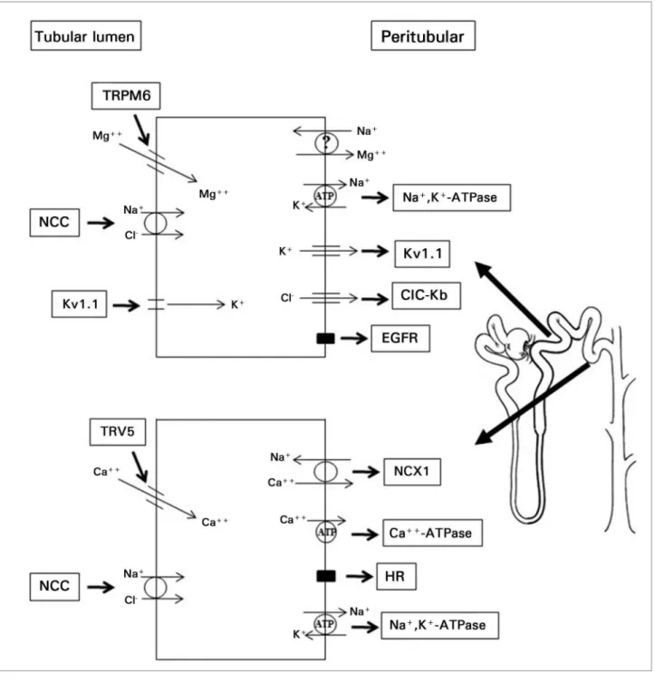
Magnesium is reabsorbed in the initial portion of the DCT through the TRPM6 expressed in the apical membrane of the DCT cells. In the final portion of this segment of the nephron and in the connecting tubule, TRPV5 allows calcium to be reabsorbed via the transcellular pathway (Figure 3).35
MAINCONDITIONS AFFECTING THE DISTAL CONVOLUTED TUBULE
GITELMANSYNDROME
Individuals with Gitelman syndrome have hypokalemic metabolic alkalosis combined with hypomagnesemia and hypocalciuria. The clinical manifestations and biochemical markers for Gitelman syndrome may mimic the signs of type-3 Bartter syndrome. Symptoms may start during childhood and persist throughout adult life. Some individuals are asymptomatic, while others experience muscle weakness, cramps, severe neuromuscular symptoms, paresthesia, tetany or palsy correlated with hydroelectrolytic disorders.35-38
The mutations found in most patients with Gitelman syndrome are located in gene SLC12A3.35,39
PSEUDOHYPOALDOSTERONISMTYPE II - GORDON’SSYNDROME
Gordon’s syndrome is an autosomal dominant disease associated with increased NaCl renal reabsorption and impaired distal secretion of K+
and H+. The alterations inherent to the disease
stem from mutations on the gene encoding WNK, a family of serine-threonine protein kinases.40
Some mutations remove the inhibitory effect WNK4 exerts over the NaCl cotransporter (NCC) in the distal convoluted tubule. Other mutations produce increases in the expression of WNK1. Once NCC is activated by WNK1, in such gene mutations the transport of Na+ is
Figure 3. Cells of the distal convoluted tubule showing the NaCl cotransporter (NCC) in the luminal membrane, activated after the expression of Na+-K+-ATPase in the basement membrane. The cells in the initial portion of the distal convoluted tubule also express Mg2+ channel (TRPM6) on the luminal face. This channel is modulated by NCC efficiency and the transcellular movement of ions K+ and Cl-. TRPM6 channels are also modulated by the activation of the epithelial growth factor receptor (EGFR) expressed on the basement membrane. The mechanism by which Mg2+ leaves these cells is yet unknown. The cells at the end of the distal convoluted tubule and of the connecting tubule express Ca2+ channel (TRPV5) on the luminal face. TRPV5 is also modulated by NCC efficiency. Ca2+leaves the cell with the aid of Ca2+-ATPase and Na+-Ca2+ cotransporter (NCX1) expressed on the basement membrane. These cells also have receptors to multiple hormones (HR), which regulate Ca2+ reabsorption - the cases of estrogen, PTH, and vitamin D.
The clinical manifestations of Gordon’s syndrome include hyperkalemia, mild metabolic acidosis, suppressed plasma renin activity, and normal or elevated aldosterone levels.41
Lower levels of sodium in the collecting duct cells impair the generation of a potential difference and
cause hyperkalemia and metabolic acidosis. The secretion of K+ and H+ is consequently decreased.
EAST/SESAME SYNDROME
mental retardation, and sensorineural hearing loss occur in association with a salt wasting syndrome described by the acronym EAST. The disorders include the activation of the renin-angiotensin-aldosterone system, hypokalemic metabolic acidosis, and hypomagnesemia with hypocalciuria. Patient urine concentrating ability remains unaffected.28
The EAST/SeSAME syndrome is an autosomal recessive disease caused by mutations and loss of function of gene KCNJ10 encoding Kir 1.4, a potassium channel expressed in the basement membrane of the distal convoluted tubule. Kir 1.4 and Na+-K+-ATPase are thought to jointly
aid in the local recirculation of potassium ions.35
HYPOMAGNESEMIAWITHSECONDARYHYPOCALCEMIA (HSH)
Gene TRPM6 encoding the TRPM6 channel was identified as the culprit for HSH, an autosomal recessive disease.28,35
Clinical reports indicate hypomagnesemia is caused by reduced Mg2+ absorption in the bowel,
and not necessarily by urinary loss of magnesium. Hypocalcemia is a secondary condition, given that patients benefit from the administration of Mg2+. Calcium homeostasis has been associated
with Mg2+ blood levels.28,35
The clinical manifestations of HSH appear soon after birth in the form of spasms, tetany, and generalized seizures.28,35
ISOLATEDAUTOSOMALRECESSIVEHYPOMAGNESEMIA
This rare form of hypomagnesemia was initially described in two siblings with low Mg2+ blood
levels caused by increased magnesium urinary excretion and normal Ca2+ blood levels. These
individuals had seizures and psychomotor retardation. Genetic studies revealed a mutation in the epidermal growth factor (EGF) gene, which impaired the autocrine/paracrine secretion of EGF. EGF receptors regulate the insertion of the TRPM6 channels in the luminal membrane of the distal convoluted tubule. Therefore, the inhibition of the EGF receptors for lack of a substrate leads to magnesiuria secondary to diminished TRPM6 channel expression.28,35
ISOLATEDDOMINANTHYPOMAGNESEMIA
Patients diagnosed with this type of hypomagnesemia experience renal losses of Mg2+ and hypocalciuria due to mutations in the
structure of Na+-K+-ATPase. Na+-K+-ATPase
has three described subunits: α, β, and γ; α and β are catalytic subunits and γ is a modulating subunit. Gene FXYD2 encodes subunit γ and G41R mutation causes changes in the affinity with Na+ and K+ and in the polarization of the
apical membrane, which may decrease Mg2+
transport. However, the precise role of subunit γ in Mg2+ tubular transport regulation has not
been entirely uncovered.28,35
C
OLLECTINGDUCTThe collecting duct is characterized by the heterogeneity of its cells. This segment of the nephron contains principal cells and intercalated cells of types A, B, and non-A non-B.44
In the principal cells, sodium is reabsorbed separately from chloride via the transcellular pathway through the amiloride-sensitive epithelial sodium channel (ENaC). Sodium reabsorption is driven by the activity of Na+-K+
-ATPase expressed in the basement membrane. Transcellular transport of sodium favors the secretion of a cation - potassium or hydrogen - as a consequence of the principle of electroneutrality. Principal cells also express ROMK channels in their apical membranes through which K+ is
secreted.1,45
Urine acidification occurs in type-A intercalated cells due to the secretion of H+.
The transport of this cation is made possible by the activity of H+-ATPase expressed in the
face of the lumen of these cells and facilitated by the potential difference generated when the neighboring principal cells reabsorb sodium. H+
originates from the catalytic reaction between carbonic anhydrase II and carbonic acid. The latter is formed when CO2 is hydrated.
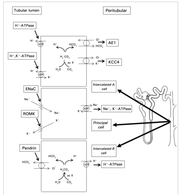
Figure 4. Representation of three types of collecting duct cells: intercalated-A, principal, and intercalated-B cells. Intercalated-A cells express H+ -ATPase on the luminal membrane and Cl-HCO3- (AE1) transporter on the basement membrane. This arrangement favors the secretion of acids. The expression of enzyme H+-K+-ATPase on the luminal face helps conserve K+ in situations of potassium depletion. Intercalated-B cells express H+-ATPase on the basement membrane and Cl-HCO
3 transporter, called pendrin in these cells, on the luminal face. Thus, the individual can retain H+ and eliminate bicarbonate in situations of alkalemia. Principal cells express amiloride-sensitive epithelial sodium channel (ENaC) on the luminal face. Na+,K+-ATPase expression on the basement membrane generates an electrochemical gradient that allows the reabsorption of Na+ and the secretion of K+ by ROMK channels.
through the Cl--HCO 3
- (AE1) cotransporter
expressed in the basement membrane of type-A intercalated cells (Figure 4). The H+ ions secreted
in the tubular lumen combine with anions such
as phosphate and ammonia (NH3), resulting in the formation of ammonium (NH4+). Ammonia
secretion reduces urine acidification and the excretion of ammonium. Type-A intercalated cells also express H+-K+-ATPase in their luminal
membranes.1,9 Despite the numerous studies on
the matter, there is no consensus in the literature over the role of H+-K+-ATPase in acid-base
balance, only in potassium homeostasis.
Type-B intercalated cells are involved in the secretion of bicarbonate and play an important role in the regulation of the acid-base balance in contexts of alkalemia. Type-B intercalated cells express H+-ATPase in the basement membrane
and chloride-bicarbonate exchange protein pendrin in the luminal membrane. Pendrin transports Cl- into the intracellular medium and
HCO3- to the lumen of the tubule.1,44
Non-A non-B intercalated cells express both H+-ATPase and pendrin in their apical
membranes.44
While the role of type-A and type-B intercalated cells has been established, the role of non-A non-B intercalated cells still requires some clarification.
MAINCONDITIONSAFFECTING THECOLLECTINGDUCT
DISTALRENALTUBULARACIDOSIS
Distal renal tubular acidosis, also known as type-1 RTA, is characterized by hyperchloremic metabolic acidosis arising from failure to secrete hydrogen ions in the collecting duct. Inherited type-1 RTA has two variants: a) autosomal dominant type-1 RTA with mild clinical manifestations and b) autosomal recessive type-1 RTA with severe symptoms during childhood, including hearing loss in some cases. Symptoms of early onset include polyuria, vomiting, dehydration, lower weight and height, hypokalemia, urinary pH above 6.0, hypercalciuria, hypocitraturia, and rickets.15,22 Hypercalciuria combined with a
urinary pH above 6.0 favors the deposition of calcium in the kidneys and the establishment of nephrocalcinosis. Diagnosis is classically defined when higher urinary pH and systemic metabolic acidosis are identified. In some cases, patients with the disease may have normal blood pH and slightly increased urinary pH levels, making the diagnosis of the condition more challenging. In such circumstances, urine acidification tests with
oral administration of ammonium chloride (NH4Cl) or furosemide combined with fludrocortisone are needed.46
Mutations on gene AE1 (SLC4A1) have been associated with autosomal dominant type-1 RTA. The gene, located in chromosome 17q21-22, is a member of the anion exchanger family expressed in the basement membrane of type-A intercalated cells.8,47,48 Genome analysis has
found two recessive genes associated with type-1 RTA: a) one in chromosome 2p13 encoding subunit B1 of H+/ATPase (ATP6V1B1) and b)
another in the 7q33-34 locus encoding a specific renal proton pump subunit (ATP6V0A4).8
LIDDLESYNDROME
Individuals diagnosed with Liddle syndrome have severe hypertension, metabolic alkalosis, and hypokalemia, accompanied by low renin and aldosterone blood levels as a consequence of the mutations in the ENaC subunits.49
The ENaC is made up of subunits α, β, and γ. In the kidney, the ENaC allows Na+ to enter through
the luminal membrane, while maintaining the homeostasis of the extracellular fluid and proper blood pressure levels.45 Mutations on
genes SCNN1B and SCNN1G respectively affect subunits β and γ of the ENaC and cause Liddle syndrome. Patients diagnosed with the syndrome have significantly increased ENaC activity, which causes them to retain sodium and experience high blood pressure unrelated to renin or aldosterone levels. Additionally, increased sodium reabsorption stimulates potassium secretion and favors the onset of hypokalemia.49
PSEUDOHYPOALDOSTERONISMTYPE I (PHAI)
the ENaC, which results in loss of function in the Na+ transport in aldosterone target tissues.
The clinical manifestations seen in patients diagnosed with PHA1 include neonatal renal sodium wasting, dehydration, life-threatening hypotension, hyperkalemia, metabolic acidosis, and growth failure.50-52
CONGENITALHYPOALDOSTERONISM
Congenital hypoaldosteronism is a rare inherited autosomal recessive disease correlated with gene CYP11B2 in chromosome 8q24.3. Gene CYP11B2 encodes aldosterone synthase (CYP11B2), an enzyme responsible for the synthesis of aldosterone in the adrenal cortex. When mutated, gene CYP11B2 blocks the synthesis of aldosterone. Consequently, patients with congenital hypoaldosteronism experience recurring episodes of hypovolemia with hyponatremia, hyperkalemia, and metabolic acidosis.53
CONGENITALADRENALHYPERPLASIA
Gene CYP11B1 is located adjacently to gene CYP11B2 in chromosome 8q22. Gene CYP11B1 encodes enzyme 11β-hydroxylase, which mediates the synthesis of cortisol. Mutations to this gene decrease the synthesis of cortisol, thus increasing the secretion of ACTH. Consequently, steroid precursors are overproduced, resulting in virilization of the female genitalia in neonates, precocious pseudopuberty, accelerated somatic growth, premature epiphyseal closure in individuals of both genders, and high blood pressure in approximately two thirds of the individuals carrying the mutation. The mutation on gene CYP11B1 is the second most common cause of congenital adrenal hyperplasia.54
APPARENTMINERALOCORTICOIDEXCESS
Enzyme 11β-hydroxysteroid dehydrogenase type 2 (11βHSD2), expressed in the cytoplasm of the principal cells, acts on steroid degradation and prevents the activation of mineralocorticoid receptors by glucocorticoids. Mutations on gene HSD11B2 encoding the enzyme may
cause a rare hypertensive syndrome, referred to as apparent mineralocorticoid excess, whose clinical manifestations include salt-dependent hypertension, hypokalemia, and metabolic alkalemia.55 Numerous case reports of apparent
mineralocorticoid excess published in the literature have linked the disease to injudicious use of medicinal herbs. Glycyrrhiza glabra, also known as licorice, inhibits 11βHSD2 and consequently stimulates constant mineralocorticoid receptor due to cortisol overproduction.55-57
NEPHROGENICDIABETESINSIPIDUS
The kidneys of individuals with nephrogenic diabetes insipidus cannot concentrate urine in response to the antidiuretic hormone. Children with nephrogenic diabetes insipidus have polydipsia, polyuria, hyposthenuria, dehydration, hypernatremia, and growth failure. Approximately 90% of the individuals diagnosed with nephrogenic diabetes insipidus have the X-linked recessive form of the disease caused by mutations on gene AVPV2 in chromosome Xq28. Autosomal dominant and recessive nephrogenic diabetes insipidus has been associated with mutations on gene AQP2 in chromosome 12q13.58,59
F
INALCONSIDERATIONSInherited tubulopathies can affect the growth and development of children and may be accompanied or not by neurological disorders. A significant share of the children diagnosed with inherited tubulopathies develops pediatric chronic kidney disease.
of ailments with new and better treatments, thus improving the prognosis of the children affected by them.
Despite the growing knowledge around the gene mutations associated with renal diseases, genetic tests are available only at a few centers. In Brazil, genetic tests are performed at a handful of teaching hospitals, such as the Hospital of the Federal University of Paraná, and the Central Institute and the Children’s Institute of the Hospital of the University of São Paulo.
R
EFERENCES1. Seguro AC, Kudo L, Helou CMB. Função Tubular. In: Riella M, ed. Princípios de Nefrologia e Distúrbios Eletrolíticos. 5a ed. Rio de Janeiro: Guanabara Koogan; 2010. p.38-49. 2. Kleta R, Bockenhauer D. Bartter syndromes and other
salt--losing tubulopathies. Nephron Physiol 2006;104:73-80. DOI: http://dx.doi.org/10.1159/000094001
3. Rumballe B, Georgas K, Wilkinson L, Little M. Molecular ana-tomy of the kidney: what have we learned from gene expression and functional genomics? Pediatr Nephrol 2010;25:1005-16. 4. Emma F, Montini G, Salviati L, Dionisi-Vici C. Renal
mitochon-drial cytopathies. Int J Nephrol 2011;2011:609213. PMID: 21811680 DOI:http://dx.doi.org/10.4061/2011/609213 5. Emma F, Bertini E, Salviati L, Montiniet G. Renal involvement
in mitochondrial cytopathies. Pediatr Nephrol 2012;27:539-50. DOI: http://dx.doi.org/10.1007/s00467-011-1926-6 6. Kelly CR, Landman J. Anatomia do trato urinário. In Kelly CR,
Landman J, eds. Coleção Netter de ilustrações médicas. 2a ed. Rio de Janeiro: Saunders-Elsevier; 2014. p.24-7.
7. Bobulescu IA, Moe OW. Luminal Na(+)/H (+) exchange in the proximal tubule. Pflugers Arch 2009;458:5-21. PMID: 18853182 DOI: http://dx.doi.org/10.1007/s00424-008-0595-1 8. Fry AC, Karet FE. Inherited renal acidoses. Physiology (Be-thesda) 2007;22:202-11. DOI: http://dx.doi.org/10.1152/phy-siol.00044.2006
9. Helou CMB. Distúrbios do equilíbrio ácido-base. In: Martins MA Carrilho FJ, Alves VA, Castilho EA, Cerri GG, Wen CL, eds. Clínica Médica. 1a ed. Barueri: Manole; 2009. p.614-25. 10. Cooper GJ, Zhou Y, Bouyer P, Grichtchenko II, Boron
WF. Transport of volatile solutes through AQP1. J Phy-siol 2002;542:17-29. PMID: 12096045 DOI:http://dx.doi. org/10.1113/jphysiol.2002.023218
11. Baum M. Developmental changes in proximal tubule NaCl transport. Pediatr Nephrol 2008;23:185-94. DOI: http:// dx.doi.org/10.1007/s00467-007-0569-0
12. San-Cristobal P, Dimke H, Joost GJ, Bindels RJ. Novel molecu-lar pathways in renal Mg2+ transport: a guided tour along the nephron. Curr Opin Nephrol Hypertens 2010;19:456-62. DOI: http://dx.doi.org/10.1097/MNH.0b013e32833caf61
13. Amatschek S, Haller M, Oberbauer R. Renal phosphate handling in human--what can we learn from hereditary hypophosphatae-mias? Eur J Clin Invest 2010;40:552-60. PMID: 20412291 14. Gattineni J, Baum M. Genetic disorders of phosphate
regula-tion. Pediatr Nephrol 2012;27:1477-87. DOI: http://dx.doi. org/10.1007/s00467-012-2103-2
15. Pereira PC, Miranda DM, Oliveira EA, Silva AC.
Molecular pathophysiology of renal tubular acido-sis. Curr Genomics 2009;10:51-9. DOI:http://dx.doi. org/10.2174/138920209787581262
16. Karet FE. Disorders of water and acid-base homeosta-sis. Nephron Physiol 2011;118:28-34. DOI: http://dx.doi. org/10.1159/000320885
17. Pache de Faria Guimaraes L, Seguro AC, Shimizu MH, Lopes Neri LA, Sumita NM, de Bragança AC, et al. N-acetyl-cysteine is associated to renal function improvement in patients with nephropathic cystinosis. Pediatr Nephrol 2014;29:1097-102. 18. Taranta A, Wilmer MJ, van den Heuvel LP, Bencivenga P,
Bello-mo F, Levtchenko EN, et al. Analysis of CTNS gene transcripts in nephropathic cystinosis. Pediatr Nephrol 2010;25:1263-7. DOI: http://dx.doi.org/10.1007/s00467-010-1502-5
19. Vaisbich MH, Koch VH. Report of a Brazilian multicenter study on nephropathic cystinosis. Nephron Clin Pract 2010;114:c12-8. DOI:http://dx.doi.org/10.1159/000245065
20. Özkan B, Çayır A, Koşan C, Alp H. Cystinosis presenting with findings of Bartter syndrome. J Clin Res Pediatr Endocrinol 2011;3:101-4.
21. Şimşek E, Şimşek T, Dallar Y, Can Ö, Willems PJ. A novel pathogenic DNA variation in the OCRL1 gene in Lowe syndro-me. J Clin Res Pediatr Endocrinol 2011;3:29-31. DOI: http:// dx.doi.org/10.4274/jcrpe.v3i1.06
22. Stechman MJ, Loh NY, Thakker RV. Genetic causes of hyper-calciuric nephrolithiasis. Pediatr Nephrol 2009;24:2321-32. DOI: http://dx.doi.org/10.1007/s00467-008-0807-0
23. Devuyst O, Thakker RV. Dent’s disease. Orphanet J Rare Dis 2010;5:28. DOI: http://dx.doi.org/10.1186/1750-1172-5-28 24. Devuyst O. Dent’s disease: chloride-proton exchange
con-trols proximal tubule endocytosis. Nephrol Dial Transplant 2010;25:3832-5. DOI:http://dx.doi.org/10.1093/ndt/gfq556 25. Morey M, Castro-Feijóo L, Barreiro J, Cabanas P, Pombo M,
Gil M, et al. Genetic diagnosis of X-linked dominant Hypo-phosphatemic Rickets in a cohort study: tubular reabsorption of phosphate and 1,25(OH)2D serum levels are associated with PHEX mutation type. BMC Med Genet 2011;12:116. DOI: http://dx.doi.org/10.1186/1471-2350-12-116
26. Zivičnjak M, Schnabel D, Billing H, Staude H, Filler G, Quer-feld U, et al.; Hypophosphatemic Rickets Study Group of Ar-beitsgemeinschaft für Pädiatrische Endokrinologie and Gese-llschaft für Pädiatrische Nephrologie. Age-related stature and linear body segments in children with X-linked hypophospha-temic rickets. Pediatr Nephrol 2011;26:223-31. DOI: http:// dx.doi.org/10.1007/s00467-010-1705-9
27. Vaisbich MH, Koch VH. Hypophosphatemic rickets: results of a long-term follow-up. Pediatr Nephrol 2006;21:230-4. DOI: http://dx.doi.org/10.1007/s00467-005-2077-4
28. Seyberth HW, Schlingmann KP. Bartter- and Gitelman-like syndromes: salt-losing tubulopathies with loop or DCT defects. Pediatr Nephrol 2011;26:1789-802. PMID:21503667 DOI: http://dx.doi.org/10.1007/s00467-011-1871-4
29. Fahlke C, Fischer M. Physiology and pathophysiology of ClC--K/barttin channels. Front Physiol 2010;1:155. DOI: http:// dx.doi.org/10.3389/fphys.2010.00155
30. Günzel D, Yu AS. Function and regulation of claudins in the thick ascending limb of Henle. Pflugers Arch 2009;458:77-88. PMID: 18795318 DOI:http://dx.doi.org/10.1007/s00424-008-0589-z 31. Riccardi D, Brown EM. Physiology and pathophysiology of the
calcium-sensing receptor in the kidney. Am J Physiol Renal Phy-siol 2010;298:F485-99. PMID: 19923405DOI: http://dx.doi. org/10.1152/ajprenal.00608.2009
32. Chen YH, Lin JJ, Jeansonne BG, Tatum R, Lu Q. Analysis of claudin genes in pediatric patients with Bartter’s syndrome. Ann N Y Acad Sci 2009;1165:126-34. PMID:19538297 DOI: http://dx.doi.org/10.1111/j.1749-6632.2009.04031.x
33. Deschênes G, Fila M. Primary molecular disorders and secon-dary biological adaptations in bartter syndrome. Int J Nephrol 2011;2011:396209. PMID: 21941653 DOI:http://dx.doi. org/10.4061/2011/396209
34. Fremont OT, Chan JC. Understanding Bartter syndrome and Gitelman syndrome. World J Pediatr 2012;8:25-30. DOI: http://dx.doi.org/10.1007/s12519-012-0333-9
36. Knoers NV, Levtchenko EN. Gitelman syndrome. Orphanet J Rare Dis 2008;3:22. DOI: http://dx.doi.org/10.1186/1750-1172-3-22
37. Bansal T, Abeygunasekara S, Ezzat V. An unusual presentation of primary renal hypokalemia-hypomagnesemia (Gitelman’s syndrome). Ren Fail 2010;32:407-10. DOI:http://dx.doi. org/10.3109/08860221003632873
38. Graziani G, Fedeli C, Moroni L, Cosmai L, Badalamenti S, Ponticelli C. Gitelman syndrome: pathophysiological and cli-nical aspects. QJM 2010;103:741-8. PMID:20650971 DOI: http://dx.doi.org/10.1093/qjmed/hcq123
39. Urbanová M, Reiterová J, Stěkrová J, Lněnička P, Ryšavá R. DNA analysis of renal electrolyte transporter genes among pa-tients suffering from Bartter and Gitelman syndromes: summa-ry of mutation screening. Folia Biol (Praha) 2011;57:65-73. 40. Mu S, Shimosawa T, Ogura S, Wang H, Uetake Y,
Kawakami--Mori F, et al. Epigenetic modulation of the renal β -adrenergic-WNK4 pathway in salt-sensitive hypertension. Nat Med 2011;17:573-80. DOI: http://dx.doi.org/10.1038/nm.2337 41. Furgeson SB, Linas S. Mechanisms of type I and type II
pseudo-hypoaldosteronism. J Am Soc Nephrol 2010;21:1842-5. DOI: http://dx.doi.org/10.1681/ASN.2010050457
42. Dimke H. Exploring the intricate regulatory network
controlling the thiazide-sensitive NaCl cotransporter (NCC). Pflugers Arch 2011;462:767-77. PMID: 21927811 DOI:http://dx.doi.org/10.1007/s00424-011-1027-1 43. McCormick JA, Ellison DH. The WNKs: atypical protein kinases
with pleiotropic actions. Physiol Rev 2011;91:177-219. PMID: 21248166 DOI:http://dx.doi.org/10.1152/physrev.00017.2010 44. Roy A, Al-bataineh MM, Pastor-Sole NM. Collecting duct
in-tercalated cell function and regulation. Clin J Am Soc Nephrol 2015;10:305-24. DOI:http://dx.doi.org/10.2215/CJN.08880914 45. Hamm LL, Feng Z, Hering-Smith KS. Regulation of sodium
transport by ENaC in the kidney. Curr Opin Nephrol Hyper-tens 2010;19:98-105. PMID: 19996890 DOI:http://dx.doi. org/10.1097/MNH.0b013e328332bda4
46. Walsh SB, Shirley DG, Wrong OM, Unwin RJ. Urinary acidification assessed by simultaneous furosemide and fludrocortisone treatment: an alternative to ammonium chloride. Kidney Int 2007;71:1310-6. PMID: 17410104 DOI: http://dx.doi.org/10.1038/sj.ki.5002220 47. Vasuvattakul S. Molecular Approach for Distal Renal Tubular
Aci-dosis Associated AE1 Mutations. Electrolyte Blood Press 2010;8:25-31. DOI:http://dx.doi.org/10.5049/EBP.2010.8.1.25
48. Anacleto FE, Bruce LJ, Clayton P, Hegde S, Resontoc LP, Wrong O. Distal renal tubular acidosis in Filipino children, caused by mutations of the anion-exchanger SLC4A1 (AE1, Band 3) gene. Nephron Phy-siol 2010;114:19-24. DOI: http://dx.doi.org/10.1159/000274484 49. Bogdanović R, Kuburović V, Stajić N, Mughal SS, Hilger A, Ninić
S, et al. Liddle syndrome in a Serbian family and literature review of underlying mutations. Eur J Pediatr 2012;171:471-8. DOI: http:// dx.doi.org/10.1007/s00431-011-1581-8
50. Rolim AL, Lindsey SC, Kunii IS, Fujikawa AM, Soares FA, Chiamolera MI, et al. Ion channelopathies in endocrinolo-gy: recent genetic findings and pathophysiological insights. Arq Bras Endocrinol Metabol 2010;54:673-81. DOI: http:// dx.doi.org/10.1590/S0004-27302010000800002
51. Mora-Lopez F, Bernal-Quiros M, Lechuga-Sancho AM, Le-chuga-Campoy JL, Hernandez-Trujillo N, Nieto A. Novel mutation in the epithelial sodium channel causing type I pseudohypoaldosteronism in a patient misdiagnosed with cystic fibrosis. Eur J Pediatr 2012;171:997-1000. DOI: http://dx.doi.org/10.1007/s00431-012-1697-5
52. Fernandes-Rosa FL, Antonini SR. Mineralocorticoid re-sistance: pseudohypoaldosteronism type 1. Arq Bras En-docrinol Metabol 2007;51:373-81. DOI:http://dx.doi. org/10.1590/S0004-27302007000300004
53. Hui E, Yeung MC, Cheung PT, Kwan E, Low L, Tan KC, et al. The clinical significance of aldosterone synthase de-ficiency: report of a novel mutation in the CYP11B2 gene. BMC Endocr Disord 2014;14:29. DOI: http://dx.doi. org/10.1186/1472-6823-14-29
54. Dumic K, Yuen,T, Grubic Z, Kusec V, Barisic I, New MI. Two Novel CYP11B1 Gene Mutations in Patients from Two Croatian Families with 11 β -Hydroxylase Deficiency. Int J Endocrinol 2014;2014:185974.
55. Ferrari P. The role of 11β-hydroxysteroid dehydrogena-se type 2 in human hypertension. Biochim Biophys Acta 2010;1802:1178-87. PMID: 19909806 DOI:http://dx.doi. org/10.1016/j.bbadis.2009.10.017
56. White PC. 11beta-hydroxysteroid dehydrogenase and its role in the syndrome of apparent mineralocorticoid excess. Am J Med Sci 2001;322:308-15. PMID: 11780688DOI: http://dx.doi.org/10.1097/00000441-200112000-00003 57. Yasue H, Itoh T, Mizuno Y, Harada E. Severe hypokalemia,
rhabdomyolysis, muscle paralysis, and respiratory impair-ment in a hypertensive patient taking herbal medicines con-taining licorice. Intern Med 2001;46:575-8. DOI: http:// dx.doi.org/10.2169/internalmedicine.46.6316
58. Liberatore Junior RD, Carneiro JG, Leidenz FB, Melilo--Carolino R, Sarubi HC, De Marco L. Novel compound aquaporin 2 mutations in nephrogenic diabetes insipidus. Clinics (São Paulo) 2012;67:79-82. DOI: http://dx.doi. org/10.6061/clinics/2012(01)13