Brazilian Journal of Microbiology (2011) 42: 158-171 ISSN 1517-8382
CHARACTERIZATION OF LACTOBACILLUS FROM ALGERIAN GOAT’S MILK BASED ON PHENOTYPIC, 16S rDNA SEQUENCING AND THEIR TECHNOLOGICAL PROPERTIES
Ahmed Marroki1 *; Manuel Zúñiga2; Mabrouk Kihal3; Gaspar Pérez- Martínez2
1
Laboratory of Applied Microbiology, Department of Biotechnology (IGMO), University of Oran Es-Sénia, Oran, 31100 ,
Algeria; 2Department of Biotechnology, Instituto de Agroquímica y Tecnología de los Alimentos (C.S.I.C), polígono de la Coma
s/n, Burjassot (Valencia), Spain; 3Laboratory of Applied Microbiology, Department of Biology, University of Oran Es-Sénia,
Oran, Algeria.
Submitted: February 18, 2010; Returned to authors for corrections: February 24, 2010; Approved: August 01, 2010.
ABSTRACT
Nineteen strains of Lactobacillus isolated from goat’s milk from farms in north-west of Algeria were
characterized. Isolates were identified by phenotypic, physiological and genotypic methods and some of
their important technological properties were studied. Phenotypic characterization was carried out by
studying physiological, morphological characteristics and carbohydrate fermentation patterns using API 50
CHL system. Isolates were also characterized by partial 16S rDNA sequencing. Results obtained with
phenotypic methods were correlated with the genotypic characterization and 13 isolates were identified as
L. plantarum, two isolates as L. rhamnosus and one isolate as L. fermentum. Three isolates identified as L.
plantarum by phenotypic characterization were found to be L. pentosus by the genotypic method. A large
diversity in technological properties (acid production in skim milk, exopolysaccharide production,
aminopeptidase activity, antibacterial activity and antibiotic susceptibility)was observed. Based onthese
results, two strains of L. plantarum (LbMS16 and LbMS21) and one strain of L. rhamnosus (LbMF25)
have been tentatively selected for use as starter cultures in the manufacture of artisanal fermented dairy
products in Algeria.
Key words: Lactic acid bacteria; Lactobacillus; identification; goat’s milk; technological properties; Algeria.
INTRODUCTION
The identification of lactobacilli has been based mainly on
fermentation of carbohydrates, morphology, and Gram staining
and these methods are still being used. However, the
characterization of some Lactobacillus to species level by
biochemical methods alone is not reliable (27, 40), because of
the considerable variations in biochemical attributes between
strains currently considered to belong to the same species. In
fact, some species are not readily distinguishable in terms of
phenotypic characteristics (12). In recent years, the taxonomy
has changed considerably with the increasing knowledge of the
genomic structure and phylogenetic relationships between
Lactobacillus spp. (27, 43, 47). This novel taxonomy based on
DNA analysis offers a variety of advantages over other more
conventional typing procedures, such as the stability of the
genomic DNA analysis, the capacity to discriminate bacteria at
the strain level, and the amenability to automation and
statistical analysis (21). These methods have been employed
for differentiation or identification and typing of different
species of Lactobacillus. The species of lactobacilli most
commonly found in milk, and dairy product, especially in
goat’s milk are L. plantarum, L. rhamnosus, L. casei or L.
paracasei. For this reason, the selection of Lactobacillus
strains from goat’s milk has been considered in the search for
new industrially important cultures, in order to select those
with the highest potential for industrial applications. In Algeria,
goat’s milk plays a vital role in human consumption, most
being consumed by the rural community, while little is
available on the market (5). Algerian people make various
fermented dairy products using goat’s milk. The transformation
of goat’s milk into traditional Algerian dairy products, such as
El – Klila, a traditional cheese which is popular in the country
side and is made from unpasteurised cow or goat surplus milk
(7), Jben (local traditional fresh cheese), Raïb, and Lben (local
traditional fermented milks), is achieved through spontaneous
fermentation without the addition of any selected starter. Such
products generally present irregular sensorial qualities. The aim
of the present study was to characterize Lactobacillus isolated
from goat’s milk from north-west of Algeria, using
physiological, phenotypic and genotypic methods. There are no
previous reports concerning the genetic identification of
Lactobacillus or studies that combined the phenotypic and the
genotypic identification of Lactobacillus isolated from goat’s
milk in Algeria. Additionally, in order to select adequate
strains susceptible to be used as starter cultures for the
manufacture of artisanal fermented dairy products in Algeria,
some important technological properties, including the capacity
of acidification/coagulation of skim milk, exopolysaccharide
production, aminopeptidase, antibacterial activity, and
antibiotics resistance, were also studied.
MATERIAL AND METHODS
Milk samples
Five samples of goat’s milk collected from farms located
in the region north-west of Algeria were used in this study. The
samples were collected aseptically in sterile bottles kept in an
ice-box, and transported immediately to the laboratory.
Phenotypic characterization
One milliliter of each milk sample was homogenized with
9 ml of sterile Ringer’s solution 1:4 and mixed thoroughly for
60s. Serial dilutions were made and aliquots (100 µl) of each
dilution were streaked on MRS agar (Oxoid, UK) (17). The
MRS plates were incubated at 30 °C and 45°C for 24 to 48h
under anaerobic conditions (Anaerogen, Oxoid). Ten colonies
from plates corresponding to the highest dilutions were
randomly selected and purified by subculturing. Gram-positive,
catalase negative cultures were stored at -80 °C in MRS
supplemented with 20% glycerol. Isolates were phenotypically
assigned to the genus level on the basis of: cell morphology,
Gram-positive and catalase-negative, according to the methods
and criteria described by Sharpe (42) and Kandler and Weiss
(26); CO2 production from glucose in MRS broth containing
inverted Durham tubes (32); hydrolysis of arginine, growth at
15 °C and 45 °C, tolerance to 20, 40, 65 g L-1 NaCl. The acid
production from carbohydrates (fructose, glucose, mannitol,
lactose, mannose, rhamnose, glycerol, arabinose, sorbose,
dulcitol, amygdalin, melibiose, melezitose, starch, tagatose,
arabitol, ribose, maltose, galactose, and xylose) was evaluated
by using a miniaturized assay in microtiter plates, as described
by Jayne-Williams (25). Ability to ferment carbohydrate
substrates was studied, using the API 50 CHL system
(BioMérieux, Lyon, France), following the manufacturer
recommendations.
DNA extraction and 16S rDNA sequencing
Marroki, A. et al. Characterization of Lactobacillus and their technological properties
1.6 - 1.8 at 600 nm. A 1.5 mL aliquot of each overnight culture
was centrifuged at 10000 × g for 30 s at room temperature in
order to pellet cells. Bacterial DNA was isolated by using the
UltraCleanTM Microbial DNA isolation Kit (MoBio
Laboratories Inc., Solana Beach, CA, USA), following the
instructions of the manufacturer.
The 5′ end variable region of the 16S rDNA was
PCR-amplified with primers 27F (5′-AGAGTTTGATCCTGGCTC
AG-3′) and 558R (5′-GTATTCCGCGGCTG-3) or with the
primers 27F and 1525R (5′-AAGGAGGTGWTCCARCCG
CA-3′) using a total volume of 50 µl containing 50 ng of DNA,
25 pmol of each primer, 1.6 mM of dNTPs, 2 mM MgCl2 and
1U of Taq DNA polymerase (Biotools Lab, Spain), using the
reaction buffer supplied by the manufacturer.
Amplifications were carried out in a Thermal Cycler
(PTC-100 Peltier Themal Cycler, MJR), using the following
program: for primers 27F and 558R, the 16S rDNA was
amplified as described by Linaje et al. (30); for the primers 27F
and 1525R, the PCR mixtures were subjected to an initial
denaturing step of 95 °C for 5min, followed by 30 PCR cycles
(94 °C, 15s; 52 °C, 30s; 72 °C, 2min) and final cycle at 72 °C
for 5min. The PCR products were subjected to gel
electrophoresis in 1% agarose gel, followed by staining with
ethidium bromide and visualization under UV light. A Lambda
DNA (Biorad) digested with PstI ladder was used as a
molecular mass marker.
Polymerase chain reaction products were purified by using
the GFX PCR DNA and a gel band purification Kit (General
Electric Healthcare, Spain), following the manufacturer’s
instructions. DNA sequencing was carried out by the Central
Service of Research Support of the University of Valencia
(Spain), by using the dideoxynucleotide DNA chain
termination method.
Technological properties
Acidifying activity in skim milk was assayed as described
by Psono et al. (35). Sterile skim milk samples (100 mL; 1.0%)
were inoculated with overnight cultures which had been
previously activated by two successive transfers in milk. The
pH changes were measured with a pH meter (glass electrode,
Crison, Spain) after 6, 12, and 24 h of incubation at 30°C.
Acidification activity was measured by following the change in
the pH during time. Coagulation of milk was determined after
24 h of incubation at 30°C. Screening of exopolysaccharide
(EPS) was carried out in ruthenium red milk plates, as
described by Stingele et al. (44).
Aminopeptidase activity of the strains was determined
using the synthetic substrates L-alanine ρ-nitroaniline (Ala-ρ
-NA) (Sigma,USA), and L-leucine ρ-nitroaniline (Leu-ρ-NA)
(Sigma,USA) as described by Zotta et al. (51). Stationary
phase cells grown overnight in MRS broth were harvested by
centrifugation at 10000 × g for 5 min, washed twice with sterile
50 mM potassium phosphate buffer, pH 7.0, and re-suspended
in the same buffer to obtain cell suspensions of (A 650 = 1.0).
Aminopeptidase activity was measured, according to Macedo
et al.(31). The assay mixture contained: 30µl of 20 mM
aminoacyl ρ-nitroanilide substrates dissolved in methanol, 195
µL of 50 mM potassium phosphate buffer (pH 7.0), 95 µL of
0.05% (w/v) sodium azide solution, and 75 µL of cell
suspension. After incubation at 30 °C for 1 to 4 h, the reaction
was stopped by the addition of 900 µL of 1% (v/v) acetic acid.
The release of ρ-nitroaniline (ρ-NA) (Sigma, USA) was
measured spectrophotometrically (Hewlett Packaro, Diod
Array Spectrophotometer, Germany) at 410 nm after
centrifugation of the mixture at 10000 × g for 5min. Data
obtained were compared to a calibration curve prepared using
ρ-NA (Sigma, USA) dilutions ranging from 0.1 to 20.0 mM.
One unit of enzyme activity was defined as the amount of
enzyme required to release 1 µ mol of ρ-NA min-1 under the
assay conditions.
Antibacterial activity of Lactobacillus strains was tested
with the well diffusion method described by Linaje et al. (30).
Cells were grown overnight in MRS broth. Cell-free
supernatants were obtained by centrifugation at 10000 × g for
10 min at 4 °C, adjusted to pH 6.5 with 1N NaOH, and then
filtered through 0.22µm Durapore membrane filters
(Millipore). The supernatants were adjusted to pH 6.5, in order
organic acids. A 100 µL aliquot of an overnight culture of the
indicator bacteria Listeria monocytogenes CECT 932T, Bacillus
cereus INRA AVZ 421, Staphylococcus aureus CECT 86T and
Staphylococcus aureus UT 602, was mixed with 5 mL of soft
agar (Trypticase Soya Broth for Bacillus cereus and
Staphylococcus aureus and Brain Heart Infusion for Listeria
monocytogenes, supplemented with 0.8% agar). Aliquots (50
µl) of supernatant of overnight cultures were poured in the
wells digged in the soft agar. After 24 h of incubation at 37 °C,
inhibition zones were read. A clear zone of inhibition >1 mm
around a well was scored as positive. In order to check the
thermoresistance of the bacteriocins, cell free supernatant
samples were heated at 100°C for 10 min, prior the
antibacterial assay. The proteinaceous nature of the inhibitory
activity was tested by the addition of 0.5 µg of proteinase K
(Roche Molecular Biochemicals) to the concentrated culture
supernatants (50 µL) distributed among the wells of the assay
plates.
Antibiotic susceptibility testing
Susceptibility testing was based on the agar overlay disc
diffusion test described by Charteris et al. (10), as modified by
Aymerich et al. (4). Briefly, Lactobacillus strains were grown
overnight in MRS broth at 30°C under anaerobic conditions
(Anaerogen, Oxoid). Eight ml of MRS soft agar kept at 50°C
were inoculated with 200 µL of the grown culture. Petri dishes
containing 15 mL of MRS were overlaid with 8.2 mL of the
inoculated MRS and allowed to solidify at room temperature.
Antibiotic discs were placed onto the overlaid plates and all
plates were incubated at 30°C for 24 h under anaerobic
conditions. All isolates were screened for their susceptibility to
penicillin G (10 µg), ampicillin (10 µg), vancomycin (30 µg),
tetracycline (30 µg), erythromycin (15 µg), kanamycin (30µg)
gentamicin (10 µg), and chloramphenicol (30 µg). Inhibition
zones diameters of antibiotics were compared to those defined
by Charteris et al. (10) for lactobacilli.
RESULTS AND DISCUSSION
Phenotypic identification of isolates
Isolated strains were identified based on their
physiological and biochemical characteristics given by Kandler
and Weiss (26), Dellaglio et al. (15) and Stiles and Holzapfel
(43), to the species level, and also by using API 50 CHL test
strips (BioMérieux, Lyon, France) for confirmation of species
of selected strains.
All isolates (19 Lactobacillus strains) were rod shaped
cells, Gram-positive, catalase-negative, non motile and
facultative anaerobic bacteria. Isolates were classified as
belonging to the genus Lactobacillus. All isolates were able to
grow at 15°C, 2%, and 4% NaCl. They were divided into two
preliminary groups (I and II), according to the results for CO2
production from glucose, and NH3 production from arginine:
Group I, facultatively heterofermentative, and
arginine-negative lactobacilli (94.73%); group II strictly
heterofermentative, and arginine-positive strain (one
Lactobacillus strain).
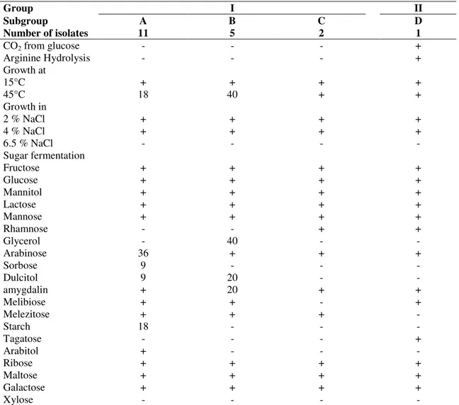
Table 1 shows the carbohydrate utilization patterns and
other physiological and biochemical characteristics of the
lactobacilli isolates. The analysis of data compared with those
of the criteria given by several authors, resulted in four
subgroups (A-D). Group I was subdivided into three subgroups
(A-C) and group II, comprised only one subgroup (D). Isolates
belonging to the four subgroups were able to ferment fructose,
glucose, mannitol, lactose, mannose, ribose, maltose, and
galactose and unable to ferment xylose.
• Subgroup A was the largest one, with 11 strains (58%)
identified as L. plantarum. The strains were able to ferment
amygdalin, melibiose, melezitose, and arabitol, but unable to
ferment rhamnose, glycerol, sorbose, dulcitol, and tagatose.
However, variations in fermentation patterns were observed for
some sugars: arabinose was fermented by 36%, starch by 18%,
Marroki, A. et al. Characterization of Lactobacillus and their technological properties
lactobacilli reported as being able to produce amylase are
strictly homofermentative (26), although some L. plantarum
have been reported as starch fermenting strains (33). Eighteen
percent of isolates from this subgroup were able to grow at
45°C. Some of the L. plantarum strains were capable of
growing at 45°C, in contrast to the characteristics given in
Bergey’s Manual (26). Other studies have also reported the
isolation of L. plantarum strains capable of growing at this
temperature (20, 39).
• Subgroup B, comprised 5 strains (26%) identified as
belonging to L .plantarum/L. pentosus species. The isolates
only differed from those of subgroup A in the inability to
ferment the starch and arabitol. The ability of some strains
(40%) to ferment glycerol resulted in their classification either
as L. plantarum or as L. pentosus (8, 50). Growth at 45°C was
observed for 40% of isolates.
• Subgroup C, included two isolates able to ferment
rhamnose, arabinose, amygdalin, and melezitose. However,
glycerol, sorbose, dulcitol, melibiose, starch, tagatose, and
arabitol were not fermented. All the isolates were able to grow
at 45°C and identified as L. rhamnosus.
• Subgroup D, with one isolate able to ferment rhamnose, arabinose amygdalin, melibiose and tagatose. In
contrast, this strain was incapable of fermenting glycerol,
sorbose, dulcitol, melezitose, starch and arabitol. This isolate
was able to grow at 45°C and was classified as L. fermentum.
Table 1. Biochemical and physiological characteristics of Lactobacillus strains isolated from Algerian goat’s milk.
Group I II
Subgroup A B C D
Number of isolates 11 5 2 1
CO2 from glucose - - - +
Arginine Hydrolysis - - - +
Growth at
15°C + + + +
45°C 18 40 + +
Growth in
2 % NaCl + + + +
4 % NaCl + + + +
6.5 % NaCl - - - -
Sugar fermentation
Fructose + + + +
Glucose + + + +
Mannitol + + + +
Lactose + + + +
Mannose + + + +
Rhamnose - - + +
Glycerol - 40 - -
Arabinose 36 + + +
Sorbose 9 - - -
Dulcitol 9 20 - -
amygdalin + 20 + +
Melibiose + + - +
Melezitose + + + -
Starch 18 - - -
Tagatose - - - +
Arabitol + - - -
Ribose + + + +
Maltose + + + +
Galactose + + + +
Xylose - - - -
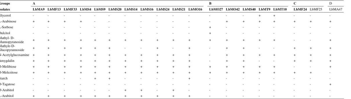
To establish the final phenotypic identification, all strains
tested for their biochemical and physiological characteristics
(Table 1), were submitted to further biochemical
characterization, using API 50 CHL galleries (BioMérieux,
Lyon, France). The programme of identification (Cox and
Thomson, Biochemistry institute, Odense University) plus
database was used for the interpretation of the strains
fermentation profiles (Table 2). The strains of the subgroups A
and B were classified as L. plantarum and those of subgroups
C and D as L. rhamnosus and L. fermentum, respectively.
A clear identification of species, especially within the
genus Lactobacillus, based on phenotypic methods, such as
fermentation patterns, may sometimes be difficult, due to an
increasing number of lactic acid bacteria species which vary on
a small number of biochemical traits (36). Commercially
available systems based on carbohydrate fermentation should
be combined with conventional phenotypic properties other
than carbohydrate fermentation or with genotypic techniques.
Table 2. Fermentation of carbohydrates by Lactobacillus strains from Algerian goat’s milk, tested by the API 50 CHL system
+: Positive reaction; -: Negative reaction.
All isolates were able to ferment ribose, galactose, glucose,fructose,mannose, mannitol, sorbitol, arbutin, esculin, salicin, celibiose, maltose, lactose, sucrose, trehalose, raffinose, gentibiose, turanose and gluconate. None fermented erythriol, arabinose, xylose, L-xylose, D-adonitol, methyl- -D-xylopyranoside, inositol, inulin, glycogen, xylitol, D-rhamnose, D-fucose, L-fucose, 2-ketogluconate and 5-ketogluconate.
Groups A B C D
Isolates LbMA9 LbMF13 LbMF33 LbMS4 LbMS9 LbMS20 LbMS14 LbMS16 LbMS24 LbMS21 LbMO16 LbMO27 LbMO42 LbMS40 LbMT9 LbMT10 LbMF24 LbMF25 LbMA47
Glycerol - - - - - - + + - - -
L-Arabinose + + + + - - - - + + + + + + +
L-Sorbose - - - + - - - - - - -
Dulcitol - - - + - - - - - - -
Mathyl-
D-Mannopyranoside + + + + + + + + + + + + + + + + - - +
Mathyle-D-Glucopyrannoside + + + + + + - - + - + - + + - - + + +
N-Acetylglucosamine + + + + + + + + + + + - + + + + + + +
Amygdalin + + + + + + + + + + + - + + - - + + +
D-Melibiose + + + + + + + + + + + + + + + + - - +
D-Melezitose + + + + + + + + + + + + + + + + + + -
Starch - - - - + + - - - - + - - - - - -
D-Tagatose - - - - - - - - +
D-Arabitol - - - + + - + - - - - - - -
Marroki, A. et al. Characterization of Lactobacillus and their technological properties
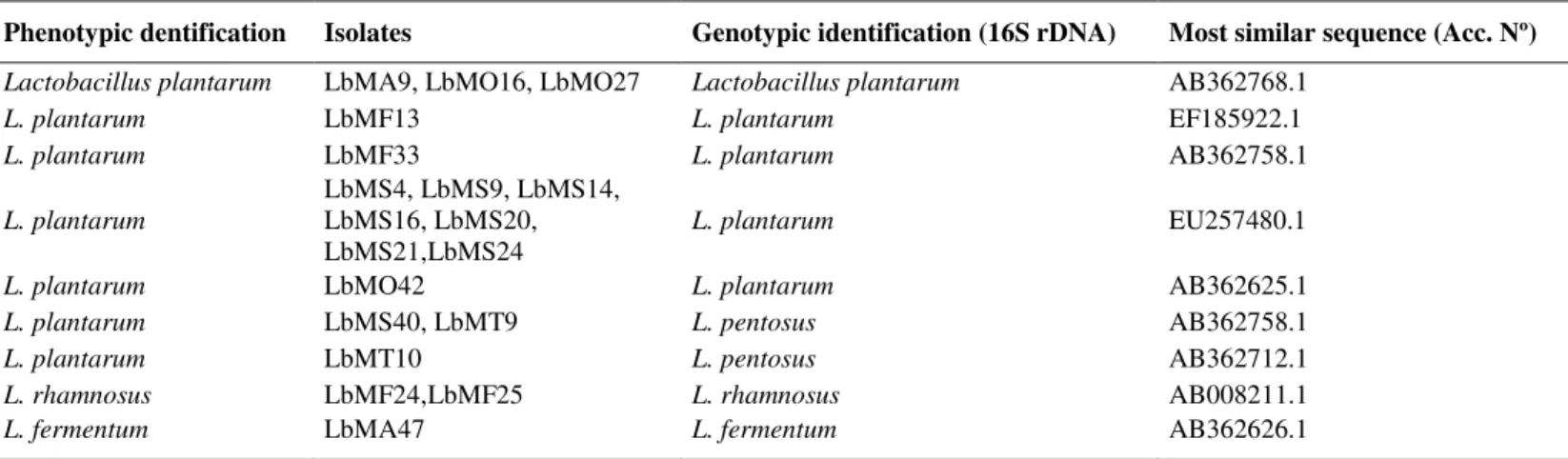
Genotypic identification of the isolates by 16S rDNA sequencing
The 16S rDNA of the 11 strains of subgroup A and 5
isolates of the subgroup B was amplified by 27F and 558R
primers, as reported by Acedo-Félix and Pérez-Martinez (1).
By partial sequencing of 16S rDNA all strains belonging to the
subgroup A were classified as L. plantarum. Of the 5 isolates
forming subgroup B, 60% were identified as L. pentosus,
(LbMS40, LbMT9 and LbMT10) and 40% as L. plantarum
(LbMO42, and LbMO27). The 16S rDNA of two isolates of
subgroup C were amplified with primers 27F and 1525R and
identified as L. rhamnosus (LbMF24 and LbMF25). The 16S
rDNA of the isolate of subgroup D was amplified by the
primers used for subgroup C and identified as L. fermentum
(LbMA 47).
Results of the PCR assay correlated with those obtained
using the API 50 CHL system for 13 isolates identified as L.
plantarum, 2 isolates as L. rhamnosus, and one isolate as L.
fermentum. However, three isolates identified as L. plantarum
by the API system were found to be L. pentosus by sequencing
16S rDNA (Table 3). This result is not surprising, given that
the two species themselves have very similar 16S rDNA
sequences that differ only by 2pb (19). In fact, it is widely
acknowledged that L. plantarum and L. pentosus belong to the
same 16S rRNA phylogenetic group and could only be
distinguished using phylogenetic analysis of sequences of the
16S-23S large spacer region (22).
The comparative evaluation of phenotypic and genotypic
results confirmed that the phenotypic test, in spite of giving
information on the biochemical and metabolic traits of LAB,
are not reliable enough for the identification of these
microorganisms, although it is a useful tool for presumptive
classification. Lactobacillus UFV H2B20, a probiotic strain,
for example, which was first identified as L. acidophilus based
on its sugar fermentation profile (38), was afterwards classified
as L. delbrueckii using molecular methods (16). One major
reason for the mismatch between phenotypic and genotypic
data might be ascribed to loosing or acquiring plasmids, which
leads to metabolite inconsistencies, as some carbohydrate
fermentation capacities are plasmid encoded (2). Genotypic
techniques are doubtlessly rapid and accurate tools for the
identification of LAB. The advantages of genotyping include
the stability of genomic DNA, its composition being
independent of cultural conditions or preparation methods, and
amenability to automation and statistical data analysis (21).
Table 3. Phenotypic and genotypic identification of Lactobacillus isolated from Algerian goat’s milk.
Phenotypic dentification Isolates Genotypic identification (16S rDNA) Most similar sequence (Acc. Nº) Lactobacillus plantarum LbMA9, LbMO16, LbMO27 Lactobacillus plantarum AB362768.1
L. plantarum LbMF13 L. plantarum EF185922.1
L. plantarum LbMF33 L. plantarum AB362758.1
L. plantarum
LbMS4, LbMS9, LbMS14, LbMS16, LbMS20, LbMS21,LbMS24
L. plantarum EU257480.1
L. plantarum LbMO42 L. plantarum AB362625.1
L. plantarum LbMS40, LbMT9 L. pentosus AB362758.1
L. plantarum LbMT10 L. pentosus AB362712.1
L. rhamnosus LbMF24,LbMF25 L. rhamnosus AB008211.1
L. fermentum LbMA47 L. fermentum AB362626.1
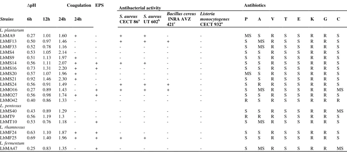
Technological characteristics of strains
Results on acidifying activity of Lactobacillus strains
isolated from goat’s milk, after 6, 12 and 24 h of growth in
skim milk are shown in Table 4. Milk pH after 24 h of
incubation varied between 4.40 and 5.54 for all cultures. All
(6.12 - 6.43) after 6 h of growth, except for strains
LbMA47,LbMS21 and LbMS16 which, respectively, were able
to decrease milk pH to values 5.31, 5.78 and 5.97. After 12 h,
the pH values ranged from 5.24 (LbMS21) to 5.94 (LbMT10),
while after 24 h, 47.37% of the strains had lowered milk pH to
4.40 - 4.96. According to their ability to reduce the pH more or
less rapidly, three clusters of L. plantarum isolates were
observed. (i) Slow acidifying strains (cluster I, 23% of L.
plantarum strains) showed a slow rate of acidifying ability
during the first 6 h of incubation ( pH ranging between
0.27-0.40 pH units). These L. plantarum strains lowered the pH
values between 0.86-1.01 pH units and 1.33-1.60 pH units after
12 h and 24 h of incubation, respectively. (ii) Medium
acidifying L. plantarum strains (cluster II) showed a faster rate
of acidifying ability after 6 h of incubation, with a pH ranging
between 0.50 and 0.57 pH units. In this cluster, two subgroups
were observed with different behaviours in their acidifying
capacity. The first subgroup A (31% of L. plantarum strains) in
which the pH values was achieved, respectively, 0.78-0.98
and 1.16-1.74 pH units, after 12 h and 24 h of incubation. On
the other hand, the second subgroup B (31% of L. plantarum
strains) showed a faster acidification rate. This subgroup
included L. plantarum strains showing a similar acidifying
activity until 6 h but capable of achieving pH values ranging
between 1.05-1.13 and 1.96-2.14 pH units after 12 h and 24 h
of incubation.(iii) Fast acidifying L. plantarum strains (15% of
L. plantarum strains) showed the highest acidifying capacity.
This two L. plantarum strains, LbMS16 and LbMS21, showed
a fast rate of acidifying ability until 6 h (0.73 and 0.92 pH units
respectively). This high capacity of acidifying milk by strains
of this cluster was also shown after 12 h and 24 h of
incubation: pH values were achieved, respectively, 1.31 to
2.20 pH units for LbMS16 and 1.46 to 2.30 pH units for
LbMS21. Result of acidifying capacity of L. plantarum strains
after 6 h and 12 h of incubation is in agreement with several
authors (14, 23). In contrast, the acidifying rate of isolates after
24 h of incubation was lower than those reported in the former
studies. The possibility to find groups of strains characterized
by different acidifying ability is frequent in L. plantarum
species (49).
L. pentosus and L. rhamnosus strains showed a more
homogenous acidifying behaviour. Strains of the first species
had a medium acidifying capacity with pH ranging between
0.43 and 0.58 pH units, after 6 h of incubation and achieved
1.18-1.30 pH units until 24 h. Furthermore, the two L.
rhamnosus strains showed a fast acidifying activity, with
values of pH 0.63 and of 0.69 pH units after 6 h. After 24 h of
incubation the pH reached values of 1.87 and 1.96 pH units.
L. fermentum strain showed the lowest rate of acidifying: pH
0.25 pH units after 6 h and resulted in pH 1.35 pH units until
24 h of incubation. Generally, L. plantarum strains produce
acid more rapidly, when compared to other lactobacilli (41,
14). The fast acidifying strains (mostly L. plantarum and L.
rhamnosus isolates) should be selected as part of a starter
preparation.
Seven isolates of L. plantarum strains and two L.
rhamnosus were able to coagulate skim milk, when inoculated
in skim milk after 24 h at 30 °C (Table 4). None of the L.
pentosus and L. fermentum strains were able to coagulate milk.
Coagulation of milk by some strains of L. plantarum and two
strains of L. rhamnosus revealed their potential as starters or
adjunct cultures in production of fermented dairy food products
(34).
Many strains of LAB produce exopolysaccharide (EPS),
which might be a capsule, closely attached as slim (9). The
production of EPS by Lactobacillus strains was examined in
milk culture and on ruthenium red milk plates. Ruthenium red
stains the bacterial cell wall, thus producing red colonies for
nonropy strains. Production of EPS prevents this staining, and
hence ropy colonies appear white on the same plates (44). The
results revealed EPS-production by isolated Lactobacillus
strains (Table 4). Among the Lactobacillus strains tested, eight
showed EPS-production. Four L. plantarum (LbMS14,
LbMS16, LbMO16 and LbMO27 strains), two L. rharmnosus
(LbMF24 and LbMF25 strains), one L. pentosus (LbMT10
strain) and one L. fermentum (LbMA47 strain) produced EPS.
Exopolysaccharides play a major industrial role in the
Marroki, A. et al. Characterization of Lactobacillus and their technological properties
production of yoghurt, drinking yoghurt, cheese, fermented
cream and milk-based desserts (18).
Results on aminopeptidase (AP) activity of 19 strains of
Lactobacillus strains tested using (Ala-ρNa and Leu-ρNa) are
shown in Figure 1. Amino acids released from peptides derived
from hydrolysis of casein may contribute directly or indirectly
for the development of flavour during ripening of cheese (48).
The proteolytic activity of dairy LAB is essential for the
bacterial growth in milk and it is involved in the development
of sensory properties of different fermented milk products (11).
Tested strains exhibiting aminopeptidase activity ranging
between 1.02 and 14.10 U for L-alanine-ρNA and 0.95 to
11.37 U for L-leucine-ρNA. The AP was divided, according to
the activity of each strain to high, medium and low activity.
Eleven percent of strains presented high activity to release
Ala-ρNA (13.12-14.10 U) and 16% of strains showed high ability
to release Leu-ρNA (10.10-11.37 U). Twenty six percent of
tested strains presented medium Ala-aminopeptidase activity
(6.14-8.35 U) and 26% Leu-aminopeptidase (5.15-9.70 U).
However, 63% of isolates revealed low activity to degrade Ala
-ρNA (1.02-4.93 U) and 58% to release Leu-ρNA (0.95-4.32
U). All tested strains of L. pentosus, L. rhamnosus, and L.
fermentum exhibited low AP activity, except for L. pentosus
LbMT10 strain, which had medium Leu-aminopeptidase
activity. Concerning the results obtained for L. plantarum
strains, two strains (LbMF13 and LbMO16) had high
Ala-aminopeptidase activity and four strains (LbMS14, LbMS16,
LbMS24 and LbMO16) had high Leu-aminopeptidase activity.
Figure1. Aminopeptidase activity of strains of the Lactobacillus tested in this study.
Well diffusion assay was used to screen 19 Lactobacillus
strains isolated from goat’s milk for their antibacterial activities
against several pathogenic indicator bacteria (Staphylococcus
aureus CECT 86T, Staphylococcus aureus UT 602, Bacillus
cereus INRA AVZ421 and Listeria monocytogenes CECT
932T) (Table 4). Bacteriocin like antimicrobial compound
production was indicated by azone of clearing of more than 1
mm against at least one of the indicator bacteria tested. The
cell-free supernatants of five L. plantarum strains (38.46 %,
an inhibition zone on agar against S. aureus CECT 86T and S.
aureus UT 602 and three L. plantarum strains (23,07%,
LbMF13, LbMS24, and LbMO16) against Bacillus cereus
AVZ 421. Antibacterial activity is a relatively frequent feature
of L. plantarum natural isolates (46). However, one strain of L.
rhamnosus strain (LbMF25) showed antimicrobial activity
against S. aureus CECT 86T and S. aureus UT 602.
Nevertheless, none of the tested strains of L. pentostus and L.
fermentum displayed inhibitory activity against any of the
indicator bacteria. In addition, all strains tested were inactive
against Listeria monocytogenes CECT 932T. Complete
inactivation of the antimicrobial activity was observed after
treatment with proteinase K, indicating the proteinaceous
nature of the antimicrobial compound, whereas treatment with
heat did not affect the inhibitory activity. Bacteriocins
produced by LAB are of particular interest because of their
potential use as natural food preservatives. The antibacterial
activity potential of some L. plantarum strains against
Staphylococcus aureus (37, 6) and Bacillus cereus (6) has been
previously reported.
Our results confirm the high incidence of
bacteriocin-producing lactic acid bacteria in milk samples, with inhibitory
activity against both pathogenic and spoilage microorganisms.
Goat’s milk may represent a source of new Lactobacillus
strains with the potential to inhibit undesirable and pathogenic
microorganisms for use in the biopreservation of dairy
products. The resistance to high temperature, the proteinaceous
nature, and the spectrum of activity of these antimicrobial
compound are advantageous for their use as biopreservatives in
food (37).
Antibiotic resistance
A key requirement for probiotic strains is that they should
not carry transferable antibiotic resistance genes. Transferable
resistance genes may pose a risk, as they can be transferred to
pathogenic bacteria (4). In this study, antibacterial
susceptibility testing of Lactobacillus strains was made
according to Charteris et al. (10). All Lactobacillus strains
isolated from goat’s milk were assayed for their susceptibility
to eight antibiotics, using the disk diffusion method. Zone
diameters were measured, and strains were classified as,
susceptible (S), moderately susceptible (MS), and resistant (R)
(Table 4). No strains of Lactobacillus were totally susceptible
to all antibiotics tested and multiple resistances to most
antibiotics were observed. Most strains were susceptible to
-lactam, inhibitors of cell wall synthesis (penicillin G and
ampicillin). Some tested strains are moderately susceptible to
the former antibiotics. However, two L. plantarum strains,
LbMO42 and LbMS24, were resistant to penicillin G or
ampicillin, respectively, and one L. pentosus strain (LbMT9)
was resistant to these two antibiotics simultaneously. Several
studies report that species of lactobacilli exhibited
susceptibility to almost all penicillins (10). Nevertheless, the
resistance of Lactobacillus strains to penicillin G and
ampicillin has beendescribed in other studies (45, 13, 24).
Our result show that all tested strains were resistant to
vancomycin, which is equally a cell wall synthesis inhibitor
(non- -lactam). Resistance of Lactobacillus species to
vancomycin is due to the presence of D-Ala-D-lactate in their
peptidoglycan, rather than the D-ala-D-ala dipeptide (28). Such
resistance is usually intrinsic, that is, chromosomally encoded
and nontransmissible (27). Concerning the protein synthesis
inhibitors, all strains tested were susceptible to tetracyclin,
erythromycin and resistant to kanamycin and gentamycin.
Lactobacilli are generally susceptible to antibiotics which
inhibit the synthesis of proteins, such as erythromycin and
tetracycline and more resistant to aminoglycosides (kanamycin
and gentamicin) (10). Chloramphenicol inhibited most tested
strains. Three Lactobacillus strains showed a moderate
susceptibility and one L. plantarum (LbMO42) strain was
resistant to this antibiotic. The high natural susceptibility of
lactobacilli to chloramphenicol (protein synthesis inhibitor) is
Marroki, A. et al. Characterization of Lactobacillus and their technological properties
Table 4. Technological characteristics and antibiotic resistance of Lactobacillus strains isolated from Algerian goat’s milk
pH Coagulation EPS
Antibacterial activity Antibiotics
Strains 6h 12h 24h 24h S. aureus
CECT 86a
S. aureus UT 602b
Bacillus cereus INRA AVZ 421c
Listeria monocytogenes CECT 932a
P A V T E K G C
L. plantarum
LbMA9 0.27 1.01 1.60 + - + + - - MS S R S S R R S
LbMF13 0.50 0.97 1.46 - - + + + - S MS R S S R R S
LbMF33 0.52 0.78 1.16 - - - S MS R S S R R S
LbMS4 0.53 1.05 2.14 - - - S S R S S R R S
LbMS9 0.51 1.13 1.97 + - - - S S R S S R R S
LbMS14 0.56 1.11 2.07 + + + + - - S S R S S R R S
LbMS16 0.73 1.31 2.20 + + - - - - S S R S S R R S
LbMS20 0.57 1.07 1.96 + - - - MS S R S S R R S
LbMS21 0.92 1.46 2.30 + - - - S S R S S R R S
LbMS24 0.56 0.91 1.49 - - + + + - S R R S S R R S
LbMO16 0.27 0.89 1.43 - + + + + - S MS R S S R R MS
LbMO27 0.56 0.98 1.74 + + - - - - S S R S S R R S
LbMO42 0.40 0.86 1.33 - - - R S R S S R R R
L. pentosus
LbMS40 0.43 0.89 1.29 - - - S S R S S R R MS
LbMT9 0.56 1.19 1.3 - - - R R R S S R R S
LbMT10 0.53 0.76 1.18 - + - - - - S MS R S S R R S
L. rhamnosus
LbMF24 0.63 1.10 1.87 + + - - - - S S R S S R R S
LbMF25 0.69 1.40 1.96 + + + + - - S S R S S R R S
L. fermentum
LbMA47 0.25 0.83 1.35 - + - - - - S MS R S S R R MS
+: Positive reaction; -: Negative reaction. EPS: Exopolysaccharide production
(a) CECT: Colección Española de Cultivos Tipo, Valencia, Spain (b)U.T: University of Tlemcen laboratory collection, Algeria
(c)INRA AVZ : Station de Technologie des Produits Végétaux, Institut national de la Recherche Agronomique, Avignon, France (INRA) PG: penicillin (10µg); A: ampicillin (10µg); V: vancomycin (30µg); T: tetracycline (30µg); E: erythromycin (15µg);
With regard to antibiotic susceptibility profiles, L.
plantarum strain (LbMO42) and L. pentosus strain (LbMT9)
showed the highest resistance to 5 of the 8 antibiotics tested. At
present, multiresistance seems to be uncommon among LAB
species, but an increasing number of strains displaying atypical
resistance levels to some antibiotics are being isolated (3).
Development of antibiotic resistance in bacteria is mainly
based on two factors, the presence of resistance genes and the
selective pressure through the use of antibiotics (29). The
overuse of antibiotics in veterinary medicine as therapeutic
agents, prophylactics and animal growth promoters on farms
may favour the development of resistance to antibiotics among
LAB. Therefore, strains intended to be used in feed and food
systems should be systematically monitored for resistances, in
order to avoid their inclusion as starters and probiotics (3).
In conclusion, as far as we know, this is the first report on
the genetic identification of Lactobacillus strains using 16S
rDNA sequence of Algeria goat’s milk isolates. This molecular
method distinguishes the closely related species L. plantarum
and L. pentosus identified using physiological features and API
50 CHL system as L. plantarum. Results on technological
properties suggest the pontential of some of L. plantarum
(LbMS16 and LbMS21) and L. rhamnosus (LbMF25) strains
as starter cultures in the manufacture of artisanal fermented
dairy product in Algeria. Moreover, these strains should be
tested in mixed cultures, in order to obtain fermented dairy
product with sensorial characteristics similar to those of
artisanal products.
ACKNOWLEDGEMENTS
A.Marroki expresses his gratitude to the “Ministère de
l’Enseignement Supérieur et de la Recherche Scientifique
Algérien" and "University of Oran- Es-Sénia-Oran" for a
research grant. This work was partially funded by a C.S.I.C.
project (Ref. 2006 7 0I 097) and the Spanish Ministry of
Science and Innovation (Consolider Fun-C-Food CSD2007-
00063).
REFERENCES
1. Acedo-Félix, E.; Pérez-Martínez, G. (2003). Significant differences between Lactobacillus casei subsp. casei ATTCC 393T and a commonly used plasmid-cured derivative revealed by a polyphasic study. Int. J. Syst. Evol. Microbiol., 53: 67-75.
2. Ahrné, S.; Molin, G.; Stahl, S. (1989). Plasmids in Lactobacillus strains isolated from meat and meat products. Syst. Appl. Microbiol., 11: 320-325.
3. Ammor, M.S.; Flóre, A.B.; Mayo, B. (2007). Antibiotic resistance in non-enterococcal lactic acid bacteria and bifidobacterium. Food Microbiol., 24: 559-570.
4. Aymerich, T.; Martin, B.; Garriga, M.; Vidal-Carou, M.C.; Bover-Cid, S.; Hugas, M. (2006). Safety properties and molecular strain typing of lactic acid bacteria from slightly fermented sausages. J. Appl. Microbiol., 100: 40-49.
5. Badis, A.; Guetarni, D.; Moussa-Boudjema, B.; Henni, D.E.; Tornadijo, M.E.; Kihal, M. (2004). Identification and technological properties of lactic acid bacteria isolated from raw goat’s milk of four Algerian races. Food Microbiol., 2: 579-588.
6. Ben Omar, N.; Abriouel, H.; Lucas, R.; Martínez-Cañamero, M.; Guyot, J-P.; Gálvez, A. (2006). Isolation of bacteriocinogenic Lactobacillus plantarum strains from ben saalga, a traditional fermented gruel from Burkina Faso. Int. J. Food Microbiol., 112: 44-50.
7. Boubekri, K.; Ohta, Y. (1996). Identification of lactic acid bacteria from Algerian traditional cheese, El-Klila. J. Sci. Food Agric., 70: 501-505. 8. Bringel, F.;Curk, M-C.; Huert, J-C. (1996). Characterization of
Lactobacillus by Southern-Type hybridation with a Lactobacillus plantarumpyr DFE probe. Int. J. Syst. Bacteriol., 46: 588-594.
9. Cerning, J. (1990). Exocellular polysaccharides produced by lactic acid bacteria. FEMS Microbiol. Rev., 87: 113-130.
10. Charteris, W.P.; Kelly, P.M.; Morelli, L.; Collins, J.K. (1998). Antibiotic susceptibility of potentially probiotic Lactobacillus species. J. Food. Prot., 61: 1636-1643.
11. Christensen, J.E.; Dudley, E.G.; Pederson, J.A.; Steele, J.L. (1999). Peptidases and amino acid catabolism in lactic acid bacteria. Antonie van Leeuwenhoek., 76: 217-246.
12. Coeuret, V.; Dubernet, S.; Bernardeau, M.; Gueguen, M.; Vernoux, J.P. (2003). Isolation, characterisation angbd identification of lactobacilli focusing mainly on cheeses and other dairy products. Lait., 83: 269-306. 13. Coppola, R.; Succi, M.; Tremonte, P.; Reale, A.; Salzano, G.; Sorrentino,
E. (2005). Antibiotic suscebtibility of Lactobacillus rhamnosus strains isolated from Parmigiano Reggiano cheese. Lait., 85: 193-204.
14. Dagdemir, E.; Ozdemir, S. (2008). Technological characterization of the natural lactic acid bacteria of artisanal Turkish white pickled cheese. Int. J. Dairy Technol., 61: 133-140.
Marroki, A. et al. Characterization of Lactobacillus and their technological properties
(1994). Caractéristiques générales des bactéries lactiques. In : de Roissart, H.; Luquet, F.M. (eds) Les bactéries lactiques : aspects fondamentaux et technologiques. Lorica, Uriage, p. 25-116.
16. De Magalhães, J.T.; Uetanabaro, A.P.T.; de Moraes, C.A. (2008). Identification of Lactobacillus UFV H2B2 (Probiotic strain) using DNA-DNA hybridation. Braz. J. Microbiol., 39: 524-546.
17. De Man, J.C.; Rogosa, M.; Sharp, M.E. (1960). A medium for the cultivation of lactobacilli. J. Appl. Bacteriol., 23: 130-135.
18. De Vuyst, L.; Degeest, B. (1999). Heteropolysaccharides from lactic acid bacteria. FEMS Microbiol. Rev., 23: 153-177.
19. Ennahar, S.; Cai, Y.; Fujita, Y. (2003). Phylogenetic diversity of lactic acid bacteria associated with paddy rice silage as determined by 16S ribosomal DNA analysis. Appl. Environ. Microbiol., 69: 444-451. 20. Estepar, J.; Del Mar Sánchez, M.; Alonso, L.; Mayo, B. (1999).
Biochemical and microbiological characterization of artisanal ‘Peñamellera’ cheese: analysis of its indigenous lactic acid bacteria. Int. Dairy J., 9: 737-746.
21. Fitzsimons, N.A.; Cogan, T.M.; Condon, S.; Beresford, T. (1999). Phenotypic and genotypic characterization of non-starter lactic acid bacteria in mature Cheddar cheese. Appl. Environ. Microbiol., 65: 3418-3426.
22. Hammes, W.P.; Vogel, R.F. (1995). The genus Lactobacillus. p. 19-54. In: Wood, B.J.B.; Holzapfel, W.H (eds), the genera of lactic acid bacteria, vol. 2. The genera of lactic acid bacteria. Blackie Academic and Professional. London, United Kingdom.
23. Herreros, M.A.; Fresno, J.M.; González Prieto, M.J.; Tornadijo, M.E. (2003). Technological characterization of lactic acid bacteria from Armada cheese (a Spanish goat’s milk cheese). Int. Dairy. J., 13: 469-479.
24. Herreros, M.A.; Sandoval, H.; González, L.; Castro, J.M.; Fresno, J.M.; Tornadijo, M.E. (2005). Antimicrobial activity and antibiotic resistance of lactic acid bacteria isolated from Armada cheese (a Spanish goats’ milk cheese). Food Microbiol., 22: 455-459.
25. Jayne-Williams, D.J. (1976). The application of miniaturized methods for characterization of various organisms isolated from the animal gut. J. Appl. Microbiol., 40: 189-200.
26. Kandler, O.; Weiss, N. (1986). Genus Lactobacillus Beijerinck 1901, 212 AL. In: Sneath, P.H.A.; Mair, N.S.; Sharpe, M.E.; Holt, J.G. (eds.) Bergey’s Manual of Systematic Bacteriology. vol 2, Baltimore, Williams & Wilkins , p. 1209-1234.
27. Klein, G.; Pack, A.; Bonapartes, C.; Reuter, G. (1998). Taxonomy and physiology of probiotic lactic acid. Int. J. Food Microbiol., 41: 103-125. 28. Klein, G.; Halmann, C.; Casas, I.A.; Abad, J.; Louwers, J.; Reuter, G.
(2000). Exclusion of vanA, vanB and vanC type glycopeptides resistance in starters of Lactobacillus reuteri and Lactobacillus rhamnosus used as probiotics by polymerase chain reaction and hybridation methods. J. Appl. Microbiol., 89: 815-824.
29. Levy, S.B. (1992). The antibiotic paradox: How Miracle Drugs are Destroying the Miracle. Plenum Press, New York, p 279.
30. Linaje, R.; Coloma, M.D.; Pérez-Martinez, G.; Zúñiga, M. (2004). Characterization of faecal enterococci from rabbits for the selection of probiotic strains. J. Appl. Microbiol., 96: 161-771.
31. Macedo, A.C.; Viera, M.; Poças, R.; Malcata, F.X. (2000). Peptide hydrolase system of lactic acid bacteria isolated from Serra de Estrela cheese. Int. Dairy J., 10: 769-774.
32. Müller, T. (1990). Comparaison of methods for differentiation between homofermentative and heterofermentative lactic acid bacteria. Zentralbl. Mikrobiol., 145: 363-3666.
33. Nigatu, A. (2000). Evaluation of numerical analyses of RAPD and API 50 CH patterns to differentiate Lactobacillus plantarum, Lact. fermentum, Lact. rhamnosus, Lact sake, Lact. parabuchneri, Lact. gallinarum, Lact.
Gallinarum, Lact. casei, Weissella minor and related taxa isolated from kocho and tef. J. Appl. Microbiol., 89: 969-978.
34. Olasupo, N.A.; Shillinger, U.; Holzapfel, W.H. (2001). Studies on some technological properties of predominant lactic acid bacteria isolated from Nigerian fermented food. Food Microbiol., 15: 157-167.
35. Psono, L.; Kotzamanides, C.; Andrighetto, C.; Lombardi, A.; Tzanetakis, N.; Litopoulou-Tzanetaki, E. (2006). Genotypic and phenotypic heterogeneity in Enterococcus isolated from Batzos, a raw goat milk cheese. Int. J. Food Microbiol., 109: 109-120.
36. Quere, F.; Deschamps, A.; Urdaci, M.C. (1997). DNA probe and PCR-specific reaction for Lactobacillus plantarum. J. Appl. Microbiol., 82: 783-790.
37. Rodríguez, E.; Gonzalez, B.; Gaya, P.; Nuñez, M.; Medina, M. (2000). Diversity of bacteriocins produced by lactic acid bacteria isolated from raw milk. Int. Dairy Journal., 10: 7-15
38. Sabioni, N.S.S.; Pinheiro, A.J.R.; Teixeira, M.A. (1998). Acidophilus milk: Isolation and characterization of Lactobacillus acidophilus from breast-fed children and from calves’ feces. Braz. J. Microbiol., 19: 393-398.
39. Sánchez, I.; Palop, L.; Ballesteros, C. (2000). Biochemical characterization of lactic acid bacteria isolated from spontaneous fermentation of ‘Almagro’ eggplantes. Int. J. Food Microbiol., 59: 9-17. 40. Schleifer, K.H.; Ehrmann, M.; Beimfohr, C.; Brokmann, E.; Ludwig, W.;
Amann, R. (1995). Application of molecular methods for classification and identification of lactic acid bacteria. Int. Dairy J., 5: 1081-1094. 41. Seseña, S.; Sánchez, I.; Palop, L. (2005). Characterization of
Lactobacillus strains and monitoring by RAPD-PCR in controlled fermentations of “Almagro” eggplants. Int. J. Food Microbiol., 104: 325-335.
42. Sharpe, M.E. (1979). Identification of the lactic acid bacteria. In: Skinner, F.A.; Lovelock, D.W. (eds.) Identification Methods for Microbiologists, Academic Press, London, p. 233-259.
43. Stiles, M.E.; Holzapfel, W.H. (1997). Lactic acid bacteria of food and their current taxonomy. Int. J. Food Microbiol., 36: 1-29.
45. Temmerman, R.; Pot, B.; Huys, G.; Swings. J. (2003). Identification and antibiotic susceptibility of bacterial isolates from probiotic products. Int. J. Food Microbiol., 81: 1-10.
46. Todorov, D.S. (2009). Bacteriocins from Lactobacillus plantarum -production, genetic organization and mode of action. Braz. J. Microbiol., 40: 209-221.
47. Vandamme, P.; Pot, B.; Gillis, M.; De Vos, P.; Kersters, K.; Swings, J. (1996). Polyphasic taxonomy, a consensus approach to bacterial systematic. Microbiol. Rev., 60: 407-438.
48. Williams, A.G.; Felipe, X.; Banks, J.M. (1998). Aminopeptidase and dipeptidyl peptidase activity of Lactobacillus spp. And non-starter lactic acid bacteria (NSLAB) isolated from Chaddar cheese. Int. Dairy J., 8:
255-266.
49. Xanthopoulos, V.; Hatzikamari, M.; Adamidis, T.; Tzaneetakis, N.; Tsakalidou, E.; Litopoulou-Tzanetaki, E. (2000). Heterogeneity of Lactobacillus plantarum isolated from Feta cheese throughout ripening. J. Appl. Microbiol., 88: 1056-1064.
50. Zanoni, P.; Farrow, J.A.E.; Phillips, B.A.; Collins, M.D. (1987). Lactobacillus pentosus (Fred, Peterson, and Anderson) sp. nov., nom. rev. Int. J. Syst. Bacteriol., 37: 339-341.