J of Evolution of Med and Dent Sci/ eISSN- 2278-4802, pISSN- 2278-4748/ Vol. 4/ Issue 77/ Sept 24, 2015 Page 13411
CHANGES IN SERUM CALCIUM AND SERUM GLUCOSE LEVELS IN
ASPHYXIATED SGA
Seema Rai1, Sharanjit Kaur2, Abdul Hamid3
HOW TO CITE THIS ARTICLE:
Seema Rai, Sharanjit Kaur, Abdul Hamid. Changes in Serum Calcium and Serum Glucose Levels in Asphyxiated SGA. Journal of Evolution of Medical and Dental Sciences 2015; Vol. 4, Issue 77, September 24;
Page: 13411-13416, DOI: 10.14260/jemds/2015/1924
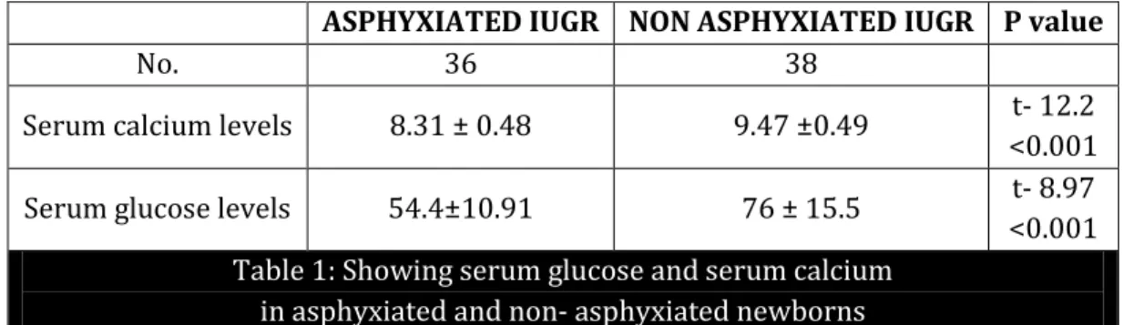
ABSTRACT: OBJECTIVE: To study effect of birth asphyxia on serum calcium and serum glucose in serum samples of asphyxia small for gestational age (SGA) of different severity and compare with controls. METHODS: Serum calcium and glucose levels were estimated in the serum samples of asphyxiated SGA newborns of different grades and non-asphyxiated control group at 24 hours of age. RESULTS: Mean serum calcium in SGA of study group (8.31±0.48mg/dl) was lower than control SGA (9.47±0.49mg/dl) p<0.01). Mean serum glucose in SGA of study group (54.4±10.91 mg/dl) was lower than control SGA (76±15.5 mg/dl) p<0.01). The present study showed that there was significant negative correlation of serum calcium and glucose with severity of asphyxia (p<0.01). CONCLUSION: Among cases, hypocalcaemia and hypoglycaemia developed early and decrease in their serum levels was directly proportional to degree of asphyxia.
KEYWORDS: Hypocalcemia, Hypoglycemia, Apgar score, Perinatal Asphyxia, SGA.
INTRODUCTION: Small for gestational age is defined as less than 10% of predicted birth weight for that gestational age which can lead to significant mortality and morbidity if not properly managed.[1]
A fetus affected by IUGR forms a subset of cases of Small for Gestational Age (SGA) infants.[2] In SGA,
the estimated weight of the fetus is below the 10th percentile for its gestational age and abdominal circumference (AC) is below the 2.5th percentile.[3] In accurately dated pregnancies, approximately
80-85% of fetuses identified as being IUGR are constitutionally small but healthy, 10-15% are 'true' IUGR cases, and the remaining 5-10% of fetuses are affected by chromosomal/structural anomalies or chronic intrauterine infections.[4] IUGR can complicate 10% to 15% of all physiologic
pregnancies.[5] However, it must be remembered that the incidence of such cases varies depending on
the population, geographic location being scrutinized and the standard growth curves used as reference.[3]
Birth asphyxia is a common neonatal problem and contributes significantly to neonatal morbidity and mortality. Globally, hypoxia of the newborn (Birth asphyxia) or the fetus ("Fresh stillbirth") is estimated to account for 23% of the 4 million neonatal deaths and 26% of the 3.2 million stillbirths each year1.Data from National Neonatal Perinatal database (NNPD) suggests that perinatal asphyxia contributes to almost 20% of neonatal deaths in India.2. Neonatal hypocalcemia and hypoglycaemia are predominant metabolic causes of seizures especially in IUGR infants.[6]
J of Evolution of Med and Dent Sci/ eISSN- 2278-4802, pISSN- 2278-4748/ Vol. 4/ Issue 77/ Sept 24, 2015 Page 13412
After birth due to abrupt cessation of placental transfer of calcium hence levels starts falling to 8-9 mg/dl and ionized calcium to 4.4-5.4 mg/dl at 24 hours of age. Serum calcium then starts rising to reach levels comparable to older children and adults by two weeks of age.[8]
Glucose is an essential nutrient for the brain. Abnormally low level can cause encephalopathy and have the potential to produce long term neurological injury. Serum glucose levels decline after birth until 1-3 hours of age, when levels spontaneously increase in normal infants. In healthy term infants, serum glucose values are rarely less than 35 mg/dl between 1 and 3 hours of life, less than 40 mg/dl from 3-24 hours and less than 45 mg/dl after 24 hours of life.[9] In birth asphyxia,
hypoglycaemia is due to glycogen depletion secondary to catecholamine release and to an unexplained hyperinsulinemic state. An initial phase of hyperglycemia and hypoinsulinemia (5-10 minutes following an acute event due to a catecholamine surge which inhibits insulin release and stimulates glucagon release) may be followed within 2-3 hours by profound hypoglycaemia.[10]
This study was undertaken to detect incidence of hypocalcemia and hypoglycaemia in asphyxiated SGA babies as to prevent the adverse effects of these biochemical abnormalities in the newborns.
MATERIAL AND METHODS: This was a hospital based case control study following simple random sampling with equal number of cases and controls, a convenient number of 74 SGA newborns were selected, 36 were asphyxiated and 38 were non-asphyxiated. This study was approved by the Institutional Ethical Committee, and informed consent was obtained from the parents of each subject. In this study, 100 asphyxiated neonates (Apgar score at one minute 7 or less) were taken as cases of study. 35 normal neonates (Apgar score at 1 minute more than 7) were taken as control. Total serum calcium and serum glucose levels were determined at 24 hours of life in all the newborns. Serum calcium was estimated by O- Cresolphthalein complexone (O-CPC) end point (kit) method (Connerty and Briggs, 1966).[6] Blood glucose estimation was done by Asatoor and King Method (Varley,
2004).[7] Babies with congenital malformations, serum creatinine levels more than 1.5mg/dl,
suspected metabolic disease, treated with diuretics and those born to mothers having hypertension, diabetes mellitus, toxaemia of pregnancy were excluded from the study.
J of Evolution of Med and Dent Sci/ eISSN- 2278-4802, pISSN- 2278-4748/ Vol. 4/ Issue 77/ Sept 24, 2015 Page 13413
ASPHYXIATED IUGR NON ASPHYXIATED IUGR P value
No. 36 38
Serum calcium levels 8.31 ± 0.48 9.47 ±0.49 t- 12.2 <0.001
Serum glucose levels 54.4±10.91 76 ± 15.5 t- 8.97 <0.001 Table 1: Showing serum glucose and serum calcium
in asphyxiated and non- asphyxiated newborns
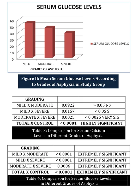
GRADING NO. SERUM CALCIUM LEVELS SERUM GLUCOSE LEVELS MILD 22 7.349±0.3399 54.785±1.771 MODERATE 11 7.157±0.09922 47.045±2.712 SEVERE 3 6.817±0.2290 39.267±1.823 TOTAL 36 7.249±0.3202 52.121±5.069 CONTROL 38 8.474±0.3640 57.409±2.302
Table 2: Serum Calcium Levels In Study And Control According To Grades Of Asphyxia
J of Evolution of Med and Dent Sci/ eISSN- 2278-4802, pISSN- 2278-4748/ Vol. 4/ Issue 77/ Sept 24, 2015 Page 13414
GRADING
MILD X MODERATE 0.0922 > 0.05 NS MILD X SEVERE 0.0157 < 0.05 S MODERATE X SEVERE 0.0025 < 0.0025 VERY SIG
TOTAL X CONTROL < 0.0001 HIGHLY SIGNIFICANT
Table 3: Comparison for Serum Calcium Levels in Different Grades of Asphyxia
GRADING
MILD X MODERATE < 0.0001 EXTREMELY SIGNIFICANT MILD X SEVERE < 0.0001 EXTREMELY SIGNIFICANT MODERATE X SEVERE 0.0006 EXTREMELY SIGNIFICANT TOTAL X CONTROL < 0.0001 EXTREMELY SIGNIFICANT
Table 4: Comparison for Serum Glucose Levels in Different Grades of Asphyxia
DISCUSSION: This study was aimed to determine effect of birth asphyxia on serum calcium and serum glucose in small for gestational age newborns. This was done by comparing 36 small for gestational asphyxiated newborns with 38 non-asphyxiated small for gestational newborns.
Hypocalcemia in birth asphyxia is due to low serum calcium intake, functional hypoparathyroidism due to hypoxia and excessive bicarbonate therapy which is further exaggerated in small for gestational newborns.[11] In general, higher the calcium concentration in the umbilical
cord blood, grater its decrease during the first two days of life. In asphyxiated newborns, decrease in the serum level of both total calcium and ionized calcium surpass that in non-asphyxiated newborns by approximately a third. The most striking cause for the early form of hypocalcemia are likely to be a transient hypoparathyroidism or a failure of end organ responsiveness.[12] In Present study, mean
J of Evolution of Med and Dent Sci/ eISSN- 2278-4802, pISSN- 2278-4748/ Vol. 4/ Issue 77/ Sept 24, 2015 Page 13415
serum calcium in SGA of study group (8.31±0.48) was lower than control non-asphyxiated SGA group (9.47±0.49) which was significant statistically (p<0.001). This is similar to that reported by Tsang et al (1975) found that total serum calcium (7.82 mg/dl) significantly lower in asphyxiated SGA babies at 24 hours of life as compared to their control group.[13] Jajoo et al (1995) also found total calcium
levels (8 mg/dl) significantly low in asphyxiated SGA babies at 24 hours of as compare to their control (9.5 mg/dl).[14]
In Present study, mean serum glucose in SGA of study group (54.4±10.91) was lower than control non-asphyxiated SGA group (76±15.5) which was significant statistically (p<0.001). With increase in severity of asphyxia there was significant (p<0.001) fall in mean serum glucose levels. Birth asphyxia can lead to exaggerated fall in serum glucose in small for gestational newborns due to low glycogen stores in these babies.[15] Another contributory factor for hypoglycaemia in asphyxiated
SGA is transient hyperinsulinemic state, deficient glycogenolysis, deficient gluconeogenesisis, deficiency of oxidative enzymes for fatty acid oxidation abnormalities of counterregulatrory hormoneal mechanisms like glucogon adrenalin etc. which causes further fall in the serum glucose levels.[16]
CONCLUSION: Among cases, hypocalcaemia and hypoglycaemia developed early and decrease in their serum levels was directly proportional to degree of asphyxia.
REFERENCES:
1. Battaglia FC, Lubchenco LO. A practical classification of newborn infants by weight and gestational age. J Pediatr. 1967; 71(2):159.
2. Sheridan C. Intrauterine growth restriction-diagnosis and management. Aust Fam Physician. 2005. pp. 717–23.
3. Peleg FD, Kennedy CM, Hunter SK. Intrauterine Growth Restriction: Identification and Management. Am Fam Physician. 1998. pp. 453–60. 466-7.
4. Manning FA. General principles and applications of ultrasonography. Maternal-fetal medicine: principles and practice. Philadelphia: Saunders; 2004.
5. Florio P, Marinoni E, DiIorio R, Bashir M, Ciotti S, Sacchi R. Urinary S100B Protein Concentrations Are Increased in Intrauterine Growth-Retarded Newborns. Pediatrics. 2006; 118:e747–54. Doi: 10.1542/peds.2005-2875.Romo A.1, Carceller R, Tobajas J. Intrauterine
growth retardation (IUGR): epidemiology and etiology. Pediatr Endocrinol Rev. 2009 Feb; 6 Suppl 3:332-6.
6. NNPD Network. National neonatal Perinatal Database- report for the year 2002-2003.NNF NNPD network. New Delhi: 2005.
7. Tsang R.C., Danovan E.F., Steinchen J.J. Calcium physiology in the neonates. Pediatric Clinic North Am. 1976; 23:611–726.
8. Tsang R.C., Light I.J., Sutherland J.M., Klenman L.I. Possible pathogenesis factors in neonatal hypocalcemia of prematurity. Journal of Pediatrics. 1973; 82:423–429. Doi: 10.1016/S0022-3476(73)80115-5.
9. Stoll BJ, Kleigmann RM. The endocrine system.Nelson Textbook of Pediatrics. Behrman RE, Kleigman RM, Jenson HB, editors. 20th ed.philadelphia: Elsevier Publisher; 2014.613-616.
J of Evolution of Med and Dent Sci/ eISSN- 2278-4802, pISSN- 2278-4748/ Vol. 4/ Issue 77/ Sept 24, 2015 Page 13416
11.David L, Anast C. Calcium metabolism in newborn infants. J. Clin. Invest. 1974; 54:287–290. Doi: 10.1172/JCI107764.
12.Manzke H, Kruse K. Physiology and pathology of calcium and phosphate metabolism in newborn infants.Personal study results and literature references. Monatsschr kinderheilkd 1984; 132(4):203-9.
13.Tsang R.C., Giger M.Ch. W., Brown D.R. Studies in calcium metabolism in infants with intrauterine growth retardation. Journal of Pediatrics. 1975; 86:936–941.
Doi: 10.1016/S0022-3476(75)80232-0.
14.Jajoo D., Kumar A., Shankar R., Bhargava V. Effects of birth asphyxia on serum calcium levels in neonates. Indian Pediatrics. 1995; 62:455–459.
Doi: 10.1007/BF02755067.
15.Giroux JD.1 Vernotte E, Gagneur A, Metz C, Collet M, de Parscau L. [Transitory hyperinsulinism
with hypoglycemia in asphyxia neonatorum]. Arch Pediatr. 1997 Dec; 4(12):1213-6.
16.F. Nili. Transient hyperinsulinismin asphyxiated small for gestation infant.Acta Medica Iranica. 1999:37(4); 204-06.
AUTHORS: 1. Seema Rai 2. Sharanjit Kaur 3. Abdul Hamid
PARTICULARS OF CONTRIBUTORS: 1. Assistant Professor, Department of
Pediatrics, MMC & H, Solan. 2. Associate Professor, Department of
Pharmacology, MMC & H, Solan. 3. Senior Resident, Department of
Pediatrics, MMC & H, Solan.
FINANCIAL OR OTHER
COMPETING INTERESTS: None
NAME ADDRESS EMAIL ID OF THE CORRESPONDING AUTHOR: Dr. Seema Rai,
Dev Bhoomi Green Valley D-15, Deonghat, Solan-173212, Himachal Pradesh.
E-mail: [email protected]