Cop
yright
© ABE&M t
odos os dir
eit
os r
eser
vados
.
Evaluation of parathyroid function
and mineral metabolism in
psychiatric patients using lithium salts
Avaliação da função paratireoidiana e metabolismo mineral em pacientes psiquiátricos usuários de sais de lítio
Thiago Costa de Oliveira1, Ivo Alves de Campos Neto1, Manuel Hermínio de Aguiar-Oliveira1, Francisco de Assis Pereira1
ABSTRACT
Objective: To evaluate parathyroid function and mineral metabolism in psychiatric patients users of lithium salts. Materials and methods: We measured the serum levels of calcium, io-nized calcium, inorganic phosphorus, alkaline phosphatase, albumin, parathyroid hormone (PTH), urea, creatinine, 25-hydroxy-vitamin D and lithium of 35 patients diagnosed with bipolar disorder in use of lithium carbonate (LC) for at least one year (Lithium Group – LG) and 35 healthy subjects (Control Group – CG). Results: The LG and CG were matched by sex and age. There was only statistic difference in relation to the levels of PTH and ionized calcium, with p < 0.004 and p < 0.03, respectively. Secondary form of hyperparathyroidism (HPT) was found in eight (22.8%) LG patients and in none of the CG. There was no correlation between lithemia, usage time and dosage of LC. Conclusion: Our data demonstrate that lithium may create an imbalance in the parathyroid axis, characterized by elevated levels of PTH. Arq Bras Endocrinol Metab. 2014;58(6):619-24
Keywords
Lithium; parathyroid; mineral metabolism; hyperparathyroidism
RESUMO
Objetivo: Avaliar a função paratireoidiana e o metabolismo mineral em pacientes psiquiátricos usuários de sais de lítio. Materiais e métodos: Foram avaliados os níveis séricos de cálcio to-tal, cálcio iônico, fósforo inorgânico, fosfatase alcalina, albumina, paratormônio (PTH), ureia, creatinina, 25-hidroxivitamina D e lítio de 35 pacientes diagnosticados com transtorno afetivo bipolar usuários de carbonato de lítio (CL) há pelo menos um ano (Grupo Lítio – GL) e 35 indi-víduos saudáveis (Grupo Controle – GC). Resultados: O GL e o GC foram pareados por sexo e idade. Somente se observou diferença estatística em relação aos níveis de PTH e cálcio iônico, com p < 0,004 e p < 0,03, respectivamente. Hiperparatireoidismo secundário foi encontrado em oito (22.8%) pacientes do GL e em nenhum do GC. No GL, não houve correlação entre litemia, tempo de uso e posologia do CL. Conclusão: Nossos dados demonstram que o lítio pode susci-tar um desequilíbrio no eixo paratireoideano, caracterizado por níveis elevados de PTH. Arq Bras Endocrinol Metab. 2014;58(6):619-24
Descritores
Lítio; paratireoide; metabolismo mineral; hiperparatireoidismo
1 Departament of Medicine, Universidade Federal de Sergipe (UFS), Aracaju, SE, Brazil
Correspondence to:
Francisco de Assis Pereira St. José Freire, 644
490020-410 – Aracaju, SE, Brazil [email protected]
Received on Sept/7/2013 Accepted on May/12/2014
DOI: 10.1590/0004-2730000002983
INTRODUCTION
A
bout six decades, lithium salts were introduced asmedical treatment due to their ability to modulate and stabilize mood in psychiatric patients with affective disorders (1,2). The use of lithium (Li) is the most effec-tive long-term therapy for affeceffec-tive disorders, such as
Cop
yright
© ABE&M t
odos os dir
eit
os r
eser
vados
.
being provided free of charges by the National Health System in the form of Lithium Carbonate (LC) (6,7).
Li has a very narrow therapeutic window, being clinically effective with plasma concentrations of 0.6-1.5 mEq/L (equivalent to 600-1,500 mg of LC) and producing many toxic effects when above these values, thus justifying the importance of quarterly to semian-nual monitoring of the serum levels of patients using Li (8-11).
When serum concentrations are much higher than those recommended, one can develop Li intoxica-tion, a situation favored by certain risk factors, such as higher age, overdose, chronic renal failure, nephrotic syndrome, cirrhosis, heart failure, diabetes insipidus in-duced by Li therapy and the use of anti-inlammatory drugs, angiotensin-converting enzyme inhibitors and thiazide diuretics (12,13).
Chronic use of Li can lead to a mild hyperparathy-roidism (HPT), which is usually reversible after the dis-continuation of the therapy (10). In the parathyroid glands, Li acts by inhibiting the cAMP second messen-ger system, which trigmessen-gers a compensatory mechanism that results in the elevation of PTH, thus increasing the plasma levels of calcium (1,10).
Many in vivo studies have shown that Li interferes
with the dynamics of PTH secretion by increasing the sensitivity threshold of the parathyroid calcium recep-tor and inhibiting renal calcium excretion (14). By shifting the PTH/calcium curve for the right, higher serum calcium levels are necessary to inhibit the secre-tion of PTH, thereby raising the levels of this hormone and calcium (10,12,15). The changes in the body re-sulting from HPT induced by Li may be the same as classically originated by primary hyperparathyroidism (PHP), even though it is known that the vast majority of cases evolve with few clinical signs of the mineral me-tabolism imbalance, except for kidney stone formation, which is the most frequent situation found (16,17).
In Li users who have been using it for at least 10 years, the prevalence of HPT was observed in approxi-mately 10-15%, being higher in women (4:1), with the occurrence of both hyperplasia and adenomas of the parathyroid glands (18,19). In conirming diagnoses of lithium-associated hyperparathyroidism, therapeutic decisions should be individualized, taking as an initial choice suspension of Li in symptomatic patients. In per-sistence of symptoms, parathyroidectomy is required with recommended bilateral neck exploration, in order to minimize the risk of disease recurrence (20,21).
The aim of this study was to evaluate parathyroid function and mineral metabolism in psychiatric patients using lithium salts, conirming the need for better con-trol of these parameters in clinical practice.
MATERIALS AND METHODS
This is a cross-sectional, descriptive study, with a quanti-tative approach and convenience sampling of 35 patients diagnosed with BAD, current using LC as treatment and with clinical follow-up in the following psychiatry public services in the city of Aracaju (SE): University Hospital of the Federal University of Sergipe (HU-UFS), Fran-cisco Fonseca Health Center (CSFF) and Jael Patrício de Lima Psychosocial Care Center III (CAPS-III). The research project was submitted for review and approval to the University Hospital Research Ethics Committee of the Federal University of Sergipe, and received the CAEE number 09436112.4.0000.5546. The Informed Consent term was signed after all participants had re-ceived detailed and speciic information related to the purposes and methods of this study.
The study included two groups: control group (CG), composed of 35 healthy individuals, and the Lithium Group (LG), comprising 35 patients under treatment with LC, matched for age and sex. The inclu-sion criterion for the LG was the continued use of LC for at least one year. Exclusion criteria for both groups were age (under 18); cancer, parathyroid and/or thy-roid dysfunction, chronic renal failure and nephrotic syndrome, pregnancy, use of drugs that interfere with the metabolism of calcium (thiazide diuretics, inhibi-tors of angiotensin converting enzyme, anti-inlam-matory drugs and calcium supplementation), and the refuse to participate in the study.
Cop
yright
© ABE&M t
odos os dir
eit
os r
eser
vados
.
The laboratory parameters studied were: PTH, by the chemiluminescence method (reference value: 11-67 pg/mL) – Immulite 2000 Siemens Healthcare Diag nostics Inc., USA; total calcium, by the calori-metric method (reference value: 8.8-11.0 mg/dL); ionized calcium, by the luorimetry method (reference value: 4.6-5.4 mg/dL); inorganic phosphorus, by the phosphomolybdate method (reference value: 2.5-4.5 mg/dL); albumin, by the bromocresol green method (reference value: 3.5-5.0 g/dL); alkaline phosphatase, kinetic method (reference value: 27-100 U/L); cre-atinine, enzymatic method (reference value: 0.7-1.2 mg/dL); urea, calorimetric method (reference value: 15-45 mg/dL) – Vitros 5.1 FS, Johnson & Johnson Company, Canada; 25-hydroxyvitamin D, chemilumi-nescence method (normal: 30-40 ng/mL) – Architect i1000 SR, Abbott Diagnostics, USA; and lithium, by the ion selective electrode method (reference value: 0.6-1.2 mEq/L) – 9180 Electrolyte Analyzer, Roche Diagnostics, Germany.
For statistical analysis of the obtained results, we used the GraphPad Prism, version 12.0 for Windows 2012 (San Diego, CA, USA). Numerical variables were described as mean and standard deviation. Ei-ther the chi-square test or Fisher’s exact test was used to evaluate categorical variables. We used Stu-dent’s t test for continuous variables analysis, if they had normality characteristics, and otherwise, a non-parametric Mann-Whitney test was applied. We used the Person’s coeficient of variation for parametric variables and Spearman to correlate nonparametric variables. The level of signiicance for rejecting the null hypothesis was 5% (p < 0,05).
RESULTS
Of the 41 patients interviewed to be included in the LG, 35 were eligible for the study. Four patients were excluded because of lack of continuous use of the drug and two for refusing to participate in the research.
The control group was composed of 21 women (60%) and 14 men (40%), whereas 22 women (62.8%) and 13 men (37.2%) comprised the LG. The mean chronological age was 47.5 ± 11.7 years (ranging from 25 to 66) in CG and 45.9 ± 12.2 years (ranging from 26 to 65) in LG. The serum Li level found in the LG patients was 0.56 ± 0.2 mEq/L (ranging from 0.19 to 0.92 mEq/L). The skin color, weight, height, BMI of participants, the treatment duration and the
Table 1. Clinical and demographic data of the Control Group (CG) and the Lithium Group (LG)
Variables (n = 35)CG (n = 35)LG p value
Skin color (W : NW) 16 : 19 18 : 17 0.95 Weight in kg (X ± SD) 64.7 ± 12.3 64,6 ± 12,93 0.97 Height in m (X ± SD) 1.56 ± 0.08 1,57 ± 0,05 0.34 BMI in kg/m2 (X ± SD) 26.7 ± 4.9 27,0 ± 4,8 0.46
Use time in years (X ± SD) NR 6,1 ± 4,2 NR Current dosage in mg/day (X ± SD) NR 702.9 ± 266.8 NR
W: white, NW: not white, NR: not rated.
Table 2. Laboratory data of the Control Group (CG) and the Lithium Group (LG)
Variables CG
(X ± SD)
LG
(X ± SD) p value
Total calcium (mg/dL) 8.7 ± 0.4 9.0 ± 0.8 < 0.1 Inorganic phosphorus (mg/dL) 3.8 ± 0.4 3.7 ± 0.6 0.5 Albumin (mg/dL) 4.4 ± 0.4 4.4 ± 0.5 0.7 Alkaline phosphatase (U/L) 66.1 ± 21.0 76.1 ± 28.1 < 0.1 Urea (mg/dL) 29.4 ± 5.5 26.7 ± 8.0 < 0.09 Creatinine (mg/dL) 0.7 ± 0.2 0.7 ± 0.2 0.9 25-hidroxy vitamin D (ng/mL) 41.1 ± 7.0 40.7 ± 32.4 < 0.06
LC current dosage are presented in table 1. Neither there was correlation between vitamin D levels and weight nor between vitamin D levels and BMI.
The serum level of urea and creatinine, renal function markers, showed no statistical difference among the studied groups, the same occurring with the blood levels of albumin, total calcium, alkaline phosphatase, inorganic phosphorus, 25-hydroxyvita-min D and lithium, parameters presented in table 2.
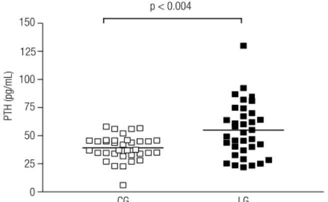
The plasma concentrations of both PTH and ion-ized calcium in LG were higher than in the CG, pre-senting statistical difference: PTH (Figure 1) - CG =
49.2 ± 11.0 pg/dL versus 54.9 ± 24.2 pg/dL (p <
0.004) for LG; ionized calcium (Figure 2) - CG = 4.2
± 0.4 mg/dL versus LG = 4.4 ± 0.4 mg/dL (p < 0.03).
Cop
yright
© ABE&M t
odos os dir
eit
os r
eser
vados
.
Among patients in the LG, it was noted that nine
patients had systemic hypertension and six had
dia-betes mellitus. These patients had these diagnoses
prior to the use of LC. Of the eight participants who had high levels of PTH, four had at least one of these comorbidities.
DISCUSSION
The HPT is a clinical syndrome characterized by ex-cessive function of the parathyroid glands, resulting in persistent PTH hypersecretion and its consequences in the body. It can be associated with elevated serum cal-cium levels or within the normal range (22). The PHP has a prevalence of approximately 1% in the adult po-pulation, with equal distribution between the genders, doubling after the age of 55, being, in this age group, two to three times more common in women than in men (23).
Currently, the signs and symptoms of PHP are becoming rarer, with 80-85% of patients being asymp tomatic. In this medical condition, PTH levels
are slightly increased and calcium level, when high, is usually only up to 1.0 mg/dL above the upper nor-mal limit. Hypophosphatemia, found in the classic form of PHP, is uncommon in patients with asymp-tomatic disease (24). At the diagnosis of PHP, the main cause of hypercalcemia in patients with nor-mal renal function, it is essential to observe the level of calcium in association with PTH levels, as there are situations in which, despite the normocalcemia, there is hormone elevation, as found in normocal-cemic PHP, a non-indolent condition, similar to an initial PHP (25).
The literature is unison in saying that, by de-creasing the calcium sensitivity in the parathyroid, Li disrupts the bone-parathyroid endocrine axis and triggers disorders in calcium homeostasis. This could lead to a classic PHP and even to a normocal-cemic HPT or hypercalcemia without PTH elevation
(9,26,27). Both in vitro studies and in vivo studies
in healthy volunteers demonstrated direct action of lithium on parathyroid cells in releasing intact PTH (28). Lithium can shift the set point of calcium-sens-ing receptors (CaSR) in parathyroid cells, leadcalcium-sens-ing onto excess release of PTH (28,29). In this work, we observed serum PTH elevated in 22.8% of patients using LC, conirming a disbalance in the physiology of the parathyroid glands caused by Li. According to data reported in most large series, the ratio for HPT in Li users between females and males is 4:1 (18,30,31). In our study, the ratio found was of 3 women for 1 man.
Despite the elevation of PTH in some patients in the LG, the levels of total calcium and ionized cal-cium of these patients remained within the normal range. In LG, it was noted, however, that the mean plasma concentrations of ionized calcium was higher compared with the CG (Figure 2). This inding is in agreement with several literature reports and it may be explained by the fact that the ionized calcium is more sensitive than the total calcium for detection of hypercalcemia in patients treated with Li (32,33).
According to the data obtained from the medical records, all patients in the LG stayed euthymic for more than six months, despite the fact that only 16 (45.7%) were with Li levels within the therapeutic range. Of the eight LG patients who presented in-creased PTH, ive had normal Li serum levels and the others had Li levels below the expected. This suggests that the HPT induced by Li was not re-Figure 1. Plasma concentration of parathyroid hormone (PTH) of the
control group (CG) and the Lithium Group (LG).
150
125
100
75
50
25
0
CG LG
p < 0.004
PTH (pg/mL)
6
5
4
3 2
0
CG
Ionized calcium (mg/dL)
LG p < 0.03
Cop
yright
© ABE&M t
odos os dir
eit
os r
eser
vados
.
lated to the values of Li in the blood, fact that could be, possibly, explained by an intrinsic property of the drug responsible for the imbalance of the para-thyroid endocrine axis and not being related to the given dose or its serum levels. This condition is in agreement with the explanation proposed by some studies, which say that the HPT induced by Li has no relation to the given therapeutic dose, but ra-ther with the presence of the medication in the body (34,35). The duration of the treatment increases the incidence of this clinical condition due to the cumu-lative power that Li has on the body (19,36).
Lithium, an inhibitor of glycogen synthase kinase 3 (GSK3), is widely used for the treatment of mood disorders. Lithium treatment signiicantly increased re-nal GSK3 phosphorylation, enhanced serum ADH and FGF23 concentrations, downregulated renal Klotho expression, stimulated renal calcium and phosphate excretion, and decreased serum 1,25(OH)2D3 and phosphate concentrations (37). Since Li is not bound to serum proteins, it is freely iltered by the kidneys and its elimination depends on the glomerular iltration rate. Consequently, Li blood levels and undesirable ef-fects caused by Li are directly related to renal function (9,10). The participants of this study were assessed by measurements of blood urea and creatinine, which were within normal limits. Such fact excluded the possibility that the HPT, found in some patients, would have been induced by renal failure (secondary HPT).
Regarding the levels of phosphorus, alkaline phos-phatase and 25-hydroxyvitamin D, there was no statis-tical difference compared to the CG. This result can be related to the presence of normocalcemic HPT, in which laboratory abnormalities are restricted to chang-es in PTH levels. Thchang-ese data are similar to those found by Khoury and cols. (33).
In our study, half of the patients with high PTH
had either hypertension or diabetes mellitus. According
to the literature, only diabetes mellitus has a positive
relation with the use of lithium salts, since Li has an inhibitor effect on the insulin secretion induced by glu-cose due to mechanisms related to the inlux of calcium in pancreatic beta cells (3,10). There was no correla-tion between the use of LC and development of these comorbidities in patients of LG, since such diagnoses were made prior to initiation of treatment with Li.
Limitations of this study were the non-evaluation of bone turnover markers, urinary calcium levels, bone mineral density and the lack of an ultrasound
evalu-ation of the kidneys and urinary tract of the patients studied in both groups. Although, Zamani and cols. found that patients on maintenance therapy with lithi-um had higher bone mineral density in both the llithi-umbar spine and the proximal femur and lower serum total alkaline phosphatase (ALP) and C-telopeptide (CTX) and osteocalcin than normal controls. The lower serum osteocalcin, CTX and ALP suggest decreased bone remodeling contrary to what is seen in primary hyper-parathyroidism, which is associated with accelerated skeletal turnover and increased osteocalcin and markers of bone resorption (38).
Guidelines recommendations for treatment with Li in patients with BAD do not mention monitoring of parathyroid function, what appears to be an important omission in the follow-up of these individuals due to the high absolute risk of developing mild, reversible hyperparathyroid state (15,38,39). Thus, our data sug-gest that the use of lithium salts, as an alternative ther-apy in psychiatric patients suffering from BAD, must be accompanied by periodic assessments of mineral metabolism in an attempt at early detection of possible imbalances in parathyroid endocrine axis, considering that 22.8% of patients using Li had elevated serum lev-els of PTH.
Disclosure: no potential conlict of interest relevant to this article was reported.
REFERENCES
1. Saunders BD, Saunders EFH, Gauger PG. Lithium therapy and hyperparathyroidism: an evidence-based assessment. World J Surg. 2009;33:2314-23.
2. Price LH, Heninger GR. Lithium in the treatment of mood disor-ders. N Engl J Med. 1994;331(9):591-8.
3. Mcknight RF, Adida M, Budge K, Stockton S, Goodwin GM, Ge-ddes JR. Lithium toxicity proile: a systematic review and meta--analysis. Lancet. 2012;379:721-8.
4. Rosa AR, Kapczinski F, Oliva R, Stein A, Barros HMT. Monito-ramento da adesão ao tratamento com lítio. Rev Psiq Clín. 2006;33(5):249-61.
5. Cipriani A, Pretty H, Hawton K, Geddes JR. Lithium in the preven-tion of suicidal behaviour and all-cause mortality in patients with mood disorders: a systematic review of randomized trials. Am J Psychiatry. 2005;162:1805-19.
6. Martin LNC, Kayath MJ. Abordagem clínico-laboratorial no diag-nóstico diferencial de hipercalcemia. Arq Bras Endocrinol Metab. 1999;43(6):472-9.
7. Ministério da Saúde. Proissional e Gestor – Medicamentos – Relação Nacional de Medicamentos Essenciais (RENAME 2012). Avaliable at: http://www.saude.gov.br. Accessed on: Jul 3, 2013. 8. Rang HP, Dale MM, Ritter JM, Flower RJ. Rang & Dale
Cop
yright
© ABE&M t
odos os dir
eit
os r
eser
vados
.
9. Collins N, Barnes TR, Shingleton-Smith A, Garret D, Paton C. Standards of lithium monitoring in mental health trusts in the UK. BMC Psychiatry. 2010;10:80.
10. Giusti CF, Amorim SR, Guerra RA, Portes ES. Endocrine distur-bances related to the use of lithium. Arq Bras Endocrinol Metab. 2012;56(3):153-8.
11. Mármol F. Litio: 55 años de historia en el tratamiento del trastorno bipolar. Med Clin (Barc). 2005;127(5):189-95.
12. Oliveira JL, Silva Júnior GB, Abreu KLS, Rocha NA, Franco LFLG, Araújo SMHA, et al. Nefrotoxicidade por lítio. Rev Assoc Med Bras. 2010;56(5):600-6.
13. Timmer RT, Sands JM. Lithium intoxication. J Am Soc Nephrol. 1999;10:666-74.
14. Bhuvaneswar CG, Baldessarini RJ, Harsh VL, Alpert JE. Adverse endocrine and metabolic effects of psychotropic drugs. CNS Dru-gs. 2009;23(12):1003-21.
15. Mak TWL, Shek C, Chow C, Wing Y, Lee S. Effects of lithium thera-py on bone mineral metabolism: a two-year prospective longitu-dinal study. J Clin Endocrinol Metab. 1998;83(11):3857-9. 16. Marx SJ. Hyperparathyroid and hyperparathyroid disorders. N
Engl J Med. 2000;343(25):1863-75.
17. Grüfeld JP, Rossier BC. Lithium nephrotoxicity revisited. Nat Rev Nephrol. 2009;5:270-6.
18. Hundley JC, Woodrum DT, Saunders BD, Doherty GM, Gauger PG. Revisiting lithium-associated hyperparathyroidism in the era of intraoperative parathyroid hormone monitoring. Surgery. 2005;138:1027-31.
19. Szalat A, Mazeh H, Freund HR. Lithium-associated hyperpara-thyroidism: report of four cases and review of the literature. Eur J Endocrinol. 2009;160(2):317-23.
20. Nair CG, Menon R, Jacob P, Babu M. Lithium-induced para-thyroid dysfunction: A new case. Indian. J Endocrinol Metab. 2013;17(5):930-2.
21. Skandarajah AR, Palazzo FF, Henry JF. Lithium-associated hyper-parathyroidism: surgical strategies in the era of minimally invasi-ve parathyroidectomy. World J Surg. 2011;35:2432-9.
22. Prospero JD, Baptista PPR, Amary MFC, Santos PPC. Paratireoides: estrutura, funções e patologia. Acta Ortop Bras. 2008;17(2):53-7. 23. Gómez JMC. Evaluación diagnóstica e diagnóstico diferencial del
hiperparatiroidismo primário. Endocrinol Nutr. 2009;56(Supl 1):14-9. 24. Bilezikian JP, Silverberg SJ. Asymptomatic primary
hyperpara-thyroidism. N Engl J Med. 2004;350(17):1746-51.
25. Marques TF, Vasconcelos R, Diniz E, Rêgo D, Griz L, Bandeira F. Normocalcemic primary hyperparathyroidism in clinical practice:
an indolent condition or a silent threat? Arq Bras Endocrinol Me-tab. 2011;55(5):314-7.
26. Komatsu M, Shimizu H, Tsuruta T, Kato M, Fushimi T, Inoue K, et al. Effect of lithium on serum calcium level and parathyroid function in maniac-depressive patients. Endocr J. 1995;42:691-5.
27. Gregoor PS, Jong GM. Lithium hypercalcemia, hyperparathyroi-dism and cinacalcet. Kidney Int. 2007;71:470.
28. Kusalic M, Engelsmann F. Effect of lithium maintenance thera-py on thyroid and parathyroid function. J Psychiatry Neurosci. 1999;24:227-33.
29. Haden ST, Stoll AL, Mccormick S, Scott J, Fuleihan GE. Alterations in parathyroid dynamics in lithium-treated subjects. J Clin Endo-crinol Metab. 1997;82:2844-8.
30. Carchman E, Ogilvie J, Holst J, Yim J, Carty S. Appropriate surgi-cal treatment of lithium-associated hyperparathyroidism. World J Surg. 2008;32:2195-9.
31. Awad SS, Miskulin H, Thompson N. Parathyroid adenomas ver-sus four-gland hyperplasia as the cause of primary hyperpara-thyroidism in patients with prolonged lithium therapy. World J Surg. 2003;27:486-8.
32. Adegboyega PA, Okorodudu AO. Intracellular ionized calcium and increasing doses of lithium chloride therapy in healthy Sprague--Dawley rats. Pharmacol Biochem Behav. 1994;49(4):1087-91. 33. Khoury AE, Petterson U, Kallner G, Aberg-Wistedt A,
Stain-Mal-mgren R. Calcium homeostasis in long-term lithium-treated wo-men with bipolar affective disorder. Prog Neuropsychopharma-col Biol Psychiatry. 2002;26(6):1063-9.
34. Rothman M. Acute hyperparathyroidism in a patient after initia-tion of lithium therapy. Am J Psychiatry. 1982;139:362-3. 35. Seely EW, Moore TJ, Leboff MS, Brown EM. A single dose of
li-thium carbonate acutely elevates intact parathyroid hormone le-vels in humans. Acta Endo (Buc). 1989;121:174-6.
36. Bendz H, Sjodin I, Toss G, Berglund K. Hyperparathyroidism and long-term lithium therapy – a cross-sectional study and the effect of lithium withdrawal. J Intern Med. 1996;240:357-65.
37. Fakhri H, Pathare G, Fajol A, Zhang B, Bock T, Kandolf R, et al. Plugers Arch. 2014;66(3):467-75.
38. Zamani A, Omrani GR, Nasab MM. Lithium’s effect on bone mine-ral density. Bone. 2009;44:331-4.