DNA Damage Induced in Glioblastoma Cells by I-131: A
!"#$%&'!()*+,-++()./#+%&"+(,$0)1$,$)$(2)3!(,+) $%0!)
Simulation
!"#$%&'(&)%'*+",-#.'/0/1*
-#1%2%&')%'#344&'(-#5/67/2
-## 8&'"9#54'&%9"8(2-#.'/0/:-## 6%;%<#+(=%%#5('<(>"#-#.'/02
?/#0%@(2)A%9)#48#+(<"4!4B;#C%7'94!4B;-#1(7D!);#48# !!"%<#E%(!)'-C%'2(9#F9">%2&");#48#5%<"7(!#67"%97%&-#C%'2(9-#G2(9 H/#0%@(2)A%9)#48#5%<"7(!#.';&"7&-#1(7D!);#48#5%<"7"9%-#C%'2(9#F9">%2&");#48#5%<"7(!#67"%97%&-#C%'2(9-#G2(9
:/#0%@(2)A%9)#48#C4I"74!4B;-#1(7D!);#48#5%<"7(!#67"%97%&-#C(2="()#54<(2%&#F9">%2&");-#C%'2(9-#G2(9
* Corresponding Address: P.O.Box: 14155-6183, Department of Radiology Technology, Faculty of Allied Health,Tehran University of Medical Sciences, Tehran, Iran
Email: [email protected]
+%7%">%<J#HKLMD!LHN??-# 77%@)%<J#OL0%7LHN?? Abstract
P=Q%7)">%J#The passage of ionizing radiation in living cells creates clusters of damaged nucleotides in DNA. In this study, DNA strand breaks induced by the beta particle of io-dine-131 (I-131), have been determined experimentally and compared to Monte Carlo
simulation results as a theoretical method of determining131I damage.
5()%2"(!&#(9<#5%)'4<&J In this experimental study, in order to create single strand breaks (SSB) and double strand breaks (DSB) in the DNA, glioblastoma (GBM) cells were
!"#$% &!% '(% )*+% ,-'.'/% % 0&% 0% $!"#% !1% 2% 345% 60)07#% !1% +880$+0&#$% 9#::"% ;#8#% #<0:=0&#$%
quantitatively by the Fast Micromethod assay. The energy spectrum of electrons released
+>%9#::"%;#8#%!?&0+>#$%?4%&@#%)098!"9! +9%A!>&#%*08:!%9!$#%BA*CDE9F%0>$%="#$%0"%0>%
input of the micro Monte Carlo code (MCDS). The percent of damage induced in cells was analyzed by Mann-Whitney test.
+%&D!)&J#G%"+7>+H90>&%8#$=9&+!>%B I(5(JF%+>%K=!8#"9#>9#%+>&#>"+&4%+>%+880$+0&#$%9#::"%9!) -pared to control cells as determined by the Fast Micromethod assay represented induced SSB and DSB damages in the DNA of irradiated cells. Comparison of experimental and theoretical results showed that the difference between the percentages of SSB per Gy
;0"%0?!=&%L5EM%0>$%6NO%;0"%0?!=&%'M% #8%345
R497!D&"49J#The differences in experimental and theoretical results may be due to the algorithm of applied codes. Since the Fast Micromethod and other experimental tech-niques do not provide information about the amount of detailed and complex damages of DNA-like base damages, the applied Monte Carlo codes, due to their capability to predict the amount of detailed damages that occur in the DNA of irradiated cells, can be used in in vitro experiments and radiation protection areas.
Keywords: Glioblastoma, Monte Carlo Method, Iodine-131, DNA Damage
Cell Journal(Yakhteh), Vol 14, No 1, Spring 2012, Pages: 25-30
R")()"49J#$%&'(&)%'*+",# -#344&'(#1-#54'&%9"8(2# -#5('<(>"#6+/#0$ #0(A(B%#"9<D7%<#"9#B!"4=!(&)4A(#7%!!&#=;# G*?:?J# #74A@(2"&49#=%)S%%9#%I@%2"A%9)(!#<()(#(9<#A49)%#7(2!4#&"AD!()"49/#R%!!#M/#HN?HT#?UV?WJ#HX*:N/
Introduction
!"#$%&%#'( #)( !'!*+,( -,( %#'%.%'+( */0%/&%#'(
leads to irreparable damages in the cell nucleous.
1'( "/*&%234/*5( 67( %$( &8!( "*%9/*,( $%&!( )#*( 0/9
-/+!(&8/&(%$(2/3$!0(-,(&8!(%'&!*/2&%#'(#)(%#'%.%'+( */0%/&%#'( :;<=( %*!2&( #*( %'0%*!2&( %'&!*/2&%#'( #)( %#'%.%'+( */0%/&%#'( %'032!$( /( >/*%!&,( #)( 9#4!23
0#3-4!( $&*/'0( -*!/?$( : @A<5( -/$!( 0/9/+!( :A <( #)( >/*%#3$(&,"!$5(/'0( 67B"*#&!%'(2*#$$(4%'?$(:C5(D<=
@#9!(#)(&8!$!(0/9/+!$(:@@A$<(2/'(-!(*!"/%*!0(-,( 2!4434/*(*!"/%*(9!28/'%$9$5(8#E!>!*(#&8!*$(: @A$<( /*!(0%)F234&(&#(*!"/%*5("#$$%-4,(4!/0%'+(&#(93&/&%#'(#*( 2!44(0!/&8(:G5(H<=(I/'2!*(2!44$(/*!(9#*!($!'$%&%>!(&#( %#'%.%'+(*/0%/&%#'(&8/'('#*9/4(2!44$5(&83$(&8!(!))!2&$(
of radiation can destroy cancerous cells. In this way,
%#'%.%'+(*/0%/&%#'(%$(/(3$!)34(&8!*/"!3&%2(&##4=
1#0%'!B;D;(:1B;D;<(%$(#'!(#)(&8!(%$#&#"!$(&8/&(8/$(
been extensively used as a beta emitter in radiation
&8!*/",=(1B;D;(%$(?'#E'(&#(2/3$!(93&/&%#'(/'0(0!/&8(
in cells that it penetrates, and in other cells at dis-tances of up to several millimeters. Since about 10% of its energy and radiation dose is via gamma
radia-&%#'5(1B;D;(%$(3$!0(%'('324!/*(9!0%2%'!(%9/+%'+(&!28
-'%J3!$=(1&(8/$(-!!'($322!$$)344,(3$!0(%'(&8!(&*!/&9!'&(
of thyroid cancer and it is considered in the treatment
#)(I6@(&39#*$($328(/$(+4%#-4/$	/(:KAL<(:M<=
1'(&8!("/$&(&E#(0!2/0!$5(&*/2?($&*32&3*!(2/4234/&%#'$(
have contributed to the understanding of mechanisms by which radiation affects cellular systems, and have been applied in nuclear medicine, scanning electron microscopy, radiation therapy, and space radiation
-%#4#+,(:N5(O<=
1'( &8%$( $&30,5( E!( 3$!0( 1B;D;( &#( %'032!( @@A( /'0( @A(#'(KAL(2!44$=(1'(#*0!*(&#(0!&!*9%'!(&8!(0%))!* -ences between the experimental data and theoretical
*!$34&$5(&8!(L#'&!(I/*4#(9!&8#0(E/$(3$!0(&#($%934/&!( &8!(-%#4#+%2/4(2#'$!J3!'2!$(#)(1B;D;(#'( 675($328( /$( &8!( /9#3'&$( #)( @@A( /'0( @A5( -,( /""4,%'+( &8!( L#'&!(I/*4#(:LI6PGI<(2#0!(/'0(&8!(9%2*#(L#'&!( I/*4#(0/9/+!($%934/&%#'(:LI @<(2#0!(/$(("*#"#$!0( -,(@!9!'!'?#(/'0(@&!E/*&(:Q5(;R<=
Materials and Methods
Cell line
SONLK5( /( 839/'( KAL( 2!44( 4%'!( )*#9( P/$&!3*(
Institute of Iran, was cultured in minimal
essen-&%/4( 9!0%39( :LTLU( K%-2#V1'>%&*#+!'5S@7<( &8/&( 2#'&/%'!0( ;RW( )!&/4( -#>%'!( $!*39( :XA@U( K%-2#V 1'>%&*#+!'5S@7<(/'0(HRR(SV94(#)("!'%2%44%'(:@%+9/5(
Germany).
Monolayer culture
KAL( 2!44$( E!*!( 234&3*!0( /$( 9#'#4/,!*$( %'( YBCH()4/$?$(:6S6I<(/&(DNZI5(HW(I[2(/'0(QHW( 839%0%&,=( 3*%'+( $3-234&3*%'+5( E!( 3$!0( "8#$
-"8/&!( -3))!*!0( $/4%'!( :PA@<( )#*( E/$8%'+( 2!44$U( ;( 9L( R=CHW( T Y7( E/$( 3$!0( )#*( &*,"$%'%.%'+(
the cells.
Cell irradiation by beta particles
X#*(#'!(\/$?(#)((9#'#4/,!*(234&3*!0(2!44$5(/($#
-43&%#'(#)(;R(9I%(1B;D;(%'(6/[](:R=C(L<(E/$(%'
-^!2&!0(%'&#(&8!(9!0%39=(Y8!(\/$?(E/$(&8!'($4#E4,( $8/?!'(&#(/28%!>!(/(8#9#+!'!#3$(0%$&*%-3&%#'(#)( 1B;D;($#43&%#'(/*#3'0(&8!(2!44$=(Y8!(\/$?(#)(KAL( 2!44$( E!*!( !_"#$!0( )#*( ;RO( 9%'3&!$( &#( 0!&!*9%'!( &8!(2#**!4/&%#'(-!&E!!'( 67(0/9/+!$(/'0(&8!(/- -sorbed dose of 2 Gy. At the end of the exposure, the medium was removed and cells were washed
/'0(2!'&*%)3+!0(-,(PA@()#*(*!9#>/4(#)(1B;D;=
Fast micromethod assay
`/0%/&%#'(%'032!0(@@A(/'0( @A(0/9/+!$(%'(&8!( 67(#)(KAL(2!44$(E!*!(!>/43/&!0(-,(&8!(X/$&(L%
-2*#9!&8#0( 67(@@A(7$$/,(/22#*0%'+(&#(&8!("*# -tocol by Schröder et al. (11). The solutions used to
0!'/&3*!( 67(E!*!(/$()#44#E$a
X43#*!$2!'&(0,!($?($#43&%#'(:$#43&%#'(7<(E/$( &8!( P%2#K*!!'( 0$ 67( J3/'&%&/&%#'( *!/+!'&( :L#
-4!234/*( P*#-!$<=( I/42%39B( /'0( 9/+'!$%39B)*!!( PA@( :I/VL+B)*!!( PA@U( $#43&%#'( A<( 2#'$%$&!0( #)( ;DN( 9L( 6/I45( C=N( 9L( bI45( G=D( 9L( 6/2]P[G,
/'0(;=H(9L(b]2P[G. Lysing solution (solution C)
2#'&/%'!0(Q=R(L(3*!/5(R=;W(@ @5(R=C(L(T Y75(/&( "](;R(E%&8(6/[]=(c,$%'+($#43&%#'($3""4!9!'&!0( E%&8( P%2#K*!!'( :$#43&%#'( <( 2#'$%$&!0( #)( CR( dc( #)(&8!(#*%+%'/4($?(0,!V9c(#)($#43&%#'(I=(T Y7( $#43&%#'( :$#43&%#'( T<( 2#'&/%'!0( CR( 9L( T Y7=( 6/[]($?($#43&%#'(:$#43&%#'(X<(2#'$%$&!0(#)(;=R( L(6/[](/'0(CR(9L(T Y7=(e#*?%'+(6/[]($# -lution (so-lution G) was prepared fresh prior to use.
7(&#&/4(#)(C(9c(#)($#43&%#'(X(E/$(/00!0(&#(;O(9c( #)($#43&%#'(T(/'0(&8!("](E/$(28!2?!0(/'0($8#340( -!(;C=GR(f(R=RC=
1'(#*0!*(&#(0!&!*9%'!(&8!(@@A(%'032!0(%'(KAL( 2!44$5(D()/42#'("%"!$(&8/&(2#'&/%'!0(;RR(g4(2#'&*#4(
group cells, (non irradiated cells), were prepared
E%&8(G(94(#)($#43&%#'( U(D(#&8!*()/42#'("%"!$(&8/&(
contained 100 µl of irradiated group cells were
/4$#("*!"/*!0(E%&8(G(94(#)($#43&%#'( =(1'(#*0!*(&#( 4,$!(&8!(2!44$5(&8!$!(+*#3"$(E!*!("4/2!0(%'(&8!(0/*?( )#*(GR(9%'3&!$=(7)&!*(GR(9%'3&!$5(&8!(\3#*!$2!'2!(
intensity of each group was measured with a
-&%#'(E/>!4!'+&8(/'0(MRR('9(!9%$$%#'(E/>!4!'+&8=
6!_&5(CHR(g4(#)($#43&%#'(K(E/$(/00!0(&#(&8!(4, -sed cells in each group (control and irradiated).
Y8!(/9#3'&(#)(@@A(E!*!(0!&!*9%'!0(/)&!*(CR(9%'
-3&!$( -,( 9!/$3*%'+( &8!( \3#*!$2!'2!( %'&!'$%&,( #)(
each group.
Preparing the calibration curve
1'(#*0!*(&#(0!&!*9%'!(&8!(/9#3'&(#)(\3#*!$2!'2!(%'
-&!'$%&,()#*(0%+!$&%#'(#)(/44(&8!(( 675(HR(g4(#)( 6/$!5( D(94(#)($#43&%#'( 5(/'0(;(94(PA@(E!*!(/00!0(&#(;RR( g4(#)('#'B%**/0%/&!0(KAL(2!44$=(7)&!*(GR(9%'3&!$(&8!( \3#*!$2!'2!( %'&!'$%&,( E/$( 9!/$3*!0( 3$%'+( /'( !_2%
-&/&%#'(E/>!4!'+&8(#)(GOH('9( /'0( !9%$$%#'( E/>!
-4!'+&8(#)(MRR('9=
Monte Carlo simulation
To estimate the initial spectrum of secondary
!4!2&*#'$("*#032!0(%'(/(-%#4#+%2/4(&/*+!&5(LI6PGI(
code was applied, and a monolayer cell culture ge-ometry established. To model a layer of cells at-tached to the bottom of a culture dish, a cylinder
E%&8(/(0%/9!&!*(#)(C=OC(29(/'0(/(8!%+8&(#)(R=G(29(
was used. Cells and culture medium were approxi-mated by water at a density of 1.0 g cmBD. To
esti-9/&!("/*&%24!(\3!'2!(%'(/(4/,!*(#)(2!44$(/&&/28!0(&#( &8!(-#&	(#)(&8!(234&3*!(0%$85(&8!("/*&%24!(\3!'2!(
was tailed in two parallel planes separated by 10 µm. To model the beta source, the probability of beta particle incidence with a variety of energies
/$($8#E'(%'(&/-4!(;(E/$(%'$!*&!0(%'&#(&8!(LI6PG2((
program. The average energy of these electrons
E/$( 2/4234/&!0( -,( !J3/&%#'( :;<5( /'0( &8%$( />!*/+!(
!'!*+,(E/$(/""4%!0(%'(&8!(LI @(2#0!(&#(!$&%9/&!(
the percent of damage.
Results
Y8!($"!2&*39(#-&/%'!0()*#9($/9"4!$(&8/&(E!*!(&/?
-!'(-,(&8!(@8%9/0.3($"!2&*#\#3*#9!&!*(/*!($8#E'(%'( F+3*!;=(Y8!(/>!*/+!(\3#*!$2!'2!(%'&!'$%&,(#-&/%'!0( )*#9(&8!(2#'&*#4(/'0(%**/0%/&!0(+*#3"$(#)(KAL(2!44$(
are shown in Table 2. This can be used to
deter-9%'!(&8!(/9#3'&(#)(@@A(/'0( @A=(Y8!(/>!*/+!(
amount of fluorescence intensity in the control and irradiated groups has shown that the ab-sorption of the 2 Gy dose of beta particles of
1B;D;( *!$34&!0( %'( )43#*!$2!'2!( *!032&%#'( %'( &8!(
irradiated groups. This reduction represented
&8!( !_%$&!'2!( #)( @@A$( /'0( @A$( %'( %**/0%/&!0( KAL(2!44$=
?NNN/N
YNN/N
KNN/N
UNN/N
HNN/N
N/N
*HNN/N
XHZ/K
[(>%#!%9B)'V9AW ABSOISSA
1GV(/DW
HNN/N UNN/N KNN/N YNN/N ?NNN/N
RDI NATE XHZ/NY
Fig 1: The spectrum obtained from samples of the irradi-ated and control groups of cells treirradi-ated with PicoGreen solution, at an excitation wave length of 485 nm and emission wave length of 600 nm (Taken by a Shimadzu
!"#$%&'&(%&)"$"%*+,
Table 1: The probability of beta decay in each energy level applied in the MCNP code in order to determine the spectrum of electrons released in medium
R=CH R=DD
R=M; R=GN
R=O;
Energy (MeV)
0.02 R=RMQ
0.9 R=RRH
R=RRM
Probability
-./0",12,-3",)". (%"4,.5"%.6",'(&%" #"7#",87$"7 8$9,&:,#&7$%&0,;<=,#"00 ,.74,$3& ",8%%.48.$"4,/9,>?,)@8,AB>C>D,$&,4"$"% -mine the amount of SSBs and DSBs
SSB DSB
!"# $%$&&#&' '()#*'$%$((#')
!"#$%"&'()#"*+",+"&-,.",*-./& (control group)
*'#!$%$&+#') +(#'"$%$&,#(,
According to table 2, the average amount of fluorescence intensity in the control group with
-.$ /01$ 234356$ 6783962$ '()#*'%((#')#$ :;6$
average amount of fluorescence intensity in the
5<.8=$ ><63>62$ ?@$ /03A6$ 6783962$ * !# %(*#!#$
According to statistical analysis, the average fluorescence intensity in these two groups (ir-radiated and control) was significantly different
B=C,#,'D#$ E628F>G.-$ .H$ H98.<6AF6-F6$ G->6-AG>@$ <6=<6A6->62$>;6$43IG484$?<63J$G-$/01$B&,,K$ ?<63JD#$ $ :;6$ F39G?<3>G.-$ F8<L6$ ;3A$ ?66-$ 2<3M-$ 3-2$GA$A;.M-$G-$N58<6$(#
!! "!! #!! $!! %!! &!! !
'()*+,-).-/01)-2344+-34546748-9:;
<=96>*;
! %! #! ! ?! &!! &%!
!"#$%#&'(!)*'+!,-#./*01#,)+'!-12#)3#+41#'5,/-+#,6#7/,*18-cence intensities of control group and DNase-treated group 9:;<=# .4'-"1# !-# '5,/-+# ,6# 7/,*18.1-.1# !-+1-8!+3># '# :?# break will occur.
O-$ .<26<$ >.$ 26>6<4G-6$ >;6$ =6<F6->$ .H$ PPQA$ 3-2$ /PQAR$M6$;3L6$8A62$>;6$G-H.<43>G.-$A;.M-$G-$SG5 -ure 2. According to this curve, the variation of
inten-AG>@$=6<$?<63J$G-$>;6$/01$A><3-2$67839A$&!+#)R$M;GF;$ 463-AR$H.<$63F;$&#!+)T&#!"$F;3-56$G-$34.8->$.H$U8.
-<6AF6-F6$G->6-AG>@R$3$&K$?<63J$MG99$.FF8<#$
The difference between average intensities in the control and irradiated groups according to table 2
GA$ *#!)$B3#8D#
*#!)
1.79 V$( #
:;8A$>;6$34.8->$.H$/PQ$GA$( # K#
The difference between the average intensities in the control and irradiated groups according to
>3?96$($GAW$ *#!$B3#8D
$$ *#!
1.79 V$( # &
:;8A$>;6$34.8->$.H$AG-596$A><3-262$?<63J$GA$( # &K# !"#$%&'()*"+"),-%.)
R, = 1
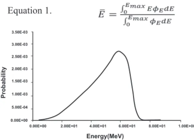
The spectrum of electrons released in the
me-2G84$ .?>3G-62$ ?@$ >;6$ XY0Z FF.26$ GA$ A;.M-$ G-$ N58<6$*R$H<.4$>;GA$A=6F><84$>;6$3L6<356$6-6<5@$.H$ 696F><.-A$F39F893>62$?@$6783>G.-$&$GA$,# !&$X6[#
\783>G.-$&#
$>"!@A!$
$>!!@A!$
%>"!@A!$
%>!!@A!$
&>"!@A!$
&>!!@A!$
">!!@A!#
!>!!@B!!
@+432C9D4E;
F3)G6G050,C
!>!!@B!! %>!!@B!& #>!!@B!& >!!@B!& ?>!!@B!& &>!!@B!!
Fig 3: Spectrum of electrons released in the medium obtained by MCNP4c code.
@')(1#A;#B1*.1-+#,6#2!661*1-+#.('88!C.'+!,-8#,6#2'5'"1#,)+'!-12# from MCDS output with an electron energy of 0.471 MeV
Damage yield (%) Clustered damage
)+#*"+ BD
(+#+&+ SSB
&#(" SSB +
,#&'" 2SSB
&#&' DSB
,#&'+ DSB +
,#,&+ DSB ++
#+,& SSBc
,# '& SSBcb
&*#(*) DSBc
'&#(*! DSBcb
Q6F38A6$?.>;$PPQF$3-2$/PQF$264.-A><3>6$4.<6$ <639GA>GF$3-2$26>3G962$<6A89>A$.H$234356$G-$>;6$/01$
hit sites, thus they are more comparable to the ex-perimental results. According to the comparison,
>;6$2GHH6<6-F6$?6>M66-$>;6$34.8->A$.H$PPQ$=6<$]@$ GA$3==<.IG43>69@$!# KR$3-2$>;6$2GHH6<6-F6$?6>M66-$ >;6$34.8->$.H$/PQ$GA$3?.8>$&K#
Discussion
Low-linear energy transfer (LET) and high-LET
<32G3>G.-$F<63>6$PPQ$3-2$/PQ$G-$3$>@=GF39$43443
-9G3-$F699$B&(D#$O-$322G>G.-R$G.-G^G-5$<32G3>G.-$F38A -es massive amounts of damage to nucleo bas-es.
/PQA$3<6$F<63>62$M;6-$3>$963A>$>M.$A><3-2$?<63JA$ 3<6$H.<462$.-$.==.AG>6$A><3-2A$.H$>;6$/01$MG>;G-$ &,$?3A6$=3G<A#$/PQA$3-2$.>;6<$F93AA6A$.H$489>G=96$
damage sites are the primary cause of
radiation-in-28F62$F699$263>;$B&*D$3-2$48>356-6AGA$B& D#$X.A>$ 6I=6<G46->39$ >6F;-G786A$ 8A62$ >.$ 26>6F>$ <32G3>G.-$ 234356$>.$/01$=<.LG26$.-9@$9G4G>62$G-H.<43>G.-$ 3?.8>$ >;6$ 6I3F>$ -84?6<$ 3-2$ A=3>G39$ F.-N58<3>G.-$ .H$ 69646->3<@$ 234356A$ >.$ >;6$ /01#$ O-A>632R$ 26 -tailed information about the spectrum of possible
234356A$ =<.28F62$ ?@$ G.-G^G-5$ <32G3>G.-$ GA$ .H>6-$ .?>3G-62$8AG-5$X.->6$Y3<9.$><3FJ$A><8F>8<6$AG48
-93>G.-$B*R$+R$&'D#$O-$>;6$=3A>$>M.$26F326AR$3==9@G-5$ G.-G^G-5$ <32G3>G.-$ >.$ G-28F6$ 234356A$ G-$ >84.<39$
cells has been considered a useful tool to cure
can-F6<#$ O_&*&$ GA$ 3-$ G4=.<>3->$ <32G.$ GA.>.=6$ .H$ G.2G-6$
mostly used in the medical and pharmaceutical
N692A#$ X.26<3>6$ 2.A6A$ .H$ O_&*&$ 3<6$ MG269@$ 8A62$
for curing thyroid cancers. Some special
proper->G6A$.H$O_&*&R$A8F;$3A$>;6$F3=3?G9G>@$.H$93?69G-5$2GH
-H6<6->$JG-2A$.H$3->G?.2G6A$BXOQ]DR$G>A$A;.<>$<3-56$ .H$?6>3$=3<>GF96A$B&$44D$B&)DR$3-2$F38AG-5$234356$ >;<.85;$F<.AA_N<6$=;6-.46-3$;3A$4326$G>$3$A8G>
-3?96$>..9$H.<$><63>G-5$]QXR$>;6$4.A>$F.44.-$3-2$
most malignant of the glial tumors. Chemotherapy, surgery and radiotherapy are common, but not
L6<@$ A8FF6AAH89R$ M3@A$ .H$ $ ><63>G-5$ ]QX$ ?6F38A6$
CNS tumors are restricted to critical organs. Thus target therapy can be a better choice.
O-$>;GA$A>82@R$]QX$F699A$F89>8<62$3A$4.-.93@
-6<A$M6<6$G<<32G3>62$?@$O_&*&R$>;6$A>3>GA>GF399@$AG5
--GNF3->$<628F>G.-$.H$U8.<6AF6-F6$G->6-AG>@$.H$>;6$ F699A$G<<32G3>62$?@$O_&*&$F.4=3<62$>.$>;6$F.-><.9$
group represented the increase in damages to
>;6G<$ /01#$ PG4893>G.-$ <6A89>A$ ?@$ XY0Z$ 3-2$ XY/P$F.26A$;3A$5GL6-$>;6$=6<F6->$.H$2GHH6<6->$
F93AA6A$.H$234356A$G-28F62$G-$/01$?@$696F><.-A$ A8F;$3A$/PQR$PPQR$/PQ`R$/PQ``R$(PPQR$6>F#$O-$
these calculations, it is assumed that a minimum
6-6<5@$.H$&!#'$6[$B&!R&+D$GA$<678G<62$H.<$G-28F
-G-5$ >;6$ PPQ$ .-$ 3$ /01$ A><3-2R$ 3-2$ G>$ GA$ 39A.$ 3A
-A8462$>;3>$3$/PQ$GA$H.<462$M;6-$>;6$?<63JA$.-$ .==.AG>6$A><3-2A$3<6$MG>;G-$a&,$?=$A6=3<3>G.-$.H$
each other. To establish criterion for the local
en-6<5@$<678G<62$>.$G-28F6$PPQR$-846<.8A$=;@AGF39$
and chemical values have been considered such
3A$ G-F982G-5$ 3L6<356$ 6-6<5@$ 9.AAR$ G.-G^3>G.-$ 6- -ergy, oscillator strength, and average excitation
=.>6->G39$ B<3-5G-5$ H<.4$ &(6[$ >.$ *,6[D$ B&+D#$ O-$ >3?96$*$$$$G>$GA$-.>62$>;3>$G-F98AG.-$.H$?3A6$234356$
substantially increases the complex proportion of
?.>;$PPQ$3-2$/PQ#$1FF.<2G-5$>.$XY/P$.8>=8>$ G-$>3?96$*R$>;6$34.8->$.H$/PQF?$BF.4=96I$/PQ$ >;3>$ G-F9826A$ ?3A6$ 234356AD$ GA$ *+K$ ;G5;6<$ >;3-$ /PQFR$3-2$>;6$34.8->$.H$PPQY?$GA$*'#)'K$;G5;
-6<$>;3-$PPQF#$PG-F6$PPQF$3-2$/PQFR$264.-A><3>6$
more realistic and detailed results of damages in
;G>$ AG>6A$ .H$ /01R$ >;6@$ 3<6$ 4.<6$ F.4=3<3?96$ >.$
experimental results. According to the
compari-A.-R$ >;6$ 2GHH6<6-F6$ ?6>M66-$ >;6$ 34.8->$ .H$ PPQ$ =6<$]@$GA$3==<.IG43>69@$!# K$3-2$>;6$2GHH6<6-F6$ ?6>M66-$>;6$34.8->$.H$/PQ$=6<$]@$GA$3?.8>$&K#$
The difference may be due to the algorithm of
>;6$ X.->6$ Y3<9.$ F.26A#$ O-$ .<26<$ >.$ 26F<63A6$ >;6$
discrepancy of the theoretical and experimental results for an individual experiment, a particular code (according to the type of radiation and ge-ometry used) must be designed.
Conclusion
:;6$26>6<4G-3>G.-$.H$G-28F62$234356A$G-$/01$ ?@$>;6$S3A>$XGF<.46>;.2$3AA3@$2.6A$-.>$=<.LG26$
information about the percent of complex
dam-356AR$ >;8A$ >;6$ X.->6$ Y3<9.$ F.26A$ 5GL6$ 4.<6$ 26 -tailed analysis of complex damages induced by
<32G3>G.-$ G-$ F699893<$ /01#$ :;GA$ G-H.<43>G.-$ F3-$
be used to estimate the amount of damage to ir-radiated cells in in vitro experiments and radiation
protection issues.
Acknowledgements
:;GA$<6A63<F;$M3A$A8==.<>62$?@$:6;<3-$b-GL6<AG
->@$.H$X62GF39$PFG6-F6A#$O-$=3<>GF893<RM6$3<6$5<63>9@$ G-26?>62$ >.$ /<$1#X.A>33<$ H.<$ ;GA$ ;69=H89$ 2GAF8A
-AG.-$ 3-2$ >6F;-GF39$ 6I=6<>GA6#$:;6<6$ GA$ -.$ F.-UGF>$
References
1. Nikjoo H. Radiation track and DNA damage . Iran J Radiat
/01%",22.3"$4$56".#$-%"
,%" Oleinick NL, Chiu SM, Ramakrisha N, Xue LY. The
789:;<=8>?" =@0><=AB;<=8>?" ;>@" 1=C>=AB;>B0" 87" DEF#G98 -tein cross-links in mammalian cells. Br J Cancer Suppl.
$('&3'6"$.)#$H2%
3. Nikjoo H, O’Neill P, Goodhead DT, Terrissol M. Compu-tational modelling of low- energy electron-induced DNA damage by early physical and chemical events. Int J
Ra-@=;<"I=8J%"$((&3&$4)56"H-&#H'.%
H%" Walters K. Modelling the probability distribution of the number of DNA double-strand breaks due to sporadic
;JK J;<=8>"87">LBJ08<=@0"M;101%"N"OP089"I=8J%",22&3",H)4$56"
161-168.
5. Van Gent DC, Hoeijmakers JH, Kanaar R. Chromosomal stability and the DNA double-stranded break connection.
E;<"/0Q"R0>0<%",22$3",4.5?$(-#,2-%
6. Goldsby RE, Fitzgerald PA. Meta[I131
]iodobenzylguani-dine therapy for patients with metastatic and unresectable pheochromocytoma and paraganglioma. Nucl Med Biol .
,22'3".)6"H(#-,%
7. Nikjoo H, Uehara S, Wilson WE, Hoshi M, GoodheadDT. Track structure in radiationM=8J8C 6" <P089 " ;>@" ;GGJ=B;
-<=8>%"S><"N"/;@=;<"I=8J%$(('3"&.4H56".))#.-H%
8. Nijkjoo H, O’Neill P, Wilson W E, Goodhead DT. Compu-tational approach for determining the spectrum of DNA damage induced by ionizing r;@=;<=8>%"/;@=;<"/01%",22$3" $)-"4)"T<",56")&&#)'.%
9. Semenenko VA, Stewart RD. Fast Monte Carlo simulation of DNA damage formed by electrons and light ions. Phys
U0@"I=8J"%,22-3"")$4&56"$-(.#$&2-%
$2%" Semenenko VA, Stewart RD. A fast Monte Carlo algorithm to simulate the spectrum of DNA damages formed by
ion-=V=>C"9;@=;<=8>%/;@=;<"/01%",22H3"$-$4H56"H)$#H)&%
11. Schrder HC,Batel R, Schwertner H, Boreiko O, Müller WE. Fast micromethod DNA single-strand-break assay.
U0<P8@1"U8J"I=8J%",22-3".$H6",'&#.2)%
$,%" Ward JF. DNA damage produced by ionizing radiation
=>":;::;J=;>"B0JJ16"=@0><=<=01?":0BP;>=1:1"87"789:;<=8>?" ;>@"90G;9;M=J=< %"T98C"ELBJ0=B"FB=@"/01"U8J"I=8J%$(''3.)6" ()#$,)%
13. W;9@"NX%"/;@=;<=8>":L<;C0>01=16"OP0"=>=<=;J"DEF"J01=8>1" 901G8>1=MJ0%"/;@=;<"/01%$(()3"$H,4.56".-,#.-'%
$H%" Ward JF, Blakely WF, Joner EI. Mammalian cells are not killed by DNA single-stran breaks caused by hydroxyl
rad-=B;J1" 798:" P @98C0>" G098*=@0%" /;@=;<" /01%$(')3" $2.4.56" .'.#.(,%
15. Holley WR, Chatterjee A. Clusters of DNA damage induced
M "=8>=V=>C"9;@=;<=8>6"X89:;<=8>"87"1P89<"DEF"79;C:0><1%"S%" OP0890<=B;J":8@0J=>C%"/;@=;<"/01%$((-3"$H)4,56"$''#$((%
16. McCluskey AG, Boyd M, Gaze MN, Mairs, RJ. [131I]
USIR";>@"<8G8<0B;>6";"9;<=8>;J0"789"B8:M=>;<=8>"<P09
-;G " 789" >0L98MJ;1<8:;%" Y;>B09" Z0<<%" ,22)3" ,,'4$#,56" ,,$-,,&%
17. Nikjoo H, Bolton CE, Watanabe R, Terrissol M, O'Neill P, Goodhead D T. Modelling of DNA damage induced by
en-09C0<=B"0J0B<98>1"4$22"0["<8"$22"K0["5%"/;@=;<"T98<"D81=:
-0<9 %,22,3"((4$#H56"&&#'2%
18. Nikjoo H, O'Neill P,Terrissol M , Goodhead DT. Quan-titative modelling of DNA damage using Monte Carlo track structure method. Radiat Environ Biophys .1999;