Arq Bras Cardiol 2003; 81: 225-8.
Paiva et al Cardiac variables in animals exposed to cigarette smoke
2 2 5
Faculdade de Medicina de Botucatu, UNESP
Mailing address: Sérgio Alberto Rupp de Paiva - Faculdade de Medicina de Botucatu UNESP - 18618-000 - Botucatu, SP, Brazil - E-mail: [email protected] English version by Stela Maris C. e Gandour
Received - 10/1/02 Accepted - 2/17/03
Arq Bras Cardiol, volume 81 (nº 3), 225-8, 2003
Sergio Alberto Rupp de Paiva, Leonardo Antonio Mamede Zornoff, Marina Politi O koshi, Katashi O koshi, Antonio Carlos Cicogna, Alvaro O scar Campana
Botucatu, SP - Brazil
Behavior of Cardiac Variables in Animals Exposed
to Cigarette Smoke
Original Article
Among health hazards caused by smoking, cardio-vascular changes stand out. Smoking influences the pre-valence of infarction through several mechanisms, such as endothelial dysfunction, greater oxidation of LDL-choles-terol, a reduction in HDL-cholesLDL-choles-terol, an increase in the le-vels of adhesion molecules and fibrinogen, an increase in platelet aggregation, and an increase in the prevalence of vascular spasm 1,2. English et al 3 were the first to report a
close relation between smoking and mortality due to ische-mic heart disease, and they were followed by other authors who stressed that association 4,5.
The proposed mechanisms, by which the constituents of cigarette smoke (carbon monoxide, nicotine, tar, and the substances contained in vapor) may cause myocardial in-farction, are as follows: thrombosis, formation of atheroscle-rotic plaque, platelet aggregation, vasoconstriction, hypo-xia, and arrhythmia induction 6. Although the association
between the smoking habit and coronary heart disease is universally accepted, little is known about the morphofunc-tional effects of smoking directly on the heart.
Thus, the objective of this study was to assess the be-havior of the variables related to myocardial mechanical function and also to in situ heart function of the animals ex-posed to cigarette smoke.
Methods
The study comprised 51 male Wistar rats weighing between 200 and 250 g divided into 2 groups as follows: control group (C) - formed by 28 animals, receiving the diet routinely used in our laboratory adjusted according to the daily ingestion observed in group S during smoke exposure; and smoking group (S) - formed by 23 animals receiving the unrestricted diet routinely used in our laboratory for 3 months and exposed to tobacco smoke in the last 30 days.
The method used was that proposed by Simani et al 7
and implemented by Wang et al 8 to expose the animals to
cigarette smoke in a modified incubator. The rats, 5 each time, were placed in the clear chamber connected to the smoking device. Smoke puffs were drawn from a cigarette
Objective - To assess the behavior of cardiac varia-bles in animals exposed to cigarette smoke.
Methods - Two groups of Wistar rats were studied as follows: control group (C), comprising 28 animals; and smoking group (S), comprising 23 animals exposed to cigarette smoke for 30 days. Left ventricular cardiac function was assessed in vivo with transthoracic echocar-diography, and myocardial performance was analyzed in vitro in preparations of isolated left ventricular papillary muscle. The cardiac muscle was assessed in isometric contractions with an extracellular calcium concentration of 2.5 mmol/L.
Results - No statistical difference was observed in the values of the body variables of the rats and in the mechani-cal data obtained from the papillary muscle between the control and smoking groups. The values of left ventricular systolic diameter were significantly greater in the smoking animals than in the control animals (C= 3.39 ± 0.4 mm and S= 3.71 ± 0.51 mm, P=0.02). A significant reduction was observed in systolic shortening fraction (C= 56.7 ± 4.2% and S= 53.5 ± 5.3%, P=0.02) and in ejection fraction (C= 0.92 ± 0.02 and S= 0.89 ± 0.04, P=0.01).
Conclusion - The rats exposed to cigarette smoke had a reduction in left ventricular systolic function, although their myocardial function was preserved.
2 2 6
Paiva et al
Cardiac variables in animals exposed to cigarette smoke
Arq Bras Cardiol 2003; 81: 225-8.
by vacuum and then blown inside the chamber. Smoke was released at a rate of 5 cigarettes/30 minutes/day, twice in the afternoon, at 5-minute rest intervals in the first week. From the second week onwards, the number of cigarettes was in-creased to a rate of 10 cigarettes/30 minutes, twice in the morning and twice in the afternoon until the end of the study period. Fifteen animals were exposed to the smoke of 40 ci-garettes/day each time. The total amount of smoke was de-rived from 940 cigarettes during the period of 4 weeks. Ac-cording to the manufacturer, each cigarette used in the expe-riment provided 14 mg of tar, 1.1 mg of nicotine, and 15 mg of carbon monoxide.
All animals underwent echocardiography, and, after an average of 3 days, they were euthanized for papillary mus-cle study. The animals were not exposed to cigarette smoke on the day of echocardiography or euthanasia.
At the end of the experimental period, the rats under-went anesthesia with ketamine hydrochloride (50 mg/kg) and xylidine hydrochloride (1 mg/kg) by the intramuscular route. Then, the anterior region of the thorax was epilated. After these procedures, the animals were placed in the left lateral decubitus position for undergoing echocardiography.
The echocardiograph used was the Sonos 2000 model (Hewlett-Packard Co.) equipped with a 7.5-MHz electronic transducer. Flow assessment was performed with the same transducer operating at 5.0 MHz. To measure the cardiac structures, M-mode images with the ultrasound beam orien-ted by the 2-dimensional image with the transducer in the pa-rasternal short-axis view were used. The image of the left ventricular cavity was obtained placing the M-mode cursor right below the plane of the mitral valve between the papil-lary muscles. The images of the aorta and of the left atrium were also obtained in the parasternal short-axis view with the M-mode cursor positioned at the level of the aortic valve. Recording of one-dimensional imaging (velocity = 100 mm/s) was performed with the UP-890MD model printer (Sony Co.). Later, the cardiac structures were manually mea-sured with the aid of a precision pachymeter according to the recommendations of the American Society of Echocar-diography 9. The cardiac structures were measured in at
least 5 consecutive cardiac cycles. Left ventricular diastolic diameter (LVDD) and left ventricular posterior wall thick-ness (LVPWT) were measured at the moment correspon-ding to its maximal diameter. Left ventricular systolic diame-ter (LVSD) was measured at the moment of the maximal sys-tolic excursion of the posterior wall. Left ventricular syssys-tolic function was assessed calculating the systolic shortening fraction [(LVDD-LVSD)/LVDD x 100] and the ejection frac-tion [(LVDD3-LVSD3)/LVDD3]. Transmitral diastolic flow (E
and A waves) was obtained with the transducer in the 4-chamber apical view. The flows were directly measured in the echocardiographic monitor.
The study of the isolated papillary flow was perfor-med according to already described procedures 3 days after echocardiography 10-12. The mechanical function was
stu-died in left ventricular papillary muscles obtained as follows: the rat was beheaded, its thorax opened, the heart rapidly re-moved and placed in Krebs-Henseleit solution previously
oxygenated (10 minutes) with 95% oxygen (O2) and 5% car-bon dioxide (CO2) at a temperature of 28oC. After dissection
of the left ventricle and sectioning of the interventricular septum, the left ventricle was divided into 2 parts, each con-taining its papillary muscle, and, then, carefully dissected in a glass chamber, constantly oxygenated and maintained at 28oC. The papillary muscles, after having their extremities
tied to 2 stainless steel rings, were rapidly transferred to the glass chamber containing the Krebs-Henseleit solution, which was constantly oxygenated with 95% O2 and 5% CO2 and maintained at 28oC due to the use of a circulating bath.
The papillary muscles were maintained in the vertical posi-tion in the glass chamber. The inferior ring was attached to a stainless steel wire of 0.031-cm-diameter connected to an outer transducer (Kyowa 120T-20B), and the superior ring was connected to a stainless steel wire similar to the former tied to the extremity of the long arm of an isotonic lever. A micrometer was placed on that extremity to control the exten-sion of the movements of the lever, providing muscle length adjustment in the muscular relaxation phase. Initial stret-ching of the cardiac fiber was performed with a light load (preload) suspended in the extremity of the short arm of the lever coupled to the 7 DCDT - 050 model length transducer (Hewlett Packard), which measured length variations during muscle contractions. The lever was rigid and light, made of aluminum with a 4:1 long arm/short arm ratio.
The muscles were stimulated 12 times per minute with platinum needle electrodes, which were coupled to an elec-tric stimulator programmed to release stimuli in 5-ms square waves, with a voltage approximately 10% greater than the minimum necessary to cause a maximal mechanical respon-se of the muscle.
The composition of the Krebs-Henseleit solution 13 in
millimoles per liter was as follows: 118.5 NaCl; 4.69 KCl; 2.52 CaCl2; 1.16 MgSO4; 1.18 KH2PO4; 5.50 glucose; and 25.88 NaHCO3. The partial pressure of oxygen in the solution was maintained between 550 and 600 mmHg.
The parameters used to characterize the papillary muscles were length (mm) and sectional area (mm2). The in
vitro length (Lmax) was obtained with a Gartner cathetome-ter. The sectional area was calculated based on the measu-res of the diameter of the central segment, considering that the muscle had a cylindrical geometrical form and specific weight of 1.0. The wet weights of the left and right ventricles (LV and RV, respectively) and atria (A), normalized to the fi-nal body weight of the rat (BW), were used as indices of atrial and ventricular hypertrophy.
The water content of the tissues was assessed by the ratio between the wet and dry weights of the following tis-sues: liver, lungs, left ventricle, and the calf muscle.
Sig-Arq Bras Cardiol 2003; 81: 225-8.
Paiva et al Cardiac variables in animals exposed to cigarette smoke
2 2 7 maStat for Windows v 2.0 statistical package (Jandel Co.,
San Rafael, CA, USA) was used for the tests. The significan-ce level of 5% was adopted for all tests.
Results
The body variables of the animals are shown in table I and the mechanical data of the isolated papillary muscles are shown in table II. No statistical difference was observed in the values of the body variables and the data obtained from the papillary muscle when comparing the control and smoking groups.
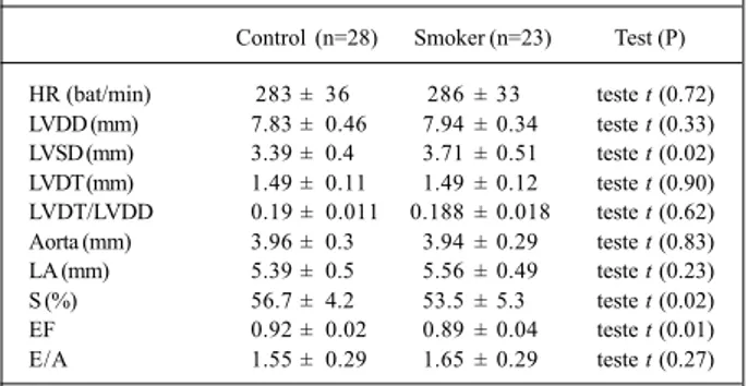
Table III shows the values obtained in the echocardio-graphic study. The values of left ventricular systolic diame-ter were significantly greadiame-ter in the smoking animals than in the controls. Exposure to cigarette smoke caused a signifi-cant reduction in left ventricular systolic function indices (systolic shortening fraction and ejection fraction). No dif-ference was observed between the groups of rats when comparing the other echocardiographic variables studied.
Discussion
This study aimed at assessing the behavior of the cardiac variables of rats exposed to cigarette smoke. The
animals underwent cigarette smoke inhalation, with all the components involved, for 30 days, and the functional changes were detected in vivo only through echocardio-graphy.
Some deleterious effects of smoking on the cardiovas-cular system are widely accepted. It has already been well documented that smoking is the major changeable risk fac-tor for acute myocardial infarction 6,15. Although the effects
of smoking on the coronary system have been intensely re-searched, the direct effects of smoke exposure on the heart are less known.
This study showed that chronic exposure to cigarette smoke resulted in effects on the rat’s heart, whose detection depended on the method used for functional analysis. Con-sidering the variables studied in the preparations with isola-ted papillary muscle, no difference could be identified bet-ween the 2 groups studied. This method allows the study of myocardial function without the interference of external fac-tors, such as preload, afterload, and heart rate. As such, our results show that no change in the myocardial mechanical performance occurred due to cigarette smoke inhalation. On the other hand, an increase in left ventricular end systolic diameter was observed in the smoking group on echocar-diography. In addition, both shortening and ejection frac-tions were statistically smaller in the smoking group. Thus, the results show that, despite the preservation of muscle function, the rats inhaling cigarette smoke had depressed left ventricular systolic function.
Some factors may explain our findings. In an experimen-tal study with dogs, the acute administration of nicotine was accompanied by an increase in inotropism 16. A significant
difference in regard to our study, however, is that we did not use nicotine, but cigarette smoke. It is worth noting that more than 4,700 different substances have already been identified in cigarette smoke 6, and some of these substances could
counteract the inotropic effects of nicotine. Another aspect to be considered is that our animals were chronically exposed to cigarette smoke and that the acute cardiovascular effects of smoking may be different from the chronic ones 17.
Table I - Body variables of the rats studied
Control (n=28) Smoker (n=23) Test (P)
Initial weight (g) 252 (250 – 260) 254 (249 -262) MW (0.90)
BW (g) 407 ± 42 398 ± 31 t (0.38)
LV (g) 0.755 ± 0.1 0.767 ± 0.095 t (0.66) RV (g) 0.214 ± 0.043 0.215 ± 0.04 t (0.91) Atrium (g) 0.091 ± 0.018 0.089 ± 0.018 t (0.73) LV/BW 1.85 ± 0.18 1.93 ± 0.17 t (0.16) RV/BW 0.52 ± 0.09 0.54 ± 0.08 t (0.53) A/BW 0.22 ± 0.04 0.22 ± 0.05 t (0.95) Liver (W/D) 3.22 ± 0.1 3.24 ± 0.09 t (0.63) Lung (W/D) 4.85 ± 0.56 4.92 ± 0.41 t (0.62) LV (W/D) 4.19 (4.12 - 4.28) 4.22 (4.19 - 4.31) MW (0.12) CSM (W/D) 4.01 (3.91 - 4.08) 4.02 (4 - 4.12) MW (0.44)
MW- Mann-Whitney test; LV- left ventricle; RV- right ventricle; A- atrium; BW- final body weight; W- wet weight; D- dry weight; CSM- calf skeletal muscle.
Table II - Variables obtained through the study of papillary muscle
Control (n=28) Smoker (n=23) Test (P)
TD (g/mm2) 7.22 ± 1.41 7.75 ± 1.7 t (0.23) TR (g/mm2) 1.08 ± 0.32 1.04 ± 0.41 t (0.71) TPT (ms) 166 ± 14 160 ± 15 t (0.11) TR50 (ms) 221 ± 29 217 ± 20 t (0.62) +dT/dt (g/mm2/s) 74.5 ± 16.5 82.0 ± 19.8 t (0.15)
-dT/dt (g/mm2/s) 21.9 ± 4.3 23.4 ± 5.5 t (0.26)
SA (mm2) 1.04 ± 0.21 1.02 ± 0.25 t (0.84)
TD- maximal tension developed; TR- resting tension; TPT- time to reach the peak of TD; TR50- time for TD to decrease 50% of its maximal value; +dT/dt-maximal velocity of temporal variation of TD; -dT/dt - +dT/dt-maximal velocity of temporal variation of relaxation; SA - sectional area.
Table III - Variables related to the dimensions of the cardiac structures and left ventricular function obtained on echocardiography
Control (n=28) Smoker (n=23) Test (P)
HR (bat/min) 283 ± 36 286 ± 33 teste t (0.72) LVDD (mm) 7.83 ± 0.46 7.94 ± 0.34 teste t (0.33) LVSD (mm) 3.39 ± 0.4 3.71 ± 0.51 teste t (0.02) LVDT (mm) 1.49 ± 0.11 1.49 ± 0.12 teste t (0.90) LVDT/LVDD 0.19 ± 0.011 0.188 ± 0.018 teste t (0.62) Aorta (mm) 3.96 ± 0.3 3.94 ± 0.29 teste t (0.83) LA (mm) 5.39 ± 0.5 5.56 ± 0.49 teste t (0.23) S (%) 56.7 ± 4.2 53.5 ± 5.3 teste t (0.02) EF 0.92 ± 0.02 0.89 ± 0.04 teste t (0.01) E/A 1.55 ± 0.29 1.65 ± 0.29 teste t (0.27)
2 2 8
Paiva et al
Cardiac variables in animals exposed to cigarette smoke
Arq Bras Cardiol 2003; 81: 225-8.
One of the most significant cardiovascular effects of smoking is vasoconstriction. Of the pathophysiological me-chanisms accepted to explain that phenomenon, the release of catecholamines by the central nervous terminations and of endothelin-1 stands out 18,19. Thus, although contractility
remains unaltered, the depression in left ventricular func-tion could result from changes in the vascular tonus, with a consequent increase in the afterload.
Furthermore, considering the hemodynamic changes caused by adrenergic stimulation and release of endothelin-1 after nicotine infusion, these substances could cause sodium and water retention, and change the load conditions to which the heart is submitted 20,21. Our study, however, identified
neither changes in the body weights of the animals of the different groups, nor changes in the relations between the wet and dry weights of the different organs. Therefore, sodium and water retention may not have played a role in the results.
References
1. Ridker PM, Genest J, Libby P. Risk factors for atherosclerotic disease. In: Heart Di-sease: a textbook of cardiovascular medicine (6th ed.), edited by Braunwald E, Zipes DP and Libby P. Philadelphia: W.B. Saunders Company, 2001, p. 1010-39. 2 Armaganijan D, Batlouni M. Impacto dos fatores de risco tradicionais. Rev Soc
Cardiol Estado de São Paulo 2000; 10: 686-93.
3. English JP, Willius FA, Berkson J. Tobacco and coronary disease. JAMA 1940; 115: 1327-9.
4. Willett WC, Green A, Stampfer MJ, et al. Relative and absolute excess risks of co-ronary heart disease among women who smoke cigarettes. N Engl J Med 1987; 317: 1303-9.
5. Doll R, Peto R. Mortality in relation to smoking: 20 years’ observations on male British doctors. Br Med J 1976; 2: 1525-36.
6. Smith CJ, Fischer TH. Particulate and vapor phase constituents of cigarette mains-tream smoke and risk of myocardial infarction. Atherosclerosis 2001; 158: 257-67. 7. Simani AS, Inoue S, Hogg JC. Penetration of the respiratory epithelium of guinea
pigs following exposure to cigarette smoke. Lab Invest 1974; 31: 75-81. 8. Wang XD, Liu C, Bronson RT, Smith DE, Krinsky NI, Russell M. Retinoid
signa-ling and activator protein-1 expression in ferrets given beta-carotene supple-ments and exposed to tobacco smoke. J Natl Cancer Inst 1999; 91: 60-6. 9. Sahn DJ, DeMaria A, Kisslo J, Weyman AE. The Committee on M-Mode
Stan-dardization of the American Society of Echocardiography. Recommendations re-garding quantitation in M-mode echocardiography: results of a survey of echo-cardiographic measurements. Circulation 1978; 58: 1072-83.
10. Cicogna AC, Padovani CR, Okoshi K, Aragon FF, Okoshi MP. Myocardial func-tion during chronic food restricfunc-tion in isolated hypertrophied cardiac muscle. Am J Med Sci 2000; 320: 244-8.
11. Cicogna AC, Padovani CR, Okoshi K, Matsubara LS, Aragon FF, Okoshi MP.
The influence of temporal food restriction on the performance os isolated cardiac muscle. Nutr Res 2001; 21: 639-48.
12. Cicogna AC, Padovani CR, Nardi SC, Okoshi MP. Efeito do envelhecimento sobre o comportamento mecânico dos músculos papilares de ratos wistar. Arq Bras Cardiol 1993; 60: 215-9.
13. Krebs HA, Henseleit K. Untersuchungen über die Harnstoff-bildung im Turkörper. Hoppe Deylers Z Physiol Chem 1932; 210: 33-66.
14. Glantz SA. Primer of Biostatistics. New York: McGraw-Hill, 1997. p473. 15. Hays JT, Dale LC, Hurt RD, Croghan IT. Trends in smoking-related diseases:
why smoking cessation is still the best medicine. Postgrad Med 1998; 104: 56-62, 5-6, 71.
16. Greenspan K, Edmands RE, Knoebel SB, Fisch C. Some effects of nicotine on cardiac automaticity, conduction, and inotropy. Arch Intern Med 1969; 123: 707-12. 17. Gidding SS, Xie X, Liu K, Manolio T, Flack JM, Gardin JM. Cardiac function in
smokers and nonsmokers: the CARDIA study. The Coronary Artery Risk Deve-lopment in Young Adults Study. J Am Coll Cardiol 1995; 26: 211-6. 18. Marano G, Ramirez A, Mori I, Ferrari AU. Sympathectomy inhibits the vasoactive
effects of nicotine in conscious rats. Cardiovasc Res 1999; 42: 201-5. 19. Haak T, Jungmann E, Raab C, Usadel KH. Elevated endothelin-1 levels after
ci-garette smoking. Metabolism 1994; 43: 267-9.
20. Packer M. The neurohormonal hypothesis: a theory to explain the mechanism of disease progression in heart failure. J Am Coll Cardiol 1992; 20: 248-54. 21. Miyauchi T, Masaki T. Pathophysiology of endothelin in the cardiovascular
sys-tem. Annu Rev Physiol 1999; 61: 391-415.
22. Tanus-Santos JE, Sampaio RC, Hyslop S, Franchini KG, Moreno H, Jr. Endothe-lin ET(A) receptor antagonism attenuates the pressor effects of nicotine in rats. Eur J Pharmacol 2000; 396: 33-7.
Another potential mechanism that could interfere with systolic performance in the animals studied could be chan-ges in heart rate. The concept that nicotine infusion or ciga-rette smoke exposure could result in an increase in heart rate has been well accepted 17,22. Our study, however, showed no
difference in heart rate between the 2 groups.
In conclusion, this study showed that rats chronically exposed to cigarette smoke for 30 days had a depression in left ventricular function. Although speculative, our results suggest that these findings could be consequent to changes in load conditions to which the heart is submitted.