Removal of Protein Capping Enhances the
Antibacterial Efficiency of Biosynthesized
Silver Nanoparticles
Navin Jain, Arpit Bhargava, Mohit Rathi, R. Venkataramana Dilip, Jitendra Panwar*
Centre for Biotechnology, Department of Biological Sciences, Birla Institute of Technology and Science, Pilani, 333 031, India
Abstract
The present study demonstrates an economical and environmental affable approach for the synthesis of“protein-capped”silver nanoparticles in aqueous solvent system. A variety of standard techniques viz. UV-visible spectroscopy, transmission electron microscopy (TEM), energy dispersive spectroscopy (EDS) and X-ray diffraction (XRD) measurements were employed to characterize the shape, size and composition of nanoparticles. The synthesized nanoparticles were found to be homogenous, spherical, mono-dispersed and covered with multi-layered protein shell. In order to prepare bare silver nanoparticles, the protein shell was removed from biogenic nanoparticles as confirmed by UV-visible spec-troscopy, FTIR and photoluminescence analysis. Subsequently, the antibacterial efficacy of protein-capped and bare silver nanoparticles was compared by bacterial growth rate and minimum inhibitory concentration assay. The results revealed that bare nanoparticles were more effective as compared to the protein-capped silver nanoparticles with varying antibac-terial potential against the tested Gram positive and negative bacantibac-terial species. Mechanistic studies based on ROS generation and membrane damage suggested that protein-capped and bare silver nanoparticles demonstrate distinct mode of action. These findings were strengthened by the TEM imaging along with silver ion release measurements using induc-tively coupled plasma atomic emission spectroscopy (ICP-AES). In conclusion, our results illustrate that presence of protein shell on silver nanoparticles can decrease their bacteri-cidal effects. These findings open new avenues for surface modifications of nanoparticles to modulate and enhance their functional properties.
Introduction
Over the past few years, worldwide escalation and augmentation of multi-drug resistance in microorganisms has been a serious concern for modern medicine [1–5]. The need for the development of new, low cost and effective antimicrobial agents independent of bacterialresis-tance has revived the interest of scientific community to explore the antimicrobial properties of
OPEN ACCESS
Citation:Jain N, Bhargava A, Rathi M, Dilip RV, Panwar J (2015) Removal of Protein Capping Enhances the Antibacterial Efficiency of Biosynthesized Silver Nanoparticles. PLoS ONE 10 (7): e0134337. doi:10.1371/journal.pone.0134337
Editor:Amitava Mukherjee, VIT University, INDIA
Received:February 21, 2015
Accepted:July 8, 2015
Published:July 30, 2015
Copyright:© 2015 Jain et al. This is an open access article distributed under the terms of theCreative Commons Attribution License, which permits unrestricted use, distribution, and reproduction in any medium, provided the original author and source are credited.
Data Availability Statement:All relevant data are within the paper and its Supporting Information files.
Funding:This research work was financially supported by University Grant Commission (www. ugc.ac.in), Government of India under Major Project Scheme [F. No. 42-185/2013(SR)].
silver and its compounds. In past few years, variety of new silver formulations such as silver sulfadiazine, silver citrate, silver lactate, etc. have been developed and various silver integrated formulations (Katadyn, Argyrol, Movidyn, Tetrasil, Alagon, etc.) were commercialized [6]. However, cost concern and recent reports of silver-resistant bacterial strains has limited the use of silver as a potential alternative to antibiotics [7].
Since, most of the biological processes take place at the nanoscale level, a combined applica-tion of biology and nanotechnology can perhaps meet this challenge [8]. Utilization of silver nanoparticles can be particularly advantageous as compared to their bulk counterpart as the former manifest high surface area to volume ratio which can provide better contact with micro-organisms. It has been demonstrated that silver nanoparticles restrict the microorganisms to develop resistance [9–11]. Additionally, at low concentrations silver nanoparticles have been reported to be non-toxic to human cells, and hence considered as an safe antimicrobial agent [12]. Owing to these properties, an enormous increase in the applications of silver nanoparti-cles for a wide range of medical and commercial products has been observed which includes household antiseptic sprays and antimicrobial coatings for medical devices that sterilze air and surfaces [13]. Silver nanoparticles have also expanded their horizons in textiles, cosmetics, air purifiers, food packaging, coating for refrigerators, water disinfection; in fact, in every applica-tion where bacteria may exert a harmful effect [14].
systematic sub-chronic reproduction toxicity assessment of silver nanoparticles coated with polyvinylpyrrolidone (hydrophilic) or oleic acid (amphiphilic) on soil earthworms (Eisenia fetida) showed no significant differences in silver accumulation or toxicity [26]. Hence, studies targeted to understand the dynamic behaviour of nanoparticle coatings (inorganic or organic) could be highly informative for designing efficient antibacterial formulations of silver
nanoparticles.
In the present study, we are reporting a one-step protocol to synthesize“protein-capped” silver nanoparticles usingAspergillussp. NJP02. The presence of protein shell on nanoparticles is highly advantageous as it imparts solubility and colloidal stability in aqueous system. In order to find out the role of protein shell in modulating antibacterial efficacy, the comparative antibacterial potential of protein-capped and bare silver nanoparticles were assessed. Our results clearly indicate that the presence of protein shell over the surface of silver nanoparticles negatively affect their antibacterial potential. Further mechanistic studies suggested that pro-tein-capped and bare silver nanoparticles demonstrate distinct mode of action.
Experimental
FungusAspergillussp. NJP02 (GenBank accession number: HM222932) was used for the extra-cellular synthesis of silver nanoparticles. The complete details for isolation and molecular char-acterization of the isolate can be obtained from our previous report [27].
Synthesis and characterization of protein-capped and bare silver
nanoparticles
The extracellular synthesis of silver nanoparticles was achieved using an indigenous protocol developed in our laboratory [28]. Briefly, the fungus was grown in MGYP medium for 72 h at 28°C on a rotary shaker (150 rpm) under dark conditions. After incubation, the fungal mycelia were separated and washed thrice with sterile water in order to remove all traces of media. Typ-ically, 10 g of biomass (fresh weight) was resuspended in 100 mL of sterile deionized Milli-Q water and further incubated for 72 h under the same conditions as described above. After incu-bation, biomass was separated by filtration using Whatman filter paper no. 1 and the fungal cell free filtrate containing extracellular secretions was collected. For synthesis of silver nano-particles, aqueous silver nitrate solution at a final concentration of 1.0 mM was added to the reaction vessels containing cell-free filtrate and incubated at 28°C on a rotary shaker (150 rpm) without light. The obtained protein-capped silver nanoparticles were used for further experi-ments. The particles were characterized by UV-visible spectroscopy, dynamic light scattering (DLS), transmission electron microscopy (TEM), energy dispersive spectroscopy (EDS) and selected area electron diffraction (SAED) analysis. The detailed procedures for the above analy-ses have been discussed in our previous report [28]. The crystalline phase of nanoparticles was measured by X-ray diffraction (XRD) studies using a Rigaku MiniFlex-II bench top diffractom-eter operated at a voltage of 40 kV and current of 30 mA with CuKαradiation. In order to
check the stability of protein-capped silver nanoparticles, UV-visible spectrum of three month old sample was also recorded.
SDS binding to the nanoparticles, if any. To ensure the complete removal of SDS, dialysis was carried out against Milli-Q water with four changes of water. The obtained bare silver nanopar-ticles were characterized using Fourier transform infrared spectroscopy (FTIR), Photolumines-cence (PL) spectroscopy, UV-visible spectroscopy and DLS measurements. FTIR spectra of freeze-dried samples were recorded on a Shimazdu IR Prestige-21 FTIR spectrometer. Photolu-minescence measurements were performed on a Horiba FluroMax-4 spectrofluorometer with an excitation wavelength of 280 nm using 90° illumination. In order to achieve maximal signal-to-noise ratio, excitation and emission slit width values were attuned to 2.5 and 3.0 nm, respectively. visible spectroscopy measurements were carried out on a Jasco V-630 UV-visible spectrophotometer at a resolution of 1 nm. DLS measurements were conducted using a Malvern Zetasizer Nano ZS instrument and the obtained data were analysed using Zetasizer software.
Antibacterial efficiency of silver nanoparticles
In vitrobactericidal effects of protein-capped and bare silver nanoparticles were evaluated against clinically-important bacterial pathogens procured from the Institute of Microbial Tech-nology, India. The tested strains were Gram positiveBacillus cereus(MTCC 430) and Pseudo-monas putida(MTCC 102); and Gram negativeEscherichia coli(MTCC 1652) andKlebsiella pneumoniae(MTCC 432). The selected bacterial species were exposed to protein-capped or bare silver nanoparticles (50μg nanoparticles per mL of medium) for 30 min at 37°C unless otherwise stated.
Assays for antibacterial activity. To examine the effect on bacterial growth rate, the selected bacterial species were grown separately in 100 mL of nutrient broth supplemented with 50μg of protein-capped or bare silver nanoparticles per mL of medium. The bacterial growth rates were determined by measuring the absorbance at 600 nm at different time inter-vals (0.1 absorbance corresponds to a concentration of 108cells per mL). Media without nano-particles and bacterial cells were used as positive and negative controls, respectively.
Minimum inhibitory concentration (MIC) values of protein-capped and bare silver nano-particles were determined by performing dehydrogenase assay in a 96-well plate. Bacterial inoculums were prepared by washing the overnight grown culture twice with phosphate buffer saline (pH 7.4) followed by dilution to achieve a final concentration of 108cfu mL-1. 100μL of bacterial inoculum and 20μL of triphenyl tetrazolium chloride (TTC; 3 mg mL-1solution) were added in each well. Subsequently, 20μL of 50μg mL-1silver nanoparticle (protein-capped or bare) solution was added and the plates were incubated for 18 hours at 37°C under dark con-ditions. The triphenyl formazan (TPF) formed was measured at 485 nm using an ELISA reader. The MIC assay was done in triplicate and executed thrice to validate the MIC values for the each tested bacterial species.
intracellular esterases result in hydrolysis of DCFH-DA to non-fluorescent 2,7-dichlorodihy-drofluorescin (DCFH) which in presence of ROS oxidizes to fluorescent dichlorodihydrofluor-escin (DCF) [30]. The fluorescent signal intensity of DCF was measured by Perkin Elmer VICTOR X Multilabel Plate Reader at an excitation and emission wavelength of 485 nm and 535 nm, respectively. Bacterial cells incubated in 25 mM ascorbic acid for 1 h before DCFH-DA exposure were used as positive control while cells without DCFH-DA treatment were used as negative control [31].
Peroxidase and superoxide dismutase (SOD) activities were measured in triplicate following the method of Hochman and Goldberg [32] and Arora et al. [33], respectively. After exposure, the bacterial cells were centrifuged at 8,000 rpm for 5 min and the pellet was sonicated to obtain the crude enzyme extract. Bacterial culture without nanoparticle treatment served as control.
Reaction mixture for the assay of peroxidase contained 1 mL of 0.01 M pyrogallol, 2 mL of 0.1 M phosphate buffer (pH 6.0), and 1 mL of 5 mM hydrogen peroxide [32]. The reaction was initiated by adding 1 mL of enzyme extract and the mixture was incubated at 25°C for 5 min. Subsequently, the reaction was terminated by adding 1 mL of 2.5 N H2SO4and the amount of
purpurogallin formed was estimated by measuring the absorbance at 420 nm. In blank, 1 mL of sterile water was used instead of extract. One unit of peroxidase was defined as the amount of enzyme required to form 1 mg of purpurogallin per min under the specified conditions.
The SOD assay is based on the ability of SOD to inhibit the photochemical reduction of nitroblue tetrazolium (NBT) [33]. Reaction mixture for the assay of SOD contained 13.0 mM methionine, 6.3μM NBT, 6.5μM riboflavin, 0.1 mM EDTA, and 50 mM phosphate buffer (pH 7.8). The reaction was initiated by adding 500μl enzyme extract (diluted 10 times) to 1.5 mL of reaction mixture followed by incubation at 30°C for 10 min under 6000 lux light intensity. After incubation, the tubes were immediately transferred to dark conditions and the absor-bance was measured at 560 nm. Reaction mixture containing sterile water instead of enzyme extract served as blank. The non-irradiated reaction mixture served as negative control. One unit of the SOD activity was defined as the amount of enzyme required to inhibit the reduction of NBT by 50% under the specified conditions.
Malondialdehyde (MDA) formation, a measure of membrane damage was monitored using thiobarbituric acid (TBA) as a probe molecule [34]. After exposure to protein-capped or bare silver nanoparticles, the bacterial cells were hydrated in 1 mL of 2.5% (w/v) trichloroacetic acid (TCA) and subsequently centrifuged at 12,000 rpm for 20 min at 4°C. The supernatant (100μl) was mixed with 0.5% (w/v) TBA reagent prepared in 20% (w/v) TCA. The reaction mixture was heated at 100°C for 30 min in a water bath followed by centrifugation at 12,000 rpm for 10 min at 4°C. The absorbance of the MDA-TBA adduct was measured at 532 nm. MDA con-tent was expressed as picomoles per mg of protein using a molar extinction coefficient of 1.56 mM-1cm-1[34]. The extent of membrane leakage was also determined by quantification of protein [35], total sugar [36] and nucleic acid content. The nucleic acid leakage was detected by measuring the absorbance of culture supernatant at 260 nm (1 OD unit = 50μg mL-1nucleic acid).
Inductively coupled plasma atomic emission spectroscopy (ICP-AES) measurements.
The dissolution of silver ions from protein-capped or bare silver nanoparticles was measured in nutrient medium after exposure to bacterial cells. The silver ions were separated by dialyzing the culture medium against sterile Milli-Q water for 12 h using a 12 kDa cellulose membrane and measured by Shimadzu ICPS-7500 instrument. The silver ion concentration in nutrient medium (without silver nanoparticles) was used as control.
phosphate buffer saline (pH 7.2) followed by a pre-fixation step with 2.5% glutaraldehyde (pre-pared in 0.1 M cacodylate buffer) for 2 h at 4°C. Post-fixation of the bacterial cells was per-formed with 1% OsO4treatment for 1 h at 4°C. After post-fixation, the OsO4solution was
removed by washing twice the bacterial cells with 0.1 M cacodylate buffer followed by staining with filtered uranyl acetate (1%) solution. For TEM imaging, a drop of stained bacterial sample was placed on a carbon-coated copper grid and dried overnight in a vacuum desiccator. The grids were observed on a Hitachi H-7650 TEM instrument operated at a constant voltage of 100 kV.
Results and Discussion
Characterization of protein-capped and bare silver nanoparticles
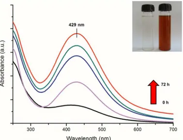
A gradual change in the colour of reaction medium (containing fungal cell-free filtrate and precursor silver ions) from colourless to reddish brown revealed a visual evidence for silver nanoparticle synthesis (Fig 1inset). UV-visible spectrum showed a gradual increase in the absorbance at 429 nm with respect to time (Fig 1). The absorption maxima at 429 nm can be attributed to the surface plasmon resonance (SPR) vibrations of synthesized silver nanoparti-cles [37]. No further increase in absorbance was observed after 72 h of reaction (data not shown), which indicated the complete reduction of precursor silver ions in reaction medium. Stability of as-synthesized silver nanoparticles was monitored periodically for more than three months. It was observed that the nanoparticle solution was extremely stable at room tempera-ture, with no evidence of particle aggregation as determined by UV-visible spectroscopy measurements.TEM micrograph (Fig 2A) revealed the presence of mono-dispersed and predominantly spherical particles with no visible aggregation. The SAED pattern (Fig 2Ainset) attests the crystallanity of silver nanoparticles. The particle size distribution histogram obtained from TEM measurements revealed that most of the particles ranged between 40–80 nm with a mean diameter of 54 ± 8.9 nm (Fig 2B). These results were in well agreement with the values obtained by DLS measurements (Fig 2C). It has been well reported that different fungi can synthesize nanoparticles of varied composition, sizes and shapes which may be due to the differences in their extracellular protein profiles [38–40].
Fig 1. UV visible spectrum of reaction medium as a function of time (0, 12, 24, 48 and 72 h).Inset shows tubes containing fungal cell-free filtrate (a) without and (b) with silver nitrate solution after 72 h of reaction.
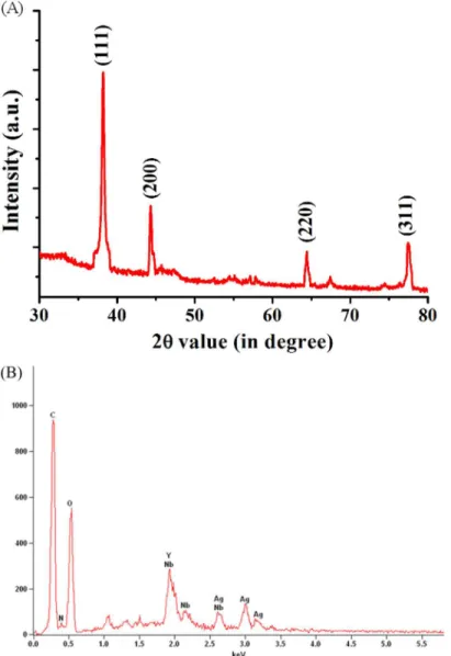
X-ray diffraction pattern recorded by preparing drop-coated film of protein-capped silver nanoparticles further validated the crystalline nature of nanoparticles. The well-defined peaks at 2θvalues of 38.03°, 46.18°, 64.60°, and 77.18° corresponds to (111), (200), (220) and (311) planes of silver, respectively (Fig 3A). These values were in complete agreement with the face-centered cubic (fcc) lattice structure of crystalline silver (JCPDS file no. 04–0783). A similar pattern of XRD spectrum has been reported for metallic silver nanoparticles synthesized by other fungi [41]. EDS was carried out to determine the elemental composition of as-synthe-sized nanoparticles and capping molecules. An intense optical absorption band at 3.0 KeV
Fig 2. (A) A representative transmission electron micrograph showing spherical shaped silver nanoparticles (scale bar equivalent to 50 nm). Inset showing SAED pattern recorded from a single nanoparticle. Particle size distribution histogram of silver nanoparticles as determined using (B) transmission electron microscope and (C) dynamic light scattering measurements.
doi:10.1371/journal.pone.0134337.g002
Fig 3. (A) XRD spectrum of as-synthesized protein-capped silver nanoparticles with Bragg’s diffraction values shown in parentheses. (B) EDS spectrum showing the elemental composition of silver nanoparticles.
confirmed the presence of pure metallic silver nanoparticles (Fig 3B). Other peaks observed for C, N and O atoms indicated the presence of proteins as capping molecules.
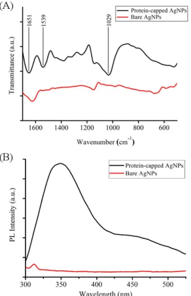
UV-visible absorption spectrum of supernatant obtained after SDS treatment of as synthe-sized silver nanoparticles showed a broad absorption peak near 280 nm which corresponds to aromatic amino acids of proteinsS1 Fig[28]. FTIR and photoluminescence measurements of protein-capped and bare silver nanoparticles were carried out to confirm the removal of cap-ping proteins from the surface of silver nanoparticles. FTIR spectrum of protein-capped silver nanoparticles (Fig 4A) exhibited characteristic bands of amide I and amide II at wavenumbers 1651 and 1539 cm-1, respectively and C-N stretching vibration band of aliphatic amine at 1029 cm-1[28]. The disappearance of these characteristic protein bands in the FTIR spectrum of bare silver nanoparticles clearly indicated the removal of proteins from nanoparticles. The PL spectrum (Fig 4B) of protein-capped silver nanoparticles showed a distinct emission peak at 340 nm, which could be attributed to the tyrosine residues of capping proteins. In contrast,
Fig 4. (A) FTIR spectra and (B) Photoluminescence spectra of protein-capped and bare silver nanoparticles.
absence of emission peak in case of bare silver nanoparticles confirmed the complete removal of protein molecules.
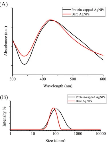
The removal of protein shell by SDS treatment reduced the size of nanoparticles as reflected by the blue shift observed in the SPR peak from 429 to 425 nm in case of bare silver nanoparti-cle (Fig 5A). Calzolai et al. [42] reported a similar observation while studying interaction between human ubiquitin and gold nanoparticles. Furthermore, hydrodynamic particle size distribution and polydispersity index (PDI) analysis was carried out to investigate the occu-pancy of protein shell in case of protein-capped silver nanoparticles. The protein-capped silver nanoparticles showed a mean particle size of 90.53 nm (PDI = 0.357) which decreased to 58.39 nm (PDI = 0.396) in case of bare silver nanoparticles indicating the successful removal of pro-tein shell from nanoparticles after SDS treatment (Fig 5B). The plausible reason for multi-lay-ered shell (~32 nm thick) could be the non-specific and non-competitive binding of proteins present in the surrounding environment (reaction medium). The persistence of thick protein shell on biogenic silver nanoparticles has attracted our attention to compare the antibacterial efficacy of protein-capped nanoparticles in comparison to bare silver nanoparticles.
Antibacterial efficacy of protein-capped and bare silver nanoparticles
The time-dependent bacterial growth in presence of protein-capped and bare silver nanoparti-cles was monitored by measuring the absorbance at 600 nm [43]. The protein-capped as well asFig 5. (A) UV visible spectra and (B) particle size distribution of protein-capped and bare silver nanoparticles.
bare silver nanoparticles were found to adversely affect the bacterial growth with major impli-cations on the exponential phaseS2 Fig. Growth dynamics profile of both Gram positive bacte-rial species revealed that the protein-capped nanoparticles were more effective in controlling the bacterial growth as compared to bare silver nanoparticles. In contrast, bare nanoparticles were found to be more effective than protein-capped nanoparticles in case of Gram negative bacterial species. The greater efficacy of bare silver nanoparticles against Gram negative bacte-ria observed during the present study is in complete agreement with previous reports
[19,44,45]. Overall, the impact of both types of silver nanoparticles was observed to be more prominent on Gram negative bacteria as compared to Gram positive bacteria. This can be hypothesized in terms of their different cell wall composition wherein a relatively thick and continuous peptidoglycan cell wall in Gram positive bacteria could restrict the entry of bare sil-ver nanoparticles. Howesil-ver, the interactions of teichoic acid (which span the peptidoglycan layer) and side chains of amino acids of protein-capped silver nanoparticles may facilitate their possible entry in Gram positive bacterial species.
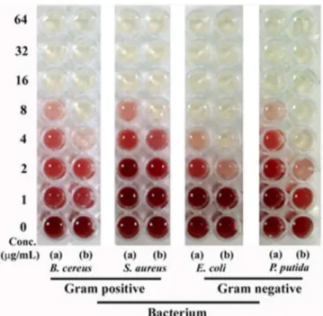
Based on the doubling time and cell size each bacterial species shows relatively different growth kinetics. Keeping this fact in mind we performed the more compelling minimum inhibitory concentration (MIC) test by dehydrogenase assay. A positive correlation was observed between the antibacterial activity and concentration of nanoparticles (Fig 6). Overall, the tested silver nanoparticles were found to be more effective against Gram negative bacteria as compared to Gram positive bacteria. The observed MIC values for protein-capped silver nanoparticles were 8μg mL-1and 4–8μg mL-1for Gram positive and Gram negative bacteria, respectively. On the other hand, MIC values for bare silver nanoparticles were found to be 2–4μg mL-1and 2μg mL-1for Gram positive and Gram negative bacteria, respectively. The obtained MIC values suggested that protein-capped silver nanoparticles were less effective as compared to bare silver nanoparticles. This can be attributed to the presence of protein shell on silver nanoparticles. It can be anticipated that the subtle changes on nanoparticle surface with protein shell can modulate their interactions with the bacterial surface, leading to
Fig 6. Dehydrogenase assay demonstrating MIC profiles of (a) protein-capped and (b) bare silver nanoparticles against selected Gram positive and Gram negative bacteria.
differential antibacterial profiles. To date, however, there is no clear understanding that surface modification of nanoparticles with protein shell can control the antibacterial properties of sil-ver nanoparticles. The modulation of antibacterial profile due to the presence of proteins on nanoparticle made it more interesting and motivated us to decipher the involved mechanism.
Bactericidal mechanism of protein-capped and bare silver nanoparticles
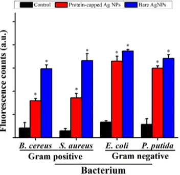
The exact mechanism of bactericidal effect of silver nanoparticles has yet not been elucidated and is open for debate [7,46]. The most commonly reported mechanism is the generation of free radicals such as peroxide, superoxide and hydroxyl ions by silver nanoparticles which could play a key role in executing bactericidal effects [47].Fig 7depicts the magnitude of ROS species formation represented as fluorescent counts due to DCF formation in bacterial cells exposed to the protein-capped and bare silver nanoparticles as compared to the control. The magnitude of ROS species generation was approximately two fold higher for bare silver nano-particles as compared to the protein-capped nanonano-particles in case of Gram positive bacterial species. The low ROS production observed in case of protein-capped silver nanoparticles may be attributed to the presence of biocompatible molecules in the shell. In contrast, absence of these biocompatible molecules on bare silver nanoparticles led to accelerated ROS production. Surprisingly, almost similar magnitude of ROS species generation was observed for both pro-tein-capped and bare silver nanoparticles in Gram negative bacterial species.It has well been reported that silver ions catalyse the decomposition of H2O2and
subse-quently results in the formation of free radicals (OH and/or O2-) [48]. Hence, the role of ROS
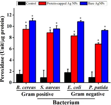
generation and its consequences on the antioxidant system was evaluated by measuring the activity of ROS related enzymes viz. peroxidase (Fig 8) and SOD (Fig 9). In general, an increase in the activities of both these enzymes was observed in all the tested bacterial species when exposed to bare silver nanoparticles. It can be inferred that the removal of protein shell not
Fig 7. Relative fluorescence intensity (with respect to H2O2) showing the cellular ROS formation
capability of protein-capped and bare silver nanoparticles as compare to control.Vertical bars represent standard errors. Significant differences from control (p0.05) are marked with asterisk.
only generates higher amount of ROS molecules but also leads to the formation of cellular oxi-dants which also interfere with the respective antioxidant system [49].
The generation of free radicals such as peroxide, superoxide and hydroxyl ions by silver nanoparticles have been reported to exert lipid peroxidation and damage membrane integrity [9,50–52]. Furthermore, biochemical and proteomic studies strengthened the fact that silver nanoparticles resulted in an immediate dissipation of the proton motive force which causes de-energization of cell membrane and consequently cell death [53]. Keeping these facts in mind,
Fig 8. Levels of peroxidase in untreated and treated bacterial cells.The data are expressed as mean±standard error of three independent experiments (p<0.05).
doi:10.1371/journal.pone.0134337.g008
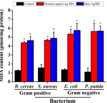
the membrane damage studies were performed by measuring MDA content. In general, an increased MDA content was observed in all the tested bacterial species when exposed to silver nanoparticles (Fig 10). However, marginal difference was observed between protein-capped and bare silver nanoparticles.
To corroborate the membrane damage results, we further estimated the release of bio-mole-cules (protein, carbohydrate and nucleic acid) from bacterial cells after silver nanoparticle treatment. As expected, a higher membrane leakage was observed in the bacterial cells treated with bare silver nanoparticles as compared to those treated with protein-capped silver nano-particlesS1 Table. Moreover, it was clearly evident that the bactericidal action of silver nano-particles was more effective against Gram negative bacterial species. Damage to the cell membrane led to cell distortion causing release of carbohydrates, proteins and nucleic acids has also been reported previously [54,55].
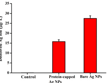
The release of silver ions from silver nanoparticles determines their antibacterial activity [46,47,56–58]. It has been demonstrated that aerobic conditions can readily oxidized silver nanoparticles in aqueous solutions resulting in the release of silver ions [59]. The positive charge generated due to release of Ag+ions from silver nanoparticles develops an electrostatic interaction with the negatively charged bacterial cell membrane [60]. Moreover, the reaction of silver ions with carbohydrates, hydroxyls and thiols of bacterial cell wall and nuclear mem-brane leads to cell distortion and death [12,61]. In the present study, the release of silver ions from protein-capped and bare silver nanoparticles was estimated using ICP-AES. The dis-solved silver ion concentration for protein-capped and bare silver nanoparticles was measured as 15.8 and 27.5μg L-1, respectively (Fig 11). It clearly indicates that the presence of protein shell acts as a barrier preventing the release of silver ions from the nanoparticles. These results are in complete accordance with a previous study which demonstrated that surface coating play a key role in dissolution of silver ions from nanoparticle surface and the toxicity of silver nanoparticles depends on the surface coating [62].
Fig 10. Malondialdehyde (MDA) assay demonstrating the difference in membrane damage capability of protein-capped and bare silver nanoparticles.Vertical bars represent standard errors. Significant differences from control (p0.05) are marked with asterisk.
TEM measurements were carried out to determine the distribution and location of the silver nanoparticles, as well as morphology of the bacteria after treatment with protein-capped or bare silver nanoparticles. TEM micrographs of representative Gram positive (B.Cereus) and Gram negative (E.coli) bacterium showed that protein-capped silver nanoparticles resulted in
Fig 11. ICP-AES analysis of silver dissolution profiles of protein-capped and bare silver nanoparticles.
doi:10.1371/journal.pone.0134337.g011
Fig 12. Transmission electron micrographs depicting the morphological changes in Gram positiveB. cereusand Gram negativeE.coliafter exposure to protein-capped and bare silver nanoparticles.
lesser membrane damage. In contrast, bare silver nanoparticles caused the complete loss of membrane integrity and resulted in the release of cytoplasm (Fig 12).
Conclusions
The present study demonstrates an economical and environmentally affable approach for the synthesis of protein-capped silver nanoparticles. The extracellular nature of synthesis makes the present method simple, straightforward and allows ease in downstream processing. The as-synthesized nanoparticles were spherical, mono-dispersed and coated with protein molecules. To the best of our knowledge, the present investigation first time demonstrates that the pres-ence of capping molecules significantly influpres-ences the activity of biologically synthesized nano-particles. Removal of capping molecules (protein shell) from the surface of silver nanoparticles showed significant increase in their antibacterial activity against both Gram-positive and Gram-negative bacterial species. The present findings strongly postulate that surface modifica-tions of nanoparticles with proteins can significantly modulate their functional properties.
Supporting Information
S1 Fig. UV-visible absorption spectrum of supernatant showing presence of proteins.
(TIF)
S2 Fig. Growth dynamics of tested bacterial species in presence of protein-capped or bare silver nanoparticles as compared to controls.
(TIF)
S1 Table. Magnitude of membrane leakage from bacterial cells after exposure to silver nanoparticles.
(DOC)
Author Contributions
Conceived and designed the experiments: NJ JP. Performed the experiments: NJ MR. Analyzed the data: NJ AB RVD JP. Contributed reagents/materials/analysis tools: JP. Wrote the paper: NJ AB JP.
References
1. Neu HC. The crisis in antibiotic resistance. Science. 1992; 257: 1064–1073. PMID:1509257 2. Stewart PS, Costerton JW. Antibiotic resistance of bacteria in biofilms. The Lancet. 2001; 358: 135–
138.
3. Allen HK, Donato J, Wang HH, Cloud-Hansen KA, Davies J, Handelsman J. Call of the wild: Antibiotic resistance genes in natural environments. Nat Rev Microbiol. 2010; 8: 251–259. doi:10.1038/ nrmicro2312PMID:20190823
4. Andersson DI, Hughes D. Persistence of antibiotic resistance in bacterial populations. FEMS Microbiol Rev. 2011; 35: 901–911. doi:10.1111/j.1574-6976.2011.00289.xPMID:21707669
5. Andersson DI, Hughes D. Antibiotic resistance and its cost: is it possible to reverse resistance? Nat Rev Microbiol. 2010; 8: 260–271. doi:10.1038/nrmicro2319PMID:20208551
6. Pradeep T, Anshup. Noble metal nanoparticles for water purification: A critical review. Thin Solid Films. 2009; 517: 6441–6478.
7. Mijnendonckx K, Leys N, Mahillon J, Silver S, Van Houdt R. Antimicrobial silver: uses, toxicity and potential for resistance. BioMetals. 2013; 26: 609–621. doi:10.1007/s10534-013-9645-zPMID:
23771576
9. Morones JR, Elechiguerra JL, Camacho A, Holt K, Kouri JB, Ramírez JT, et al. The bactericidal effect of silver nanoparticles. Nanotechnology. 2005; 16: 2346–2353. doi:10.1088/0957-4484/16/10/059
PMID:20818017
10. Sheehy K, Casey A, Murphy A, Chambers G. Antimicrobial properties of nano-silver: A cautionary approach to ionic interference. J Colloid Interface Sci. 2015; 443: 56–64. doi:10.1016/j.jcis.2014.11. 074PMID:25531416
11. Suchomel P, Kvitek L, Panacek A, Prucek R, Hrbac J, Vecerova R, et al. Comparative study of antimi-crobial activity of AgBr and Ag nanoparticles (NPs). PLoS ONE. 2015; 10: e0119202. doi:10.1371/ journal.pone.0119202PMID:25781988
12. Marambio-Jones C, Hoek EMV. A review of the antibacterial effects of silver nanomaterials and poten-tial implications for human health and the environment. J Nanopart Res. 2010; 12: 1531–1551.
13. Rai M, Yadav A, Gade A. Silver nanoparticles as a new generation of antimicrobials. Biotechnol Adv. 2009; 27: 76–83. doi:10.1016/j.biotechadv.2008.09.002PMID:18854209
14. Bartlomiejczyk T, Lankoff A, Kruszewski M, Szumiel I. Silver nanoparticles-allies or adversaries? Ann Agric Environ Med. 2013; 20: 48–54. PMID:23540211
15. Neal AL. What can be inferred from bacterium–nanoparticle interactions about the potential conse-quences of environmental exposure to nanoparticles? Ecotoxicology. 2008; 17: 362–371. doi:10.1007/ s10646-008-0217-xPMID:18454313
16. Xu H, Qu F, Xu H, Lai W, Wang YA, Aguilar ZP, et al. Role of reactive oxygen species in the antibacte-rial mechanism of silver nanoparticles onEscherichia coliO157: H7. BioMetals. 2012; 25: 45–53. doi:
10.1007/s10534-011-9482-xPMID:21805351
17. Holt KB, Bard AJ. Interaction of silver(I) ions with the respiratory chain ofEscherichia coli: an electro-chemical and scanning electroelectro-chemical microscopy study of the antimicrobial mechanism of micromo-lar Ag+. Biochemistry. 2005; 44: 13214
–13223. PMID:16185089
18. Li WR, Xie XB, Shi QS, Zeng HY, OU-Yang YS, Chen YB. Antibacterial activity and mechanism of silver nanoparticles onEscherichia coli. Appl Microbiol Biotechnol. 2010; 85: 1115–1122. doi:10.1007/ s00253-009-2159-5PMID:19669753
19. Feng QL, Wu J, Chen GQ, Cui FZ, Kim TN, Kim JO. et al. A mechanistic study of the antibacterial effect of silver ions onEscherichia coliandStaphylococcus aureus. J Biomed Mater Res. 2000; 52: 662–668. PMID:11033548
20. Xiu ZM, Zhang QB, Puppala HL, Colvin VL, Alvarez PJ. Negligible particle-specific antibacterial activity of silver nanoparticles. Nano Lett. 2012; 12: 4271–4275. doi:10.1021/nl301934wPMID:22765771 21. Panáček A, Kvítek L, Prucek R, KolářM, Večeřová R, Pizúrova N, et al. Silver colloid nanoparticles:
Synthesis, characterization, and their antibacterial activity. J Phys Chem B. 2006; 110: 16248–16253. PMID:16913750
22. Kvitek L, Panaček A, Soukupova J, Kolar M, Večerova R, Prucek R, et al. Effect of surfactants and
poly-mers on stability and antibacterial activity of silver nanoparticles. J Phys Chem C. 2008; 112: 5825–
5834.
23. Ahamed M, Karns M, Goodson M, Rowe J, Hussain SM, Schlager JJ, et al. DNA damage response to different surface chemistry of silver nanoparticles in mammalian cells. Toxicol Appl Pharmacol. 2008; 233: 404–410. doi:10.1016/j.taap.2008.09.015PMID:18930072
24. Li Z, Greden K, Alvarez PJ, Gregory KB, Lowry GV. Adsorbed polymer and NOM limits adhesion and toxicity of nano scale zerovalent iron toE.coli. Environ Sci Technol. 2010; 44: 3462–3467. doi:10. 1021/es9031198PMID:20355703
25. Somasundaran P, Chakraborty S, Qiang Q, Deo P, Wang J, Zhang R. Surfactants, polymers and their nanoparticles for personal care applications. J Cosmet Sci. 2004; 55: S1–S17. PMID:15645085 26. Shoults-Wilson WA, Reinsch BC, Tsyusko OV, Bertsch PM, Lowry GV, Unrine JM. Effect of silver
nano-particle surface coating on bioaccumulation and reproductive toxicity in earthworms (Eisenia fetida). Nanotoxicology. 2011; 5: 432–444. doi:10.3109/17435390.2010.537382PMID:21142839 27. Jain N, Bhargava A, Tarafdar JC, Singh SK, Panwar J. A biomimetic approach towards synthesis of
zinc oxide nanoparticles. Appl Microbiol Biotechnol. 2013; 97: 859–869. doi: 10.1007/s00253-012-3934-2PMID:22382164
28. Jain N, Bhargava A, Majumdar S, Tarafdar JC, Panwar J. Extracellular biosynthesis and characteriza-tion of silver nanoparticles usingAspergillus flavusNJP08: A mechanism perspective. Nanoscale. 2011; 3: 635–641. doi:10.1039/c0nr00656dPMID:21088776
29. Wang H, Joseph JA. Quantifying cellular oxidative stress by dichlorofluorescein assay using microplate reader. Free Radic Biol Med. 1999; 27: 612–616. PMID:10490282
31. Rico CM, Hong J, Morales MI, Zhao L, Barrios AC, Zhang JY, et al. Effect of cerium oxide nanoparticles on rice: A study involving the antioxidant defense system andin vivofluorescence imaging. Environ Sci Technol. 2013; 47: 5635–5642. doi:10.1021/es401032mPMID:23662857
32. Hochman A, Goldberg I. Purification and characterization of a catalase-peroxidase and a typical cata-lase from the bacteriumKlebsiella pneumoniae. Biochim Biophys Acta. 1991; 1077: 299–307. PMID:
2029529
33. Arora S, Jain J, Rajwade JM, Paknikar KM. Cellular responses induced by silver nanoparticles:In vitro
studies. Toxicol Lett. 2008; 179: 93–100. doi:10.1016/j.toxlet.2008.04.009PMID:18508209 34. Buege JA, Aust SD. Microsomal lipid peroxidation. Methods Enzymol. 1978; 52: 302–310. PMID:
672633
35. Bradford MM. A rapid and sensitive method for the quantitation of microgram quantities of protein utiliz-ing the principle of protein-dye bindutiliz-ing. Anal Biochem. 1976; 72: 248–254. PMID:942051
36. Fales FW. The assimilation and degradation of carbohydrates by yeast cells. J Biol Chem. 1951; 193: 113–124. PMID:14907695
37. Lu X, Rycenga M, Skrabalak SE, Wiley B, Xia Y. Chemical synthesis of novel plasmonic nanoparticles. Annu Rev Phys Chem. 2009; 60: 167–192. doi:10.1146/annurev.physchem.040808.090434PMID:
18976140
38. Vigneshwaran N, Kathe AA, Varadarajan P, Nachane RP, Balasubramanya R. Biomimetics of silver nanoparticles by white rot fungus,Phaenerochaete chrysosporium. Colloids Surf B. 2006; 53: 55–59.
39. Gericke M, Pinches A. Biological synthesis of metal nanoparticles. Hydrometallurgy. 2006; 83: 132–
140.
40. Shankar SS, Ahmad A, Pasricha R, Sastry M. Bioreduction of chloroaurate ions by geranium leaves and its endophytic fungus yields gold nanoparticles of different shapes. J Mater Chem. 2003; 13: 1822–
1826.
41. Shaligram NS, Bule M, Bhambure R, Singhal RS, Singh SK, Szakacs G, et al. Biosynthesis of silver nanoparticles using aqueous extract from the compactin producing fungal strain. Process Biochem. 2009; 44: 939–943.
42. Calzolai L, Franchini F, Gilliland D, Rossi F. Protein-nanoparticle interaction: identification of the ubiqui-tin-gold nanoparticle interaction site. Nano Lett. 2010; 10: 3101–3105. doi:10.1021/nl101746vPMID:
20698623
43. Zhou Y, Kong Y, Kundu S, Cirillo JD, Liang H. Antibacterial activities of gold and silver nanoparticles againstEscherichia coliandBacillus Calmette-Guérin. J Nanobiotechnol. 2012; 10: 1–9.
44. Fayaz AM, Balaji K, Girilal M, Yadav R, Kalaichelvan PT, Venketesan R. Biogenic synthesis of silver nanoparticles and their synergistic effect with antibiotics: A study against Gram-positive and Gram-neg-ative bacteria. Nanomed Nanotech Biol Med. 2010; 6: 103–109.
45. Amato E, Diaz-Fernandez YA, Taglietti A, Pallavicini P, Pasotti L, Cucca L, et al. Synthesis, characteri-zation and antibacterial activity against Gram positive and Gram negative bacteria of biomimetically coated silver nanoparticles. Langmuir. 2011; 27: 9165–9173. doi:10.1021/la201200rPMID:21736306 46. Cui L, Chen P, Chen S, Yuan Z, Yu CP, Ren B, et al.In situstudy of the antibacterial activity and
mech-anism of action of silver nanoparticles by surface-enhanced raman spectroscopy. Anal Chem. 2013; 85: 5436–5443. doi:10.1021/ac400245jPMID:23656550
47. Reidy B, Haase A, Luch A, Dawson KA, Lynch I. Mechanisms of silver nanoparticle release, transfor-mation and toxicity: A critical review of current knowledge and recommendations for future studies and applications. Materials. 2013; 6: 2295–2350.
48. Chang Q, Yan L, Chen M, He H, Qu J. Bactericidal mechanism of Ag/Al2O3againstEscherichia coli. Langmuir. 2007; 23: 11197–11199. PMID:17902710
49. Dimkpa CO, Calder A, Gajjar P, Merugu S, Huang W, Britt DW, et al. Interaction of silver nanoparticles with an environmentally beneficial bacterium,Pseudomonas chlororaphis. J Hazard Mater. 2011; 188: 428–435. doi:10.1016/j.jhazmat.2011.01.118PMID:21339046
50. Lima R, Seabra AB, Durán N. Silver nanoparticles: a brief review of cytotoxicity and genotoxicity of chemically and biogenically synthesized nanoparticles. J Appl Toxicol. 2012; 32: 867–879. doi:10. 1002/jat.2780PMID:22696476
51. Sayes CM, Gobin AM, Ausman KD, Mendez J, West JL, Colvin VL. Nano-C60cytotoxicity is due to lipid peroxidation. Biomaterials. 2005; 26: 7587–7595. PMID:16005959
52. Farmer EE, Mueller MJ. ROS-mediated lipid peroxidation and RES-activated signaling. Annu Rev Plant Biol. 2013; 64: 429–450. doi:10.1146/annurev-arplant-050312-120132PMID:23451784
54. Choi O, Deng KK, Kim NJ, Ross L Jr, Surampalli RY, Hu Z. The inhibitory effects of silver nanoparticles, silver ions, and silver chloride colloids on microbial growth. Water Res. 2008; 42: 3066–3074. doi:10. 1016/j.watres.2008.02.021PMID:18359055
55. Travan A, Pelillo C, Donati I, Marsich E, Benincasa M, Scarpa T, et al. Non-cytotoxic silver nanoparti-cle-polysaccharide nanocomposites with antimicrobial activity. Biomacromolecules. 2009; 10: 1429–
1435. doi:10.1021/bm900039xPMID:19405545
56. Brett DW. A discussion of silver as an antimicrobial agent: Alleviating the confusion. Ostomy Wound Manage. 2006; 52: 34–41.
57. Chen X, Schluesener H. Nanosilver: A nanoproduct in medical application. Toxicol Lett. 2008; 176: 1–
12. PMID:18022772
58. Love SA, Maurer-Jones MA, Thompson JW, Lin YS, Haynes CL. Assessing nanoparticle toxicity. Annu Rev Anal Chem. 2012; 5: 181–205.
59. Liu J, Sonshine DA, Shervani S, Hurt RH. Controlled release of biologically active silver from nanosilver surfaces. ACS Nano. 2010; 4: 6903–6913. doi:10.1021/nn102272nPMID:20968290
60. Kim JS, Kuk E, Yu KN, Kim JH, Park SJ, Lee HJ, et al. Antimicrobial effects of silver nanoparticles. Nanomedicine. 2007; 3: 95–101. PMID:17379174
61. Hwang ET, Lee JH, Chae YJ, Kim YS, Kim BC, Sang BI, et al. Analysis of the toxic mode of action of sil-ver nanoparticles using stress-specific bioluminescent bacteria. Small. 2008; 4: 746–750. doi:10.1002/ smll.200700954PMID:18528852