INTERNATIONAL RESEARCH JOURNAL OF PHARMACY
www.irjponline.com ISSN 2230 – 8407
Research Article
ANTIMICROBIAL ACTIVITY OF BIOSYNTHESIZED SILVER NANOPARTICLES PREPARED FROM THE
LEAF EXTRACT OF LANTANA CAMARA
Kumarasamyraja D*1, Jeganathan N. S.2
1
Department of pharmacy, Annamalai University, Annamalai Nagar, Chidambaram, India
2
Periyar College of Pharmaceutical Sciences, Trichy, India
*Corresponding Author Email: [email protected]
Article Received on: 16/03/13 Revised on: 07/04/13 Approved for publication: 11/05/13
DOI: 10.7897/2230-8407.04541
IRJP is an official publication of Moksha Publishing House. Website: www.mokshaph.com
© All rights reserved.
ABSTRACT
Lantana camara commonly known as weed or red sage is found throughout India. It was reported to be used in traditional medicine system for infectious diseases. The present study designed to evaluate antimicrobial activity of aqueous extract of Lantana camara andbio synthesized silver Nanoparticles from aqueous extract of Lantana camara. When the extract of the plant was treated with silver nitrate (1mM) solution, a yellowish brown color developed indicating the formation of silver Nanoparticles. The anti microbial activity of biosynthesized silver Nanoparticles of the extract and aqueous extract of
Lantana camara was investigated against two gram positive and two gram negative human pathogenic bacteria and fungi by using disc diffusion method. The biosynthesized silver Nanoparticles showed promising anti bacterial activity at 300µg/ml concentration compared with aqueous extract of Lantana camara. Similarly the silver Nanoparticles at the same level of concentration also exhibited significant antifungal activity against Candida albicans and Aspergillus niger. Conclusion: The results indicate that the biosynthesized silver Nanoparticles from Lantana camara have better antimicrobial activity than with that of aqueous extract of Lantana camara and almost equal antimicrobial activity when compared with the standard.
Key words: Antibacterial activity, Antifungal activity, Silver nanoparticles (Ag-Np’s), TEM, UV- Vis spectrum.
INTRODUCTION
The medicinal uses of silver have been documented since 1000 B.C. Silver is a health additive in traditional Chinese and Indian Ayurvedic system of medicine. Silver together with copper, is commonly used to inhibit bacterial and fungal growth in chicken farms and in post harvested cleaning of oysters. Silver resistance is important to monitor because modern technology has developed a wide range of products that depend on silver as a key microbial component. In late 1970s, Robert O. Becker has discovered that silver ions promote bone growth and kill surrounding bacteria 1. Silver
kills some 650 different disease causing organisms. Silver based topical dressing has been widely used as a treatment for infections in burns, open wounds and chronic ulcers. Nanomaterials are the leading requirement of the rapidly developing field of Nanomedicine, bio nanotechnology. Nanoparticles are being utilized as therapeutic tools in infections, against microbes and thus understanding the properties of Nanoparticles and their effect on microbes is essential for clinical applications 2. Recently, there has been a great deal of interest surrounding the discovery that silver Nanoparticles (Ag-Np’s) are significantly more effective antimicrobial agents in terms of the minimum effective concentration than their Ag+ counterparts 3. Biological approach for the synthesis of Ag-Np’s (Green synthesis) is an eco-friendly and cost-effective method as compared to the other chemical and physical methods. This has promoted research in the well known activity of silver ions and silver-based compounds, including Ag-Np’s 4. The synthesis of Ag-Np’s using chemical and physical methods requires high pressure, energy, temperature and toxic chemicals. The results of chemical synthesis method showed some unfavorable impact in the medical applications. When compared with chemical and physical methods, green synthesis method provides a low cost, environment friendly, easily scale up for large scale synthesis, and no need to use high pressure, energy, temperature and toxic chemicals 5.
Hence, Ag-Np’s have been applied to a wide range of healthcare products, such as scaffold, water purification systems and medical devices. The most important application of silver and Ag-Np’s is in pharmaceutical industry for the preparation of topical ointments to prevent infection against burn and open wounds. The toxic effects of silver on bacteria have been investigated for more than 60 years and the acting mechanism of silver has been known to some extent. Therefore; the preparation of uniform nanosized Ag-Np’s with specific requirements in terms of size, shape, and physical and chemical properties is of great interest in the formulation of new pharmaceutical products 6. Lantana camara commonly known as weed or red sage or
“Unnichedi” in Tamil, “Pulikampa” in Telugu and
“Caturang” in Hindi is a significant weed commonly found throughout India. Since, very long time Lantana camara
reported in traditional medicine for the treatment of infectious diseases 7. Hence, the aim of the present study was
to develop a novel approach for the green synthesis of Ag-Np’s using aqueous extract of Lantanacamara and exploring its antimicrobial activity against some selected Gram positive and Gram negative organisms and comparing their antimicrobial activity with the leaf extract of Lantana camara.
MATERIALS AND METHODS Plant collection
Preparation of aqueous extract of Lantana Camara The dried plant material was powdered using a disintegrator to get a coarse powder. The powdered samples were kept in sealed containers for extraction purposes. About 10gm of the sample was shaken with 40 ml of double distilled sterilized water in a 100ml Erlenmeyer stopper flask for 15 min and then the mixture was boiled for 5 min. The extract was cooled and filtered through Whatman no 1 filter paper. The resultant filtrate was kept in a refrigerator 7.
Collection of Microorganism
The microorganisms used in this experiment were Bacillus subtilis (10877), Staphylococcus aureus (29838),
Pseudomonas aeruginosa (27854), Escherichia coli (1130) and fungus culture Candida albicans and Aspergillus niger.
They were obtained from Boss Laboratories, Madurai, Tamil Nadu and India.
Bio synthesis of silver Nanoparticles
In a typical reaction procedure, 10 ml of refrigerated filtrate was treated with 90ml of AgNo3 (1mM) solution. The
resulting solution was incubated in dark (to minimize the photo activation of silver nitrate), at 37◦C under static condition. The development of yellowish brown color solution indicated the formation of Ag-Np’s (Figure 1). the colored Ag-Np’s solution was centrifuged at 10,000 rpm for 10 min, the supernatant liquid was decanted. The resulting suspension was re dispersed in 10 ml sterile distilled water and centrifugation process was repeated for three times. Thereafter, the purified suspension was used for characterization of Ag-Np’s 8.
Characterization of silver Nanoparticles
The reduction of pure Ag+ ions was monitored by measuring the UV-Vis spectrum of the reaction medium at 5 h after diluting a small aliquot of the sample into distilled water. The color change in reaction mixture (metal ion solution +
Lantana Camara extract) was recorded through visual observation. UV-Vis spectral analysis was done by using UV-Vis spectrophotometer UV-2450 (Shimadzu) at the wavelength of 200– 800 nm9. The particle size range of the Ag-Np’s was determined by using particle size analyzer, Mastsizer 2000. The particle size was determined based on the Brownian motion of the nanoparticles. Transmission Electron Microscope (TEM) was used to view the morphology and size of bio synthesized Ag-Np’s in Philips model CM 200 instrument operated at an accelerating voltage of 200 kV 10.
Assay for antimicrobial activity of Ag Nanoparticles against microorganisms
The antimicrobial activity of aqueous extract of Lantana Camara and Ag-Np’s synthesized from Lantana Camara was determined by using disc diffusion method. Two gram positive bacteria (Bacillus subtilis, Staphylococcus aureus)
and two gram negative bacteria (Pseudomonas aeruginosa, Escherichia coli) were used for this study. The organisms were sub-cultured on Mueller Hinton Agar medium (MHA), incubated at 37°C for 24 h and stored at 4°C in the refrigerator to maintain stock culture. Petri plates were prepared with 20 ml of sterile MHA medium. The test cultures were swabbed on the top of the solidified media and allowed to dry for 10 min. The tests were conducted with
three different level of concentrations at 100,200 and 300 µg /ml respectively of aqueous extract and Ag-Np’s suspension prepared from the extract. The loaded discs were placed on the surface of the medium and left for 30 min at room temperature for compound diffusion. Negative control was prepared using respective solvent. Amikacin (50 µg/ml) was used as a standard positive control. The plates were incubated for 24 h at 37°C. A zone of inhibition was recorded in millimeters 11. The results of both crude extract and Ag-Np’s from crude extract of Lantana Camara given in Table 1& 2; Figure 5 & 6.
Anti fungal screening
Fungus cultures of Candida albicans and Aspergillus niger
were used for this study. The anti fungal activity was performed according to the standard reference method. The initial concentration of aqueous extract and Ag-Np’s suspension of Lantana Camara was 100µg/ml. The initial test concentration was serially diluted twofold. Each one was inoculated with 50µg/ml of suspension containing 104spore/ml of fungi. The anti fungal agent ketokonazole was included in the assays as a standard positive control. The plates were incubated between 24 h and 72 h at 27 °C 12. The results are given in Table 3& 4; Figure 7 & 8.
RESULTS AND DISCUSSION UV-Vis spectra analysis
It is well known that Ag-Np’s exhibit yellowish brown color in aqueous solution due to excitation of surface Plasmon vibrations in Ag-Np’s. The plant Lantana camara aqueous extract when mixed with the aqueous solution of the silver nitrate, silver ion complex produced and this complex was responsible for changing the color from watery to yellowish brown due to reduction of silver ion, which may be the indication of formation of Ag-NP’s 13.The UV- spectrum of
Lantana camara Ag-Np’s was recorded from the reaction medium. The results showed maximum absorption peak was exhibited between 322 – 350 nm (Figure 2).
Transmission electron microscopy (TEM)
The pictures below obtained by TEM shows the Ag-Np’s in aqueous extract of Lantana camara leaves. TEM analysis reveals that the Ag-Np’s are predominantly spherical. The overall morphology of the Ag-Np’s produced by reduction of Ag+ ions with 1mM AgNO3 is composed of almost uniform
Nanoparticles 9.
Particle size distribution
The particle size range of Ag-Np’s synthesized from L. camara was monitored by using particle size analyzer Mastsizer 2000. The obtained graph showed that Ag-Np’s average size range is 0.772 µm.
Antimicrobial activity of biosynthesized Ag-Np’s using
Lantana camara leaf extract
Table 1: In vitro antibacterial activity of aqueous leaf extract of Lantana camara
Drug Con:µg/ml Zone of inhibition (mm)
B. subtilis S.aureus P.aeruginosa E.coli
Aqueous leaf extract
Lantana camara
100 10 10 R 10
200 11 12 R 11
300 13 13 12 13 Standard (Amikkacin) 50 20 16 18 17
Table 2: In vitro antibacterial activity of Ag-Np’susing Lantana camara leaf extract
Drug Con:µg/ml Zone of inhibition (mm)
B. subtilis S.aureus P.aeruginosa E.coli
Lantana camara Ag-Np’s 100 11 10 12 14
200 14 12 14 14
300 14 12 14 14
Standard (Amikkacin) 50 20 18 17 17
Table 3: In vitro antifungal activity of aqueous leaf extract of
Lantana camara
Drug Con:µg/ml Zone of inhibition (mm)
C.albicans A.niger
Aqueous leaf extract
Lantana camara
100 9 10
200 11 11
300 14 14 Standard
(ketokonazole)
50 16 16
Table 4: In vitro antifungal activity of biosynthesized
Lantana camara Ag-Np’s
Drug Con:µg/ml Zone of inhibition (mm)
C.albicans A.niger
Lantana camara
Ag-Np’s
100 17 25
200 18 25
300 18 25 Standard
(ketokonazole)
50 17 20
Figure1: the photograph showing color change of plant extract after adding AgNo3 (a) Lantana Camara extract (b) AgNo3 solution (c) Ag-Np’s
Figure 2: UV-Vis spectra of Ag Nanoparticles biosynthesized from leaf extract of Lantana camara
Figure 3:TEM images of Ag-Np’s synthesized using leaf extract of Lantana camara. Particle size distribution
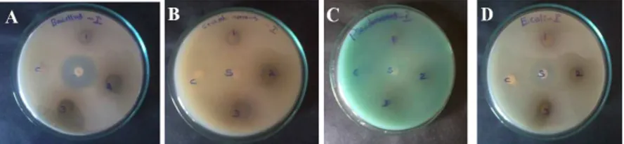
Figure 5: Anti bacterial activity of aqueous leaf extract of Lantana camara, A- B. subtilis, B - S. aureus, C- P. aeruginosa, D- E. coli
‘1’,’2’ and ‘3’ represents zone of inhibition of the extract at the concentration of 100, 200 and 300 µg/ml respectively. The centre zone ‘S’ represents zone of inhibition of standard antibacterial agent.
Figure 6: Anti bacterial activity of Ag-Np’sbiosynthesized from leaf extractof Lantana camara, A- B. subtilis, B - S. aureus, C- P. aeruginosa, D- E. coli; ‘1’,’2’ and ‘3’ represents zone of inhibition of Ag-Np’s using L. camara leaf extract at the concentration of 100, 200 and 300 µg/ml
respectively. The centre zone ‘S’ represents zone of inhibition of standard antibacterial agent
Figure 7: Inhibition of fungal growth of aqueous leaf extract of Lantana camara, E-C.albicans, F-A. niger
‘1’,’2’ and ‘3’ represents zone of aqueous leaf extract of L. camara at the concentration of 100, 200 and 300 µg/ml respectively. The centre zone ‘S’ represents zone of inhibition of standard antifungal agent (ketokonazole) at the concentration of 50 µg/ml and ‘C represents zone of control
Figure 8: Inhibition of fungal growth by Lantana camara Ag-Np’s, E-C. albicans, F-A. niger
‘1’,’2’ and ‘3’ represents zone of inhibition of biosynthesized Ag-Np’susing L. camara leaf extract at the concentration of 100, 200 and 300 µg/ml respectively. The centre zone ‘S’ represents zone of inhibition of standard antifungal agent (ketokonazole) at the concentration of 50 µg/ml and ‘C
represents zone of control
Another possible contribution to the bactericidal properties of Ag-Np’s is the release of silver ions from particles15. Here the antimicrobial activity of Ag-Np’s prepared from leaf extract of Lantana camara have been investigated against B. subtilis,
S. aureus, P. aeruginosa, E. coli and fungus cultures C. albicans and A. niger by Disc diffusion method on Mueller-Hinton broth. The antibacterial activity of aqueous extract of
Lantana camara and Ag-Np’s from Lantana camara showed great response against all investigated microorganism. The diameters of growth inhibition zone were analyzed by different concentration of aqueous extract of Lantana camara
and Ag-Np’s. The inhibition zone ranged from 100 to 300µg/ ml with highest inhibition zone values observed in 300µg/ ml in both prescribed samples, when compared with values from Table 1 & 2; Ag-Np’s from Lantana camara had greatest level of resistance showed B. subtilis, P.aeruginosa, E.coli
(the zone inhibition 14 mm), but aqueous extract of Lantana camara showed (13mm is the zone of inhibition). It is clearly revealed that aqueous leaf extract of Lantana camara
Ag-Np’s at the concentration of 300µg/ ml showed highest activity on Gram-positive and Gram-negative bacteria which are comparable with the aqueous extract of Lantana camara
CONCLUSION
In the present study, it was disclosed that aqueous leaf extract of Lantana camara can be converted into Ag-Np’s by green synthesis method. The synthesized Ag-Np’s from Lantana camara aqueous leaf extract was compared withaqueous leaf extract of Lantana camara for antibacterial activity with various concentrations. Based on the results we conclude Ag-Np’s from Lantana camara showed better antimicrobial activity then Lantana camara aqueous leaf extract on pathogenic microorganisms. The reported results suggested that green synthesized Lantana camara -Ag-Np’s could be used in the medical field for their efficient antimicrobial activity after undertaking proper clinical trial.
REFERENCES
1. Vedpriya Arya, Ratika Komal, Manbir Kaur and Anita Goyal. Silver nanoparticles as a potent antimicrobial agent A Review; Pharmacologyonline. 2011; 3: 118-124.
2. Kim, Soo-Hwan, Hyeong-Seon Lee and Dong-Seok Lee. Antibacterial Activity of Silver-Nanoparticles against Staphylococcus Aureus and
Escherichia coli.Korean J. Microbiol. Biotechnol. 2011; 39(1): 77–85. 3. Peter Irwin, Justin Martin and Ly-Huong Nguyen. Antimicrobial activity
of spherical silver nanoparticles prepared using a biocompatible macromolecular capping agent. Evidence for induction of a greatly prolonged bacterial lag phase; Journal of Nanobiotechnology 2010; 8(1)34. http://dx.doi.org/10.1186/1477-3155-8-34
4. M Gnanadesigan, M Anand, S Ravikumar. Antibacterial potential of biosynthesized silver Nanoparticles using Avicennia marina mangrove plant. Appl Nanosci 2012; 2(2): 143–147. http://dx.doi.org/ 10.1007/s13204-011-0048-6
5. Padmanaban sivakumar, Chandran nethradevi, Sahadevan renganathan. Synthesis of silver Nanoparticles using Lantana camara fruit extract and its effect on pathogens. Asian journal of Pharmaceutical and Clinical Research. 2012; 5(3): 97-101.
6. A Nasrollahi, Kh Pourshamsian and P Mansourkiaee. Antifungal activity of silver nanoparticles on some of fungi. Int.J.Nano.Dim. 2011; 1(3): 233-239.
7. Kumarasamyraja D, Jeganathan NS, Manavalan R. Pharmacological Review of Lantana Camara .L Review Article. Int. J. Pharm & Ind. Res. 2012; 2(1): 1-5.
8. Ponarulselvam S, Panneerselvam C, Murugan K. Synthesis of silver nanoparticles using leaves of Catharanthus roseus Linn. G. Don and their antiplasmodial activities. Asian Pacific Journal of Tropical Biomedicine. 2012; 2 (7): 574-580. http://dx.doi.org/10.1016/S2221-1691(12)60100-2
9. Veera babu Nagati, Rama Koyyati, Manisha R Donda. Green Synthesis and characterization of Silver nanoparticles from Cajanus cajan leaf extract and its antibacterial activity. International Journal of Nanomaterials and Biostructures. 2012; 2(3) 39-43.
10. Prasad TNVKV, Elumalai EK, Khateeja S. Evaluation of the antimicrobial efficacy of phytogenic silver nanoparticles. Asian Pacific Journal of Tropical Biomedicine 2011; 1(5): S82-S85. http://dx.doi.org /10.1016/S2221-1691(11)60130-5
11. Asra Parveen, Aashis S Roy and Srinath Rao . Biosynthesis and Characterization of Silver Nanoparticles from Cassia Auriculata Leaf Extract and In Vitro Evaluation of Antimicrobial Activity. International Journal of Applied Biology and Pharmaceutical Technology. 2012; 3(2): 222- 228.
12. Saranraj, PD Stella and Sajani Samuel. Antibacterial potentiality of ethanol and ethyl acetate extract of Acalypha indica against human pathogenic bacteria. Journal of Ecobiotechnology. 2010; 2 (7): 23 -27. 13. Fan SR, Liu XP, Li JW. Clinical characteristics of vulvo vaginal
candidiasis and antifungal susceptibilities of Candida species isolates among patients in southern China from 2003 to 2006. J Obstet Gynaecol
Res.2008;34(4):561-566.http://dx.doi.org/10.1111/j.1447-0756.2008.00817.x PMid:18937710
14. Raut rajesh W, Jaya RL, Niranjan SK, Sahebrao BMDK. Phytosynthesis of silver nanoparticles using glirricidia sepium (Jacq.). Curr Nanosci. 2009; 5(1): 117-122. http://dx.doi.org/10.2174/157341309787314674 15. Muthu A, N Rajmohan Rangasamy. Green synthesis of silver
Nanoparticles using Ixora coccinea leaves extract Mater Lett 2013; (article in press).
Cite this article as:
Kumarasamyraja D, Jeganathan N. S. Antimicrobial activity of biosynthesized silver nanoparticles prepared from the leaf extract of Lantana camara. Int. Res. J. Pharm. 2013; 4(5):203-207