Snežana Miljanić,1,* Marina Ratkaj,2 Igor Avdejev,2 Karlo Meglić,1 Adriana Kenđel1
1 Division of Analytical Chemistry, Department of Chemistry, Faculty of Science, University of Zagreb, Horvatovac 102a, HR-10000 Zagreb, Croatia 2 PLIVA Croatia, Teva Pharmaceutical Industries Ltd., Research and Development, Prilaz baruna Filipovića 29, HR-10000 Zagreb, Croatia * Corresponding author’s e-mail address: [email protected]
RECEIVED: March 15, 2015 REVISED: July 15, 2015 ACCEPTED: September 15, 2015
THIS PAPER IS DEDICATED TO DR.SVETOZAR MUSIĆ ON THE OCCASION OF HIS 70TH BIRTHDAY
Abstract: Surface-enhanced Raman scattering (SERS) enhancement factors (EF) were evaluated for RNA mononucleotides: adenosine 5'-mono-phosphate (AMP), guanosine 5'-mono5'-mono-phosphate (GMP), cytidine 5'-mono5'-mono-phosphate (CMP) and uridine 5'-mono5'-mono-phosphate (UMP), on silver nanoparticles, which differed in shape (nanospheres, nanostars) and stabilizing anionic layer (chlorides, citrates) on the metal surface. In freshly prepared silver colloids the enhanced Raman scattering was observed for all the RNA mononucleotides on the chloride coated silver nanosphe-res, Ag_Cl nsp (EF ≈ 104), for AMP only on the citrate coated silver nanospheres, Ag_cit nsp (EF ≈ 103), while not obtained at all for any of the
mononucleotides on the citrate stabilized silver nanostars, Ag_cit nst. Upon aggregation, the SERS activity of all the silver colloids increased, whereby the purine mononucleotides, AMP and GMP, more strongly scattered radiation on Ag_Cl nsp, and the pyrimidine mononucleotides, CMP and UMP, on Ag_cit nsp. Regardless of the silver nanoparticles, the higher EFs were evaluated for AMP and GMP (EF up to 5×10⁶), than
for CMP and UMP (EF ≈ 5×10⁴).
Keywords: surface-enhanced Raman scattering, enhancement factor, RNA mononucleotide, silver nanoparticles, shape, anions.
INTRODUCTION
URFACE-ENHANCED Raman scattering (SERS) spectros-copy is a powerful vibrational spectrosspectros-copy technique that allows for highly sensitive structural detection of low concentration molecules in vicinity to nanostructured me-tal surfaces.[1] Owing to recent developments in design and
fabrication of plasmonic substrates, essential for the Raman scattering enhancement, application of the SERS spectros-copy in analytical, biophysical and life sciences constantly increases.[2] The enhancement of the Raman scattering is
associated with two mechanisms: an electromagnetic mechanism and a chemical mechanism. The electromagne-tic enhancement mechanism results from the amplification of the radiation by excitation of the localized surface pla-smon resonance (LSPR), while the chemical enhancement mechanism involves charge transfer when the excitation e-nergy is resonant with the metal-molecule charge transfer
electronic states. Although electromagnetic mechanism do-minantly contributes to the overall enhancement, the total SERS enhancement factor with respect to the normal Ra-man signal, in most cases is the product of both mechani-sms, reaching up to 1010.
The prerequisite for the Raman scattering enhan-cement is adsorption of the molecules onto or near a ro-ughened metal surface. In order to enable effective surface adsorption, various SERS active substrates have been deve-loped, ranging from electrodes, metal island films, to com-monly used metal colloids.[3] Colloidal suspensions seem to
be particularly attractive due to rather simple and inexpen-sive preparation procedures, average SERS enhancement up to 106 and fair reproducibility.[4] In addition, the maximal
Croat. Chem. Acta2015, 88(4), 387–396 DOI: 10.5562/cca2720 by the surface plasmon resonance in the Vis region.
Howe-ver, experimental and theoretical studies on plasmonic substrates implied that, beside the metal type, morphology of the nanoparticles strongly affects LSPR, resulting in en-hancement of the Raman scattering upon Vis as well as NIR excitation.[5] Given that subtle changes in the nanostructure
alter the local field enhancement and lead to significant LSPR shifts, much emphasis has been put on controlling the shape of the metallic nanoparticles. Hence a huge intensifi-cation of the electromagnetic field has been observed for the anisotropic metal nanoparticles.[5] Among
non-spheri-cally shaped nanoparticles nanostars have been particularly attractive due to the tip-associated plasmon resonance, which can be synthetically tuned into the NIR region.[6] In
addition to the shape effect, the plasmon frequency for the single nanoparticle decreases as the particle size increases as well as the nanoparticles aggregate.[4,7] Except for the
change in the surface plasmon resonance, nanoparticles ag-gregation leads to generation of enormous electric field in interparticle gaps. To control nanoparticle aggregation, an aggregating agent is usually introduced into the colloidal su-spension, where either by reacting with the metal surface or by coating the nanoparticle, reduces the surface charge. Except for stabilization of the metallic nanoparticles in the suspension, the surface coatings is of the utmost impor-tance for the adsorption of the molecules on the colloidal nanoparticles.[8] If colloidal nanoparticles and molecules
had charges of the same sign, adsorption process could be strongly hindered, or even completely prevented.
Here, we report on an influence of the nanoparticle properties on the surface-enhanced Raman scattering of four RNA mononucleotides: adenosine 5'-monophosphate (AMP), guanosine monophosphate (GMP), cytidine 5'-monophosphate (CMP) and uridine 5'-5'-monophosphate (UMP) (Scheme 1). Given that majority of the effective metal nanoparticles carry a negative charge due to the layer of stabilizing anions on their surface, hence facilitating adsorption of the positively charged molecules, this work was aimed at the study of the Raman scattering of the ani-onic species from the enhancing surface of the same charge. With regard to the strongest enhancement gene-rally observed for the silver nanoparticles, species on the
metal surface, responsible for the negative surface charge, were varied on the Ag nanospheres, whereby those with the surface layer of the citrate[9,10] and chloride[11] ions were
employed. Furthermore, the influence of the nanoparticle shape was studied using silver nanospheres[10] and
nanos-tars,[6] both being stabilized by citrates on the metal
sur-face. Based on recent studies on label free detection of DNA using sulfate salts to aggregate silver nanoparticles,[12,13]
the effect of aggregation on SERS enhancement of the RNA mononucleotides was studied with sodium sulfate as an ag-gregating agent. Finally, a suitability of various Ag nanopar-ticles for the study of small, negatively charged bio-molecules was evaluated by calculation of the analytical enhancement factor.[14]
EXPERIMENTAL
Chemicals and Solutions
Silver nitrate (Kemika), sodium hydroxide (Kemika), triso-dium citrate (Kemika), hydroxylamine hydrochloride (Ke-mika) and hydroxylamine solution (50 w/w in water) (Aldrich) were of analytical reagent grade and used as supplied for the silver colloids preparation. Sodium sulfate, used for aggregation of colloidal nanoparticles, was purcha-sed from Kemika. Water was purified by passage through Milli-Q (Millipore) deionizing and filtration columns.
RNA mononucleotides: adenosine 5’-monophos-phate disodium salt, guanosine 5’-monophos5’-monophos-phate diso-dium salt, cytidine 5’-monophosphate disodiso-dium salt and uridine 5’-monophosphate disodium salt were purchased from Sigma-Aldrich. Stock solutions of the mononucleo-tides were prepared by dissolution of the substance in water of Milli-Q purity resulting in the final concentration of 1×10⁻¹ mol/L. These solutions were further on diluted
to solutions of 2.5 ×10⁻⁴, 2.5 × 10⁻⁵, 2.5 × 10⁻⁶ and
2.5×10⁻⁷ mol/L, used for preparation of the samples for
SERS measurements.
The aqueous stock solutions of the RNA mononucle-otides (1×10⁻¹ mol/L) were used for the measurement of the
DOI: 10.5562/cca2720 Croat. Chem. Acta2015, 88(4), 387–396 diluted with Milli-Q water (2 mL).
Colloid Preparation
Silver nanospheres stabilized by the surface chloride ions (Ag_Cl nsp) were prepared according to the reduction method of silver nitrate with hydroxylamine hydrochlo-ride.[11] Hydroxylamine hydrochloride (0.017 g) was
dis-solved in Milli-Q water (10 mL) with addition of sodium hydroxide (2 mol/L, 0.15 mL). A silver nitrate solution was prepared by dissolving silver nitrate (0.017 g) in Milli-Q wa-ter (90 mL). Alkaline hydroxylamine hydrochloride solution was added rapidly to the silver nitrate solution under vigor-ous stirring and kept stirring for 10 min. For the resulting grey colloidal suspension a maximum at 414 nm was ob-tained in the UV/Vis absorption spectrum. A pH value of the prepared colloid was 6.7.
Silver nanospheres stabilized by the surface citrate ions (Ag_cit nsp) were prepared according to the modified Lee and Meisel reduction method with trisodium citrate.10
Aqueous solution of 1 % (w/v) trisodium citrate (2 mL) was added to the boiling solution of 1×10⁻³ mol/L silver nitrate
(100 mL) and kept boiling for 90 minutes. The resulting col-loidal suspension was grey coloured, characterized by an absorption maximum at 426 nm and a pH value of 6.8.
Silver nanostars (Ag_cit nst) were produced by the reduction of silver nitrate using two reducing agents: hydroxylamine in the first stage and citrate in the second stage of the preparation process.[6] In the mixture of
hydroxylamine solution (0.06 mol/L, 5 mL) and NaOH (0.05 mol/L, 5 mL), solution of silver nitrate (1×10⁻³ mol/L,
90 mL) was added dropwise under agitation. After 5 min of mixing, trisodium citrate (1 % (w/v), 1 mL) was added and the mixture was stirred for the following 15 min. For the obtained light grey suspension an absorption maximum at 389 nm was observed and a pH of 7.9 was measured.
Instrumentation
The Raman and SERS spectra were measured on a Raman-RXN1 spectrometer (Kaiser Optical Systems), equipped with a laser diode emitting at 785 nm and a thermoelectrically cooled CCD detector. The samples were analyzed with a 100 m-thick probe of collection fibers. The spectra were acqu-ired using a laser power of 400 mW in the 3450−150 cm−1
spectral range at a resolution of 4 cm−1 and were averaged
over three scans, having an exposure time of 10 s.
mesh).
For the pH measurement, a Mettler Toledo pH meter (model FE20 FiveEasy) with a Mettler Toledo LE409 glass e-lectrode was used. The pH meter was calibrated with stan-dard aqueous buffer solutions of pH = 7.00 and 4.01.
RESULTS AND DISCUSSION
Silver Colloidal Suspensions
To study the enhancement efficiency of the nanoparticles varying in the stabilizing ion layer on the silver surface, which affect the adsorption of the molecules on the enhan-cing metal surface, silver nanospheres covered by chloride (Ag_Cl nsp) and citrate (Ag_cit nsp) ions were prepared. These two types of anions differed in their structural bulki-ness and binding affinity with the silver surface, chlorides being smaller and having a higher affinity for Ag than citra-tes.[15] Both anions were introduced into the colloidal
sus-pensions with the reducing agents during the colloid preparation process. They were adsorbed on the metal sur-face, providing enough charge to the nanoparticles to ma-intain them dispersed in the aqueous medium due to the inter-particle repulsion forces. Given that citrates also par-ticipated in the reduction reaction with the silver ions, it was very likely that beside citrates, their oxidation pro-ducts, such as acetoacetic acid, also existed on the silver surface.[10] Unlike the citrate ions, chlorides were not
invol-ved in the chemical reduction of silver, but only as counter ions formed the surface layer, stabilizing the produced me-tal nanoparticles in the suspension.[11]
In order to keep the same nanoparticle coating pro-perties, and to study the influence of the nanoparticle morphology, citrate stabilized star-shaped nanoparticles (Ag_cit nst) were prepared. The citrate ions were added into the colloid preparation mixture to accelerate the re-duction process in the second step of the nanostars synthe-sis, leading to the growth of long star arms. As in case of the silver nanospheres, citrates also participated in formation of the negatively charged layer on the metal surface, pre-venting the nanoparticles from precipitation.
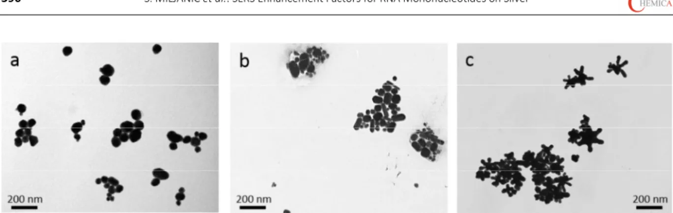
Croat. Chem. Acta2015, 88(4), 387–396 DOI: 10.5562/cca2720 the uniformly shaped silver nanospheres with the chloride
ions on their surface, more irregular forms and rods of 90100 nm length were observed in the colloid prepared by the citrate reduction method (Figure 1b). The star-shaped nanoparticles consisted of the central core and several (59), 4090 nm long arms, having an average diameter in the range of 100220 nm (Figure 1c). Except for a few mo-nodispersed nanoparticles, the majority of all the prepared silver nanoparticles formed aggregates.
In the UV/Vis/NIR extinction spectra of the colloidal suspensions a maximum around 400 nm, typical of the sil-ver plasmon resonance, was obtained (Figure 2). Regardless of the anionic coating, the spherically shaped nanoparticles, Ag_Cl nsp and Ag_cit nsp, gave rise to similar spectra with the maxima at 414 and 426 nm, respectively. Unlike the spectra of the silver nanospheres, however, in the UV/Vis/NIR spectrum of the silver nanostars, Ag_cit nst, a weak maximum at 389 nm was observed, accompanied by a strong extinction at longer wavelengths. The large extinction background was attributed to the various morp-hologies of the existing nanoparticles, which resulted from different number, shape and size of the star arms.[6]
All the prepared silver colloidal suspensions were of neutral pH value, i.e. the pH of 6.7, 6.8 and 7.9, was measu-red for Ag_Cl nsp, Ag_cit nsp and Ag_cit nst, respectively.
SERS Enhancement
The silver nanoparticles prepared by reduction methods with citrate and hydroxylamine hydrochloride have been widely used as the SERS active substrates and found very effective for the SERS measurement of various positively charged species.[3,4] The detection of the species carrying
a negative charge, however, has not been as successful due to the electrostatic repulsive interactions between the analyte and the stabilizing anionic layer, preventing an efficient adsorption of the analyte molecules onto the metallic surface. To overcome these drawbacks, an aggregating agent is usually added into the colloid,
neutralizing the surface charge on the one hand, and forming the nanoparticles aggregates with numerous “hot-spots” of extremely strong electric fields, on the other hand.
Aiming at the study of the surface enhancement of the negatively charged molecules, the RNA mononucleoti-des were chosen as the model molecules for several rea-sons. (i) All four mononucleotides had the same structural moiety responsible for the anionic character of the mole-cule, the phosphate group, but differed in the nucleic base involved in the adsorption of the biomolecule onto the me-tal surface. (ii) The adsorption mechanisms of the RNA mo-nonucleotides onto the citrate coated silver nanoparticles had been thoroughly studied and discussed in our previous work.[16] (iii) Since the mononucleotides are the building
blocks of the nucleic acids, knowing the Raman scattering enhancement of the structural elements attached to the va-rious SERS substrate would be beneficial in the future SERS measurements of biomacromolecules.
In order to compare enhancement efficiency of the studied silver nanoparticles for the SERS measurements of Figure 1. TEM micrographs of a) chloride coated silver nanospheres, Ag_Cl nsp, b) citrate coated silver nanospheres, Ag_cit nsp, and c) citrate coated silver nanostars, Ag_cit nst.
DOI: 10.5562/cca2720 Croat. Chem. Acta2015, 88(4), 387–396 the RNA mononucleotides, an analytical enhancement
fac-tor, EF, was calculated. It is usually used to evaluate an en-hancement of the Raman scattering obtained from an analyte adsorbed on a SERS active surface with respect to the normal Raman scattering of the same analyte in solu-tion.[14] The Raman and SERS measurements were
conduc-ted under the same experimental conditions, including laser wavelength, laser power, detector and spectrometer. The enhancement factor was calculated according to the following equation:
SERS SERS RS RS
I c
EF
I c
(1)
where I is the intensity of a band and c is the analyte con-centration in the solution (RS) or in the colloidal suspension (SERS), whereas subscripts RS and SERS refer to the Raman spectrum and SERS spectrum, respectively.
The Raman spectra of the RNA mononucleotides (1× 10⁻¹ mol/L) in aqueous solution were acquired and the main
vibrational bands tentatively assigned (Figure 3, Table 1). On first sight the obtained spectra reflected the complexity of the mononucleotides structure, being simpler in case of the pyrimidine mononucleotides, CMP and UMP, and more complex in case of the purine mononucleotides, AMP and GMP. Nevertheless, in all the Raman spectra dominated bands assigned to the ring modes of the nucleic bases, except for a band at 977 cm1 (978 cm1) common to the all
mononucleotides, which was attributed to the symmetrical stretching of the phosphate group.[17]
The SERS spectra of the mononucleotides were mea-sured in prepared silver colloids in the concentration range from 110−7 to110−4 mol/L. Since the Raman scattering
was poorly enhanced from the citrate stabilized silver na-nospheres and not observed at all from the silver nanostars, sodium sulfate was added into the colloidal samples to in-duce the nanoparticles aggregation, followed by the SERS enhancement. The representative SERS spectra of the RNA mononucleotides (1×10⁻⁵ mol/L) in the aggregated colloids
are shown in Figure 4, and the assignment of the main vib-rational bands is given in Table 1. Based on a detailed a-nalysis of the SERS spectra, adsorption mechanisms of the RNA mononucleotides on the silver nanoparticles had been thoroughly studied in our previous work and were discus-sed elsewhere.[16]
While selecting the appropriate band for the EF cal-culation, a distinctive, intense and isolated band was pre-ferred and as such observed in both, the Raman and SERS spectrum. Even though slightly overlapped by neighboring bands, particularly in the case of the guanine mononucleo-tide, the Raman bands at 728, 669, 782 and 783 cm1,
assi-gned to the aromatic ring breathing, were chosen for evaluating the Raman scattering enhancement of AMP, GMP, CMP and UMP, respectively. Given that involvement of the nucleic bases in the adsorption on the metal surface could affect the intensity of the studied bands, additional bands for the adenine (1338 cm1) and guanine (1322 cm1)
containing mononucleotides were also analyzed for compa-rison.
In the freshly prepared silver colloids, not containing the aggregating agent, the enhanced Raman scattering was observed for all the RNA mononucleotides on the chloride coated silver nanospheres, for AMP only on the citrate Figure 3. Raman spectra of the RNA mononucleotides
(1101 mol/L) in aqueous solution. Excitation at 785 nm. The spectra are displaced for visual clarity.
Croat. Chem. Acta2015, 88(4), 387–396 DOI: 10.5562/cca2720 Table 1. A tentative assignment of the main vibrational bands in the Raman spectra of the RNA mononucleotides in aqueous solution (1 101 mol/L) and in the SERS spectra of RNA mononucleotides on the aggregated silver colloids (1105 mol/L)
RNA mononucleotide
Wavenumber / cm−1
Assignment Raman Ag_Cl nsp Ag_cit nsp SERS Ag_cit nst
AMP
1580 w ring (6)
1509 w ring (6)
1482 w ring (6)
1458 w 1457 w 1454 w ring (6)
1377 w 1390 w 1384 w ring (C6N1)
1338 m 1329 m 1328 s 1332 s ring (5)
1306 m ring (5)
1251 w C6NH2
1218 vw ring (5)
1027 w 1029 w citrates
998 sh 1001 vw as CO (ribose)
977 m s PO32
951 w 948 w citrates
924 vw 924 w 921 w ring (6)
851 w 849 vw s CO (ribose)
728 s 740 s 738 s 741 s ring breathing
638 vw ring (6)
536 w 564 w 558 m 557 w ring (6)
237 vs AgCl
223 vs 225 vs AgN
GMP
1574 m 1579 w 1579w 1578 w ring (6)
1486 s 1508 w 1507 w 1506 w ring (6); ring (5)
1453 w 1472 w ring (5)
1414 w ring (6)
1365 m ip N1H; ring (6)
1322 m 1328 s 1326 s 1329 s ring (5)
1176 w ring (5)
1024 w 1023 w citrates
997 vw 1001 vw as CO (ribose)
977 m s PO32
952 w 951 w citrates
869 m 880 w 879 w ring (6)
815 w 812 m 801 m ring (6)
669 m 668 s 669 s 669 m ring breathing
584 w 609 w 606 w ring (6)
501 w 519 m 515 m 516 m ring (6)
234 vs AgCl
DOI: 10.5562/cca2720 Croat. Chem. Acta2015, 88(4), 387–396 coated silver nanospheres, while not obtained at all for any
of the mononucleotides on the silver nanostars. Studies on SERS measurement of the anionic species on the citrate re-duced silver nanoparticles implied that a competitive bin-ding between the stabilizing anions and the anionic analytes with the silver surface take place, when the anion with the higher affinity for silver replaces the other one.[15]
It was also found that chlorides bind to the silver surface more strongly than citrates,[15] indicating that the latter
should be displaced by the analyte molecule more easily than the former. Given the poor SERS spectrum of AMP in the citrate coated colloid and the very weak SERS spectra of
the RNA mononucleotides in the chloride coated colloid, the studied biomolecules had obviously a lower affinity for silver than both of the stabilizing surface anions. Another factor to consider was the surface coverage by the stabili-zing anionic species dependent on their concentration and molecular structure. According to the nanoparticles prepa-ration procedure, the chloride concentprepa-ration was 2.4×10⁻³
mol/L in the colloidal suspension, which was higher than those of the biomolecules. On the other hand, the concen-tration of citrates was not known due to their participation in the reduction process. It was very likely that the bulky citrate ions prevented effective adsorption of the mono-CMP
1294 w 1304 w 1295 m 1298 w ring (N3C4)
1243 m 1211 vw 1219 w 1219 w ring (N1C2N3)
1025 m 1028 w citrates
978 m s PO32
949 w 950 w citrates
871 vw 877 vw ring
782 s 790 m 787 s 787 m ring breathing
597 w 583 w 600 w ring (
C2
N3
C4
)237 vs AgCl
221 vs 219 vs AgN; AgO
UMP
1669 vw C=O; C5=C6
1624 vw 1624 vw C=O
1394 w ip N3H
1393 w 1390 w citrates
1229 s 1236 vw 1215 w ring (N1C2N3)
1029 w 1027 w citrates
978 m s PO32
950 w 949 w citrates
783 s 797 w 792 m 793 m ring breathing
645 vw ring
562 w 577 w 562 vw ring (C2N3C4)
237 vs AgCl
219 vs 223 vs AgN; AgO
Abbreviations: vs, very strong; s, strong; m, medium; w, weak; vw, very weak; sh, shoulder; , stretching; , deformation; s, symmetrical; as, antisymmetrical; ip, in plane.
Croat. Chem. Acta2015, 88(4), 387–396 DOI: 10.5562/cca2720 nucleotides onto the enhancing metal surface, hindering
their approach to the nanoparticles, while there was a larger surface available for the interaction with the mononucleotides on the chloride stabilized nano-spheres.[18]
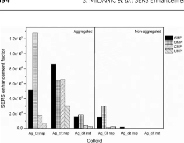
The enhancement factors calculated for the RNA mo-nonucleotides in the silver colloids (1×10⁻⁵ mol/L) with and
without the aggregating agent are compared in Figure 5. The EF values of the order of 104 were calculated for the
chloride coated silver colloid, in absence of the aggregating agent. However, the scattered radiation was 210 times en-hanced upon Ag_Cl nsp aggregation, showing a very similar trend with respect to the mononucleotides in the non-ag-gregated colloid. Interestingly, the SERS intensity of AMP in-creased as much as 40 times on the aggregated Ag_cit nsp, if compared to the colloid not containing the aggregating agent. Moreover, the Raman scattering of the other biomo-lecules on the aggregated citrated covered nanospheres was also obtained, which was for the pyrimidine mononuc-leotides, CMP and UMP, even more enhanced than on the aggregated chloride coated silver nanospheres. Consistent with the enhancement in the freshly prepared colloids, the weakest Raman scattering was observed from the aggrega-ted silver nanostars. In general, the RNA mononucleotides intensively scattered radiation in all the sodium sulfate tre-ated colloids, as a result of the nanoparticles aggregation. The addition of the aggregating agent surely affected the morphology of the nanoparticles as well as the chemical properties of the metallic surfaces, all contributing to the increased SERS activity. Owing to the formation of the lar-ger aggregates, the surface plasmon resonance shifted towards longer wavelengths, being easily excited with the used laser radiation (785 nm). While inducing the aggrega-tion of the silver nanoparticles, sodium caaggrega-tions most likely reduced the negative surface charge, leading to weaker re-pulsive forces between the silver nanoparticles and the
mononucleotides. Sulfate ions did not interfere with the measured biomolecules due to the low affinity of sulfates for the silver surface.[15]
Comparing the citrate coated nanoparticles of different shape, nanospheres and nanostars, a stronger enhancement had been expected from the star-shaped nanoparticles. A huge intensification of the electro-magnetic field on the nanostars was suggested, particularly in the spaces between the star arms.6 Besides, the large
extinction observed in the UV/Vis/NIR spectrum, implying the plasmon resonance in the NIR region, coincided with the wavelength of the laser excitation at 785 nm. Nevertheless, the maximal EF, calculated for instance for AMP (1×10⁻⁶ mol/L) on Ag_cit nanostars, was 3.4×10⁴,
which was almost one order of magnitude lower than the respective EF value in Ag_cit nanospheres, reaching 2.3× 10⁵ (Figure 6).
Considering all the studied mononucleotides, the stronger enhancement was observed for AMP and GMP, than for CMP and UMP, regardless of the silver colloid (Figure 6). This could be attributed to the larger Raman cross-section of the condensed five- and six-membered rings of the purine mononucleotides, when compared to the single ring system of the pyrimidine mononucleotides, as well as to a greater number of the functionalities in the molecular structure of the purine bases capable of binding with the silver surface. Additionally, it was interesting to note that the Raman scattering of GMP was the most enhanced from Ag_Cl nsp at all the measured GMP concentrations, reaching up the EF value of 5.8× 10⁶
(1×10⁻⁷ mol/L), while it was concentration dependent for
AMP, being more intense from Ag_cit nsp at higher AMP concentrations (1×10⁻⁵ and 1×10⁻⁴ mol/L), but
from Ag_Cl nsp at lower AMP concentrations (1×10⁻⁷
and 1 ×10⁻⁶ mol/L). A difference in the enhancement
factors, dependent on the molecular structure of the Figure 5. Comparison of the SERS enhancement factors for
the RNA mononucleotides (1105 mol/L) on the aggreg-ated and non-aggreagted silver colloids: Ag_Cl nsp, Ag_cit nsp and Ag_cit nst.
DOI: 10.5562/cca2720 Croat. Chem. Acta2015, 88(4), 387–396 tal surface, as sterically and electrostatically most
conveni-ent. Moreover, for the purine mononucleotides, AMP and GMP, adsorption through the larger aromatic ring was su-ggested, which was concentration dependent in case of AMP. Differing in applied excitation radiation, 1064 nm in our previous study and 785 nm in our current study, the ob-tained spectra did not fully coincide, as expected. Neverthe-less, the position and relative intensity of the strongest SERS bands indicated the proposed adsorption mechani-sms. Therefore, the binding with the surface of Ag_cit nst was mostly driven by interactions through the adenine N1 atom and NH2 group as well as via the guanine N1H and C=O
group. Looking closely at the SERS spectra of the purine mo-nonucleotides, obtained from Ag_Cl nsp and Ag_cit nsp, si-gnificant spectral changes were not observed (Figure 4). The position and relative intensity of the main vibrational bands in the SERS spectra obtained from different silver na-noparticles resembled each other, indicating very similar o-rientation of the molecules on both enhancing surfaces. Hence the differently enhanced scattering could be associ-ated with the distance of the molecules from the silver sur-face, whereby at concentrations of 1×10⁻⁵ and 1×10⁻⁴
mol/L AMP was placed closer to the citrate coated silver na-nospheres, and GMP to the chloride coated ones. The SERS spectra of the pyrimidine mononucleotides, CMP and UMP, were rather weak, though more enhanced from the silver nanospheres with the citrate ions on the surface (EF ≈ 5×10⁴)
than from the silver nanospheres with the surface layer of the chloride ions (EF ≈ 1×10⁴).
By decreasing the RNA mononucleotides concentra-tion, the enhancement factors increased (Figure 6). The de-pendence of the calculated EFs on the mononucleotide concentration was associated with the surface coverage and surface selection rules. Hence the intensity of the SERS spectra depended on the orientation of the molecules towards the metal surface, whereby vibrations with polari-zability change normal to the metal surface resulted in the strongest enhancement of the scattered radiation, and those parallel with the metal surface did not contribute to the scattering. At high concentration (1×10⁻⁴ mol/L) the
mononucleotide molecules were densely packed on the metal, resulting in a weaker enhancement due to either a slightly inclined position of the molecules on the metal surface or an ability to reabsorb scattered radiation from the neighboring molecules. Decreasing concentration a
lower than 1×10⁻⁵ mol/L were not obtained. The
vibratio-nal bands distinctive of the pyrimidine mononucleotides di-sappeared from the spectra measured in Ag_Cl nsp, and only the citrate bands contributed to the spectra acquired in the citrate covered colloids, Ag_cit nsp and Ag_cit nst. If compared to the Raman spectra of the aggregated citrate colloids (Figure 4), note that the weak citrates bands were also observed in the SERS spectra of the mononucleotides at the higher measured concentrations (1×10⁻⁵ and 1×10⁻⁴
mol/L), pointing to coexistence of the surface citrates and the biomolecules on the silver surface. Similarly, in the spectra measured on Ag_Cl nsp, a band around 237 cm1,
attributed to AgCl stretching vibration,19 indicated a
simul-taneous presence of the chloride ions on the silver nano-particles, which were not displaced by the mononucleotide molecules.
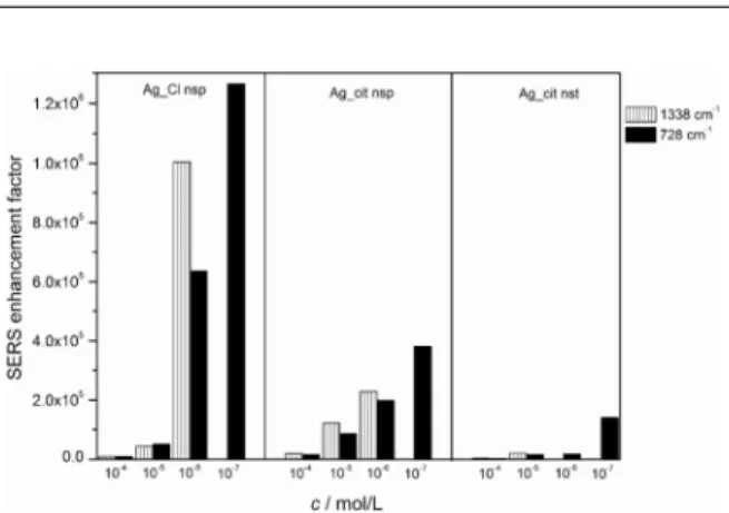
To study the influence of the selected band for the
EF calculation, the enhancement factors were evaluated u-sing two bands in the spectra of AMP and GMP. Beside the Raman bands at 728 and 669 cm1, assigned to the
six-membered ring breathing of the nucleic bases, the EFs were calculated using the intensity of the bands at 1338 and 1322 cm1, attributed to the stretching of the smaller,
five-mem-bered ring. In case of both, AMP and GMP, it was interesting to note that the slightly higher EF values were observed when calculated with the band originating from the part of the molecule, presumably not involved directly in interacti-ons with the silver surface (a column plot given only for AMP in Figure 7). However, a difference between the en-hancement factors was not significant, not exceeding the factor of 2. Hence the EFs evaluated using intensity of the ring stretching bands were 1.6 and 1.8 times higher for AMP and GMP, respectively, on Ag_Cl nsp, and 1.1 to 1.4 times on Ag_cit nsp. Due to the increase of the spectral backgro-und the intensity of the bands arobackgro-und 1330 cm–1 could not
be evaluated in the spectra measured at the lowest concen-tration of 1×10⁻⁷ mol/L.
CONCLUSION
Croat. Chem. Acta2015, 88(4), 387–396 DOI: 10.5562/cca2720 mononucleotides, pointing to suitability of the studied
silver colloids as the SERS active substrates. While a weak scattering was mostly observed from the non-aggregated silver nanoparticles, an addition of the inorganic salt into the silver colloids significantly increased their SERS activity. Hence in a series of the tested silver nano-particles, the Raman scattering of the mononucleotides was strongly enhanced from the chloride coated spheres, moderately from the citrate covered nano-spheres, and poorly from the citrate stabilized nanostars. Although characterized by plasmon resonances ranging from the blue to the near infrared region, the silver nanostars were the least effective metal substrates for the studied RNA mononucleotides. Apart from the silver nanoparticles properties, the enhancement factors dep-ended on the mononucleotide concentration and mol-ecular structure, which affected the adsorption, coverage and orientation of the molecules on the enhancing metal-lic surface. Nevertheless, the enhancement factors 105 to
106, calculated for the RNA mononucleotides on the
ag-gregated silver nanospheres, were comparable to those reported for the cationic species on silver nanoparticles, implying a successful application of the spherically shaped nanoparticles in the SERS measurements of the negatively charged biomacromolecules.
REFERENCES
[1] R. Aroca, Surface-Enhanced Vibrational Spectroscopy,
2006, John Wiley & Sons, Chichester.
[2] S. Schlücker, Surface-Enhanced Raman Spectroscopy: Analytical, Biophysical and Life Science Applications,
2011, Wiley-VCH, Weinheim.
[3] B. Sharma, R. R. Frontiera, A.-I. Henry, E. Ringe, R. P. Van Duyne, Mater. Today2012, 15, 16.
[4] R. F. Aroca, R. A. Alvarez-Puebla, N. Pieczonka, S. Sanchez-Cortez, J. V. Garcia-Ramos, Adv. Colloid. Interfac.2005, 116, 145.
[5] K. L. Kelly, E. Coronado, L. L. Zhao, G. C. Schatz, J. Phys. Chem. B 2003, 107, 668.
[6] A. Garcia-Leis, J. V. Garcia-Ramos, S. Sanchez-Cortez, J. Phys. Chem. C2013, 117, 7791.
[7] H. Bengter, C. Tengroth, S. P. Jacobsson, J. Raman Spectrosc.2005, 36, 1015.
[8] L. Mikac, M. Ivanda, M. Gotić, T. Mihelj, L. Horvat,
J. Nanopart. Res.2014, 16, 2748.
[9] P. C. Lee, D. Meisel, J. Phys. Chem.1982, 86, 3391. [10] C. H. Munro, W. E. Smith, M. Garner, J. Clarkson, P.
C. White, Langmuir1995, 11, 3712.
[11] N. Leoplod, B. Lendl, J. Phys. Chem. B2003, 107, 5723.
[12] E. Papadopoulou, S. E. J. Bell, Angew. Chem. Int. Ed. 2011, 50, 9058.
[13] E. Papadopoulou, S. E. J. Bell, Chem. Eur. J.2012,
18, 5394.
[14] E. C. Le Ru, E. Blackie, M. Meyer, P. G. Etchegoin, J. Phys. Chem. C2007, 111, 13794.
[15] S. E. J. Bell, N. M. S. Sirimuthu, J. Phys. Chem. A 2005, 109, 7405.
[16] S. Miljanić, A. Dijanošić, I. Matić, Spectrochim. Acta A2015, 137, 1357.
[17] K. Nakamoto, M. Tsuboi, G. D. Strahan, Drug-DNA Interactions: Structures and Spectra, 2008, John Wiley & Sons, New Jersey.
[18] M. V. Canamares, J. V. Garcia-Ramos, C. Sanchez-Cortes, M. Castillejo, M. Oujja, J. Colloid Interf. Sci. 2008, 326, 103.
[19] E. J. Liang, C. Engert, W. Kiefer, Vib. Spectrosc. 1995, 8, 435.