Geraniol, Alone and in Combination with
Pioglitazone, Ameliorates Fructose-Induced
Metabolic Syndrome in Rats via the
Modulation of Both Inflammatory and
Oxidative Stress Status
Sherehan M. Ibrahim, Ezzedin S. El- Denshary, Dalaal M. Abdallah*
Pharmacology and Toxicology Department, Faculty of Pharmacy, Cairo University, Cairo, Egypt
Abstract
Geraniol (GO) potent antitumor and chemopreventive effects are attributed to its antioxidant and anti-inflammatory properties. In the current study, the potential efficacy of GO (250 mg/kg) in ameliorating metabolic syndrome (MetS) induced by fructose in drinking water was elucidat-ed. Moreover, the effect of pioglitazone (5 and 10 mg/kg; PIO) and the possible interaction of the co-treatment of GO with PIO5 were studied in the MetS model. After 4 weeks of treatment, GO and/or PIO reduced the fasting blood glucose and the glycemic excursion in the intraperi-toneal glucose tolerance test. GO and PIO5/10 restrained visceral adiposity and partly the body weight gain. The decreased level of peroxisome proliferator activated receptor (PPAR)-γ
transcriptional activity in the visceral adipose tissue of MetS rats was increased by single treat-ment regimens. Though GO did not affect MetS-induced hyperinsulinemia, PIO5/10 lowered it. Additionally, GO and PIO5/10 suppressed glycated hemoglobin and the receptor for ad-vanced glycated end products (RAGE). These single regimens also ameliorated hyperurice-mia, the disrupted lipid profile, and the elevated systolic blood pressure evoked by MetS. The rise in serum transaminases, interleukin-1β, and tumor necrosis factor-α, as well as hepatic lipid peroxides and nitric oxide (NO) was lowered by the single treatments to different extents. Moreover, hepatic non-protein thiols, as well as serum NO and adiponectin were enhanced by single regimens. Similar effects were reached by the combination of GO with PIO5; however, a potentiative interaction was noted on fasting serum insulin level, while synergistic effects were reflected as improved insulin sensitivity, as well as reduced RAGE and triglycerides. Therefore, GO via the transcriptional activation of PPAR-γreduces inflammation and free radi-cal injury produced by MetS. Thereby, these effects provide novel mechanistic insights on GO management of MetS associated critical risk factors. Moreover, the co-administration of GO to PIO5 exalted the antidiabetic drug anti-MetS efficacy.
OPEN ACCESS
Citation:Ibrahim SM, El- Denshary ES, Abdallah DM (2015) Geraniol, Alone and in Combination with Pioglitazone, Ameliorates Fructose-Induced Metabolic Syndrome in Rats via the Modulation of Both Inflammatory and Oxidative Stress Status. PLoS ONE 10(2): e0117516. doi:10.1371/journal. pone.0117516
Academic Editor:Marta Letizia Hribal, University of Catanzaro Magna Graecia, ITALY
Received:July 26, 2014
Accepted:December 26, 2014
Published:February 13, 2015
Copyright:© 2015 Ibrahim et al. This is an open access article distributed under the terms of the
Creative Commons Attribution License, which permits unrestricted use, distribution, and reproduction in any medium, provided the original author and source are credited.
Data Availability Statement:The authors confirm that all data underlying the findings are fully available without restriction. All relevant data are within the paper.
Funding:The authors have no funding or support to report.
Introduction
With the global adoption of the westernized diet and increased consumption of fructose, both metabolic syndrome (MetS) and type 2 diabetes mellitus reached epidemic proportions world-wide [1,2]. A strong correlation exists between MetS and cardiovascular disease metabolic risk factors [3]. This cluster includes elevated plasma glucose and blood pressure, atherogenic dysli-pidemia, as well as central obesity [4]. In general, MetS is characterized by a pro-oxidant/proin-flammatory status that predisposes patients to major cardiovascular events [5,6]. Evidence exists that both hyperuricemia and atherogenic dyslipidemia are contributing factors in high blood pressure to augment insulin resistance [7–9].
Geraniol (GO; 3,7-dimethyl-2,6 octadien-1-ol) is a monoterpene alcohol naturally found in the essential oil of rose and lemon [10]. In rats, GO is rapidly absorbed to reach peak plasma concentration after 34 min [11]. GO is primarily concentrated in the small intestine and its me-tabolites, namely, geranic, 3-hydroxy-citronellic, and hildebrandt acids, as well as 8-hydroxy-and 8-carboxy-geraniol are eliminated in the urine [11,12]. GO possesses antioxidant activity and suppresses nitroactive stress [13,14]. This acyclic monoterpene alcohol is a potent anti-inflammatory compound that inhibits prostaglandin E2and tumor necrosis factor (TNF)-α [14–16]. Besides, GO anticancer efficacy is related to the inhibition of HMG-CoA reductase [17]. The latter action also verifies GO antiatherogenic effect, noticed recently by Jayachandran et al. [18] in hamsters.
The main goal of the present work was to verify the potential anti-MetS property of GO tar-geting its modulatory role on the inflammatory and free radical processes involved in MetS using an insulin resistant rat model induced by fructose in drinking water. Since a previous in vitro study [19] reported that GO activates several peroxisome proliferator-activated receptor (PPAR) subtypes, therefore, the aim of the present investigation was extended to validate the in vivo action of GO on the PPAR-γtranscriptional activity in the visceral adipose tissue of MetS rats. Moreover, thiazolidinediones (TZDs), such as pioglitazone (PIO), modulate specific facets of the MetS [20,21] via enhanced transcriptional activation of PPAR-γto mediate their antihy-perglycemic, antihypertensive, antihyperlipidemic, antioxidant, and anti-inflammatory proper-ties [22–26]. Therefore, the present study also assessed the possibility of a beneficial interaction between GO and PIO to reduce the TZD dose level to avoid long term undesirable effects.
Material and Methods
Ethics Statement
Experimental protocols, as well as animal handling were approved by the Research Ethical Committee of Faculty of Pharmacy, Cairo University (Cairo, Egypt) and comply with the Guide for the Care and Use of Laboratory Animals.
Animals
Induction of MetS and Experimental Design
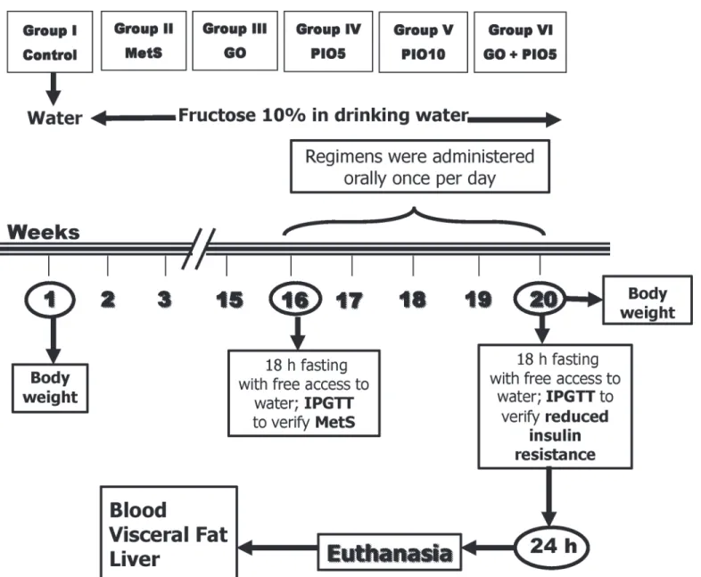
At the beginning of experimentation, rats were divided into two experimental subsets (Fig. 1). Animals had either free access to tap water (control group; n = 8 rats) or were allowed free ac-cess to freshly prepared 10% (w/v) fructose solution in tap water for 16 weeks to induce MetS [27]. Subsequently, MetS was verified by monitoring body weight changes, fasting blood glu-cose and insulin, serum triglycerides (TGs), McAuley index, and systolic blood pressure [28,29] (data not shown). Rats in both experimental subsets were exposed to the intraperitoneal
Fig 1. Adult male Wistar rats (45 days; 170±20 g) were randomly divided into 2 experimental subsets, namely, control group (free access to water for 16 weeks then vehicle for 4 weeks) or metabolic syndrome (MetS; 10% fructose solution in drinking water for 16 weeks) subset.After 16 weeks, all animals were exposed to the intraperitoneal glucose tolerance test (IPGTT) after an overnight fast; and MetS was verified in the fructose fed rats. In the MetS experimental subset, rats were subdivided into MetS (1% Tween 80; 4 weeks; p.o), geraniol (GO; 250 mg/kg/day; 4 weeks; p.o); pioglitazone (PIO; 5 and 10 mg/kg/day; 4 weeks; p.o), and GO/PIO5 groups. These animals were further supplied with 10% fructose solution in drinking water for 4 weeks. Insulin resistance was tested using IPGTT 24 h after last treatments or vehicle administration and after an overnight fast in all groups. Body weight was recorded at baseline and after 20 weeks of the beginning of the experiments. Blood was collected and then rats were euthanized to dissect the liver and visceral fat for different assessments.
glucose tolerance test (IPGTT) at week 16 (data not shown), as well as week 20. Thereafter, the MetS animals were randomly divided into 5 groups (n = 6–8 rats, each;Fig. 1) and further sup-plied for 4 weeks with the fructose solution in drinking water. In the 1stgroup, fructose fed rats were daily administered 1% Tween 80 to serve as the MetS group. The animals in the other ex-perimental groups received GO (250 mg/kg/day; p.o; Sigma-Aldrich, Saint Louis MO, USA) [30], PIO (5 and 10 mg/kg/day; p.o; MUP Company, Cairo, Egypt) in 1% Tween 80 [31], or a combination of GO with PIO5 for 4 weeks. Meanwhile, the control animals were administered the vehicle for 4 weeks. In all animals, 24 h after the last treatment or vehicle administration and subsequent to an overnight fast, IPGTT was performed at week 20. Thereafter, animals were weighed and blood was collected for plasma and serum separation; then rats were eutha-nized. In the plasma, receptor for advanced glycated end product (RAGE) and glycated haemo-globin (HbA1c) were estimated, while liver function tests, glucose homeostasis parameters, lipid profile, nitric oxide (NO), and the cytokines were verified in the serum. From each rat, the liver was dissected, blotted dry, weighed, and homogenized in ice-cold saline for the estimation of lipid peroxidation, non-protein thiols (NPSH), and NO. The visceral adipose tissue was sep-arated, weighed, and used for the nuclear extraction of PPAR-γto determine its
transcriptional activity.
Intraperitoneal Glucose Tolerance Test
IPGTT was performed at week 16 (data not shown) and week 20 using the Accu-Check gluc-ometer monitor and strips (Roche Diagnostics, West Sussex, UK) [32]. After an overnight fast, baseline blood glucose (time 0) was determined; then a glucose solution (25%; 2 g/kg, i.p) was injected, followed by the determination of blood glucose levels at 30, 60, and 120 min. The area under the curve of glucose was calculated [33].
Assessment of Body Weight Gain and Visceral Adiposity
For each rat, the body weight at the beginning and end of the experimental period was recorded and the body weight gain was determined. Moreover, the visceral fat weight was estimated at the end of the experimental period. The visceral index that signifies visceral adiposity was cal-culated as the ratio of visceral fat weight to body weight [34].
Assessment of Fasting Blood Glucose and Insulin, HOMA-IR, QUICKI,
McAuley Index, HbA1c, and RAGE
Fasting blood glucose and insulin were determined in the sera using glucose enzymatic kit (Biodiagnostic, Giza, Egypt) and rat insulin ELISA kit (ALPCO, New Hampshire, USA), re-spectively. The homeostatic model assessment-insulin resistance (HOMA-IR) and the quanti-tative insulin sensitivity check index (QUICKI) were calculated depending on fasting blood glucose and insulin levels [35,36], whereas McAuley index verification is based on fasting insu-lin and TGs levels [29]. In the plasma, HbA1c was measured using rat HbA1c ELISA kit (Mybiosource, California, USA), while RAGE was determined using rat RAGE ELISA kit (Ray Bio, Georgia, USA). Procedures were performed according to the manufacturers’instructions.
Assessment of Lipid Profile
Assessment of PPAR-
γ
Transcriptional Activity in Adipose Tissue
Procedures adapted for the adipose PPAR-γnuclear extraction and its transcriptional activity followed the provided manufacturer protocols (Abcam, Cambridge, USA). A primary poly-clonal anti-PPAR-γantibody was used for PPAR-γassessment. After addition of the horserad-ish peroxidase-conjugated antibody and the 3,3’,5,5’–tetramethylbenzidine substrate, the absorbance of the developed color was read at 450 nm using a microplate reader.Systolic Blood Pressure Assessment
Systolic blood pressure was measured by the tail cuff technique using AD instrument (MLT125R, NSW, Australia).
Serum Uric Acid Assessment
Serum uric acid was measured using colorimetric enzymatic kit (Biodiagnostic, Giza, Egypt). The experimental procedure was performed according to the manufacturer's instruction.
Assessment of Serum IL-1
β
, TNF-
α
, and Adiponectin
The proinflammatory cytokines interleukin (IL)-1βand TNF-α, as well as adiponectin were de-termined using ELISA kit (R&D, MN, USA). All procedures were performed according to the manufacturer’s instructions.
Liver Function Tests
Serum alanine aminotransferase (ALT) and aspartate aminotransferase (AST) were assessed using colorimetric enzymatic kits (Biodiagnostic, Giza, Egypt) according to the
manufacturer’s instructions.
Assessment of Hepatic Lipid Peroxides and NPSH
Liver thiobarbituric acid reactive substances were determined according to the method described by Mihara and Uchiyama [37]. o-Phosphoric acid (1%) and freshly prepared thiobarbituric acid (0.6%) solutions were added to liver homogenates. The mixture was boiled for 45 min and n-bu-tanol was added after cooling to extract the pink colored chromophore. In addition, liver NPSH was assessed using colorimetric kit purchased from Biodiagnostic (Giza, Egypt) based on the method described by Ellman [38].
Assessment of Hepatic and Serum NO
NO was measured according to the method of Miranda et al. [39]. Serum and liver homogenate were deproteinated with cold absolute ethanol (48 h; 4°C); and vanadium trichloride was used for the reduction of nitrate into nitrite, followed by the rapid addition of Griess reagent.
Statistical Analysis
Results
To detect the possible interaction between GO and PIO, the results of MetS, GO, PIO5, and GO/PIO5 groups were statistically analyzed using factorial design. A synergistic interaction oc-curs when the combined effect is greater than the sum of the effects of the two individual treat-ments. If the effect of one compound is greatly increased by the intake of the other that processes a diminished effect, the consequent interaction is described as potentiation. Nonethe-less, when the combined effect does not reach a super-additive interaction it is considered statistically insignificant.
Effect of GO, PIO, and GO/PIO5 on Glucose Homeostasis, HbA1c, and
RAGE in Fructose-induced MetS in Rats
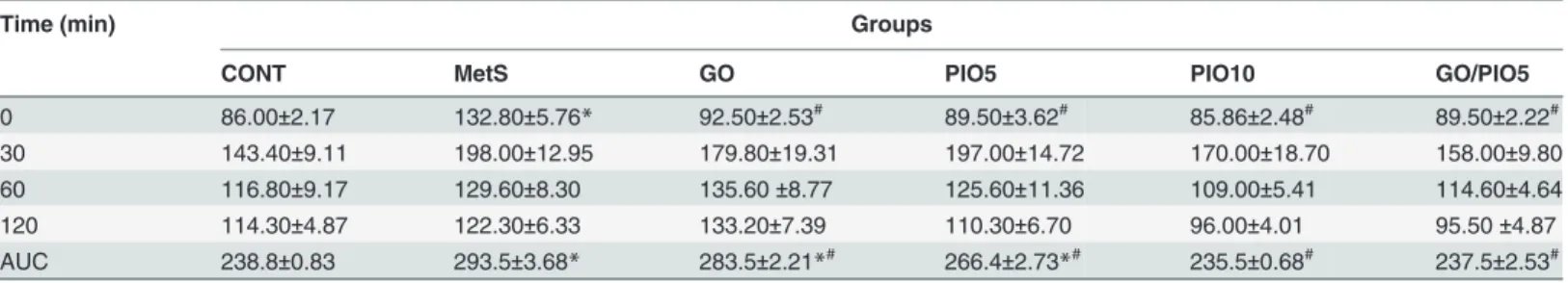
Compared to the control, a defective clearance of the intraperitoneally injected glucose load, as well as increased fasting blood glucose and insulin levels were observed in fructose fed rats (Tables1and2, respectively). Rats in GO and/or PIO groups achieved appropriate glycemic control noted by the reduced glycemic excursion in IPGTT, area under the curve of glucose, and hyperglycemia (Tables1and2). Tough both PIO dose levels decreased fasting insulin, GO did not affect it (Table 2). In the MetS rat model induced by fructose, an increment in HOMA-IR (800%) versus reductions in both QUICK (73%) and McAuley indices (53%) were observed, effects that were controlled to different extents by all treatment regimens (Table 2). GO in the combination regimen showed a potentiative interaction on the fasting insulin level that syner-gistically enhanced the McAuley insulin sensitivity index of PIO5 (Table 2). Fructose in drink-ing water increased HbA1c (2 fold;Fig. 2A) and RAGE (8.5 fold;Fig. 2B) compared to the control rats. Animals treated with GO, PIO5/10, and the combination decreased HbA1c (73, 69, 52, and 83%, respectively;Fig. 2A) and RAGE (58, 44, 13, and 10%, respectively;Fig. 2B), as compared to the fructose fed rats. The combination regimen showed a synergistic interaction on RAGE (Fig. 2B).
Effect of GO, PIO, and GO/PIO5 on Body Weight Gain, Visceral
Adiposity, Adipose PPAR-
γ
Transcriptional Activity, and Lipid Profile in
Fructose-induced MetS in Rats
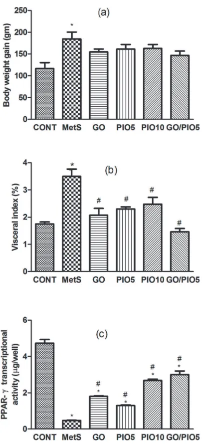
In the fructose fed rats, the body weight gain (158%;Fig. 3A) and the visceral adiposity (200%; Fig. 3B) were increased compared to control animals. Such gross changes were associated by a
Table 1. Effect of 4 weeks daily oral administration of geraniol (GO; 250 mg/kg), pioglitazone (PIO; 5 and 10 mg/kg), and GO with PIO5 on intraperitoneal glucose tolerance test (IPGTT) in fructose-induced metabolic syndrome (MetS) in rats.
Time (min) Groups
CONT MetS GO PIO5 PIO10 GO/PIO5
0 86.00±2.17 132.80±5.76* 92.50±2.53# 89.50±3.62# 85.86±2.48# 89.50±2.22# 30 143.40±9.11 198.00±12.95 179.80±19.31 197.00±14.72 170.00±18.70 158.00±9.80
60 116.80±9.17 129.60±8.30 135.60±8.77 125.60±11.36 109.00±5.41 114.60±4.64
120 114.30±4.87 122.30±6.33 133.20±7.39 110.30±6.70 96.00±4.01 95.50±4.87
AUC 238.8±0.83 293.5±3.68* 283.5±2.21*# 266.4±2.73*# 235.5±0.68# 237.5±2.53#
The fasting blood glucose titer (mg/dl) at different time intervals (0, 30, 60, and 120 min) after intraperitoneal glucose (2 g/kg) administration is presented as mean (6–8 animals)±SEM, while the calculated area under the curve (AUC) of glucose is presented.P<0.05, as compared to control (CONT) (*) and
MetS (#) groups using one-way ANOVA followed by Tukey
–Kramer test.
reduced transcriptional activity of PPAR-γ(10%) in the adipose tissue of fructose fed rats (Fig. 3C). In all treated groups, the diminished visceral adiposity (Fig. 3B) was associated by partial decreases in body weight gain (Fig. 3A). Animals receiving GO (383%), PIO5 (274%), PIO10 (576%), and GO/PIO5 (638%) enhanced the adipose PPAR-γtranscriptional activity (Fig. 3C). Concerning the lipid profile in the fructose fed rats, elevated TC (278%), TGs (175%), VLDL-TG (175%), and LDL-C (751%), as well as decreased HDL-C (82%) were noted (Table 3). The administration of GO and/or PIO modulated the deranged lipid profile markers in the serum compared to the non-treated rats (Table 3). A synergistic interaction on TGs was ob-served when GO was combined with the low dose level of PIO (Table 3).
Effect of GO, PIO, and GO/PIO5 on Systolic Blood Pressure and Serum
Uric Acid in Fructose-induced MetS in Rats
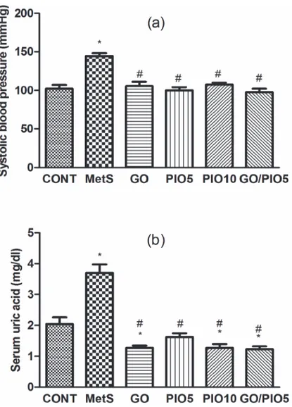
At the end of the experiment, systolic blood pressure in fructose fed rats recorded an increase to about 142%, as compared to the control rats (Fig. 4A). Animals treated with GO, PIO5/10, and the combined GO/PIO5 regimens normalized systolic blood pressure (Fig. 4A). Serum uric acid level was increased in the MetS model (181%;Fig. 4B), as compared to the control an-imals. The treatment regimens decreased uric acid level between 33 and 44%, as compared to fructose fed rats (Fig. 4B).
Effect of GO, PIO, and GO/PIO5 on Liver Function tests in
Fructose-induced MetS in Rats
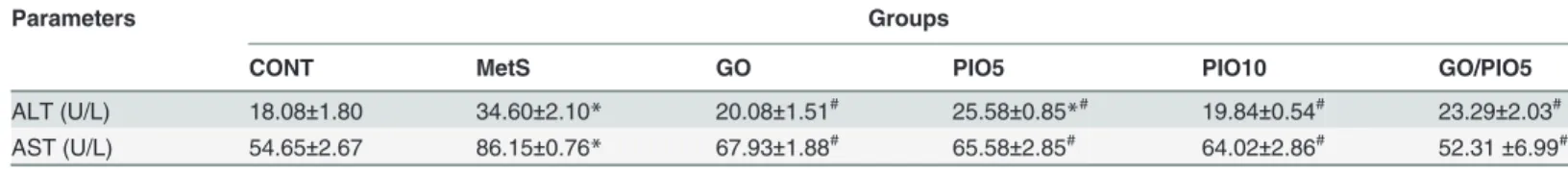
Liver enzymes, viz., ALT (2 fold) and AST (1.5 fold) were elevated in animals ingesting fruc-tose, as compared to their control counterparts (Table 4). Rats receiving GO and/or PIO low-ered these values compared to the fructose fed rats (Table 4).
Effect of GO, PIO, and GO/PIO5 on Serum IL-1
β
, TNF-
α
, and
Adiponectin in Fructose-induced MetS in Rats
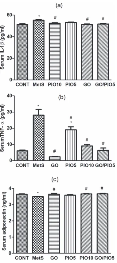
In fructose fed rats, MetS-induced inflammation manifest as elevations in IL-1β(108%; Fig. 5A) and TNF-α(462%;Fig. 5B) accompanied by the reduction in adiponectin (96%; Fig. 5C), as compared to control rats. GO and/or the TZD reduced IL-1β(Fig. 5A) and TNF-α (Fig. 5B) levels in the serum; however, PIO5 treatment did not affect IL-1β(Fig. 5A).
Table 2. Effect of 4 weeks daily oral administration of geraniol (GO; 250 mg/kg), pioglitazone (PIO; 5 and 10 mg/kg), and GO with PIO5 on plasma serum fasting blood glucose and insulin levels, as well as homeostatic model assessment-insulin resistance (HOMA-IR), McAuley index, and quantitative insulin sensitivity check index (QUICKI) in fructose-induced metabolic syndrome (MetS) in rats.
Parameters Groups
CONT MetS GO PIO5 PIO10 GO/PIO5
Fasting Blood Glucose (mg/dl) 89.63±2.47 142.00±7.10* 95.00±3.06# 84.00±1.95# 91.56±2.89# 88.38±1.86# Fasting Insulin (μIU/ml) 5.94±0.30 31.00±0.94* 29.25±0.34* 13.16±0.23*# 9.26±0.24*# 1.90±0.11*#° HOMA-IR 1.31±0.07 10.50±0.57* 6.80±0. 23*# 2.73±0.05
*# 2.09±0.05# 0.41±0.02# McAuley Index 10.31±0.18 5.44±0.01* 5.87±0.01*# 7.56±0.03*# 8.34±0.04*# 13.87±0.02*#°
QUICKI 0.37±0.00 0.27±0.00* 0.29±0.00*# 0.33±0.00*# 0.34±0.00*# 0.45±0.01*#
Values are mean (6–8 animals)±SEM. P<0.05, as compared to control (CONT) (*) and MetS (#) groups using one-way ANOVA followed by Tukey–
Kramer test. Significant synergistic interaction (°) when GO and PIO5 were combined using the factorial design test.
Meanwhile, all treatment regimens except PIO5 elevated serum adiponectin level compared to fructose fed rats (Fig. 5C).
Effect of GO, PIO, and GO/PIO5 on Hepatic and/or Serum Oxidative and
Nitroactive Stress in Fructose-induced MetS in Rats
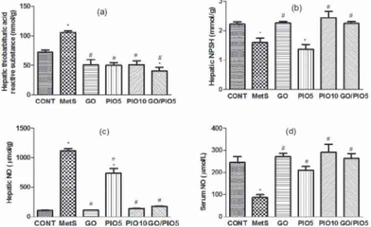
In fructose fed rats, hepatic thiobarbituric acid reactive substances reached 147% (Fig. 6A), while NPSH declined to 72% (Fig. 6B), as compared with control animals. Successfully, animals treated with GO, PIO10, and the combination prevented such alterations to different extents, as compared to the untreated rats (Figs. 6A and B). Unlike the elevated hepatic nitroactive radi-cal (1071%;Fig. 6C), the serum NO level was reduced (35%;Fig. 6D) in the fructose fed rats, as compared to the control rats. These actions were averted in GO and/or PIO-treated rats, as compared to the fructose fed rats (Figs. 6C and D, respectively).
Fig 2. Effect of 4 weeks daily oral administration of geraniol (GO; 250 mg/kg), pioglitazone (PIO; 5 and10 mg/kg), and GO with PIO5 on plasma (a) glycated haemoglobin (HbA1c) and (b) receptor for advanced glycated end product (RAGE) in fructose-induced metabolic syndrome (MetS) in rats.
Values are mean (6–8 animals)±SEM.P<0.05, as compared to control (CONT) (*) and MetS (#) groups
using one-way ANOVA followed by Tukey–Kramer test. Significant potentiative and synergistic interactions
(o) when GO and PIO5 were combined using the factorial design test.
Fig 3. Effect of 4 weeks daily oral administration of geraniol (GO; 250 mg/kg), pioglitazone (PIO; 5 and10 mg/kg), and GO with PIO5 on (a) body weight gain, (b) visceral index, and (c) adipose PPAR-γ
transcriptional activity in fructose-induced metabolic syndrome (MetS) in rats.Values are mean (6–8
animals)±SEM.P<0.05, as compared to control (CONT) (*) and MetS (#) groups using one-way ANOVA
followed by Tukey–Kramer test.
Table 3. Effect of 4 weeks daily oral administration of geraniol (GO; 250 mg/kg), pioglitazone (PIO; 5 and 10 mg/kg), and GO with PIO5 on serum total cholesterol (TC), triglycerides (TGs), very low density lipoprotein-triglycerides (VLDL-TG), low density lipoprotein-cholesterol (LDL-C), and high density lipoprotein-cholesterol (HDL-C) in fructose-induced metabolic syndrome (MetS) in rats.
Parameters Groups
CONT MetS GO PIO5 PIO10 GO/PIO5
TC (mg/dl) 82.93±8.83 230.60±9.24* 106.30±7.43# 140.20±7.11
*# 100.20±11.08# 120.80±9.93# TGs (mg/dl) 46.41±3.01 81.18±6.93* 65.01±2.42*# 60.76±4.36# 50.97±1.48# 49.33±3.35
*#° VLDL-TG (mg/dl) 9.45±0.46 16.38±1.40* 13.57±0.46*# 12.31±0.87# 10.22±0.29# 9.87±0.67# LDL-C (mg/dl) 20.70±0.20 115.20±10.52* 30.55±4.07# 55.97±2.50*# 33.44±4.60# 34.53±3.81#
HDL-C (mg/dl) 78.36±0.86 64.56±0.68* 83.86±2.23# 84.33±4.45# 82.63±0.51# 78.13±1.83#
Values are mean (6–8 animals)±SEM. P<0.05, as compared to control (CONT) (*) and MetS (#) groups using one-way ANOVA followed by Tukey–
Kramer test. Significant synergistic interaction (°) when GO and PIO5 were combined using factorial design test.
doi:10.1371/journal.pone.0117516.t003
Fig 4. Effect of 4 weeks daily oral administration of geraniol (GO; 250 mg/kg), pioglitazone (PIO; 5 and10 mg/kg), and GO with PIO5 on (a) systolic blood pressure and (b) serum uric acid level in fructose-induced metabolic syndrome (MetS) in rats.Values are mean (6–8 animals)±SEM.P<0.05, as
compared to control (CONT) (*) and MetS (#) groups using one-way ANOVA followed by Tukey
–Kramer test.
Discussion
This is the first study that shows the positive effect of GO in treating MetS and its accompanied cardiometabolic risk factors. GO ameliorated fructose-induced obesity, dyslipidemia, hyper-tension, hyperuricemia, and hyperglycemia. It also improved insulin sensitivity, noted by the reduction of HOMA-IR index, with the concomitant enhancement of QUICK and McAuley indices with a better response to IPGTT. Furthermore, the monoterpene alcohol suppressed the fructose-mediated increase in HbA1c and RAGE, as well as oxidative/nitroactive stress. Moreover, GO modulated the assessed adipokines to reduce inflammation. The administration of GO up-regulated the transcription of PPAR-γsupporting, thus, a previous in vitro study [19]. Wang et al. [40] verified that the activation of PPAR-γsuppresses the ligand-RAGE acti-vation of nuclear factor (NF)-κB and, hence, its downstream events. Following chronic hyper-glycemia, several RAGE ligands are formed including circulating advanced glycated end products and TNF-α[41,42]. In a feed forward cycle, the ligand-RAGE signaling results in the up-regulation of RAGE along with the proinflammatory mediators, viz., IL-1βand TNF-αto promote the development of insulin resistance by disturbing insulin signaling [42–44]. The ligand-RAGE induced inflammatory cascade deteriorates also endothelial function [41] to par-ticipate in MetS associated hypertension, reported herein. Additionally, the current hyperglyce-mia resulted in the elevation of the long term glycemic control index HbA1c [45]. Under a long-standing hyperglycemic condition, advanced glycated end products are formed that have been linked to the MetS mediated hypertension [46–48]. The present improved insulin sensi-tivity and antihypertensive action of GO are consequences of reduced hyperglycemia and PPAR-γactivation to suppress proinflammatory mediators, RAGE, and HbA1c. The anti-in-flammatory action of the monoterpene alcohol supports another experimental study [16].
In acquiesce with the findings of Singh et al. [49] GO decreased both of AST and ALT activi-ties, indicating the overall suppression of the inflammatory process associated with fructose in-gestion. The inhibition of ALT may be attributed to the suppression of TNF-α, up-regulation of PPAR-γ, and/or via increasing adiponectin, all of which participate in improving insulin signaling [34,50]. Adiponectin is another adipocytokine that increases peripheral glucose up-take and utilization [51,52]. It acts in contrast to TNF-α, where Liang et al. [53] displayed that this proinflammatory cytokine down-regulates adiponectin expression. In the current work, GO increased adiponectin to add to its insulin sensitizing effects. Adiponectin enhances the in-sulin receptor tyrosine phosphorylation and activates inin-sulin receptor substrate-1-mediated phosphatidylinositol-3 kinase [51,54]. GO-mediated elevation of adiponectin is partly linked to the activation of the nuclear receptor PPAR-γ, where the adiponectin promoter contains a functional PPAR element to which PPARγ/ RXR heterodimer binds [51].
Hepatic NO content was elevated in the fructose model, possibly due to the increased syn-thesis of inducible nitric oxide synthase activated by NF-κB [55,56]. In addition, the
Table 4. Effect of 4 weeks daily oral administration of geraniol (GO; 250 mg/kg), pioglitazone (PIO; 5 and 10 mg/kg), and GO with PIO5 on serum alanine amino transferase (ALT) and aspartate amino transferase (AST) levels in fructose-induced metabolic syndrome (MetS) in rats.
Parameters Groups
CONT MetS GO PIO5 PIO10 GO/PIO5
ALT (U/L) 18.08±1.80 34.60±2.10* 20.08±1.51# 25.58±0.85
*# 19.84±0.54# 23.29±2.03# AST (U/L) 54.65±2.67 86.15±0.76* 67.93±1.88# 65.58±2.85# 64.02±2.86# 52.31±6.99#
Values are mean (6–8 animals)±SEM. P<0.05, as compared to control (CONT) (*) and MetS (#) groups using one-way ANOVA followed by Tukey–
Kramer test.
Fig 5. Effect of 4 weeks daily oral administration of geraniol (GO; 250 mg/kg), pioglitazone (PIO; 5 and10 mg/kg), and GO with PIO5 on serum (a) interleukin (IL)-1β, (b) tumor necrosis factor (TNF)-α, and (c) adiponectin levels in fructose-induced metabolic syndrome (MetS) in rats. Values are mean (6–8 animals)±SEM.P<0.05, as compared to control (CONT) (*) and MetS (#) groups using one-way
ANOVA followed by Tukey–Kramer test.
imbalanced redox system was witnessed here by the increased lipid peroxidation and the de-creased NPSH. The ligand-RAGE interaction plays a role in the production of oxidative stress via the up-regulation of NF-κB with the subsequent activation of NADPH oxidase to impair endo-thelial function triggering hypertension [42]. GO, in this study, suppressed hepatic NO and lipid peroxides and enriched NPSH via the activation of both glutathione peroxidase and reductase en-zymes [49]; these results mimic those of previous studies in other models [14,49,57].
In a hyperinsulinemic state, as shown in our work in the MetS model, hyperuricemia results primarily from impaired renal excretion of uric acid that directly promotes oxidative stress and hypertension [58,59]. Moreover, serum NO was reduced in the diabetic rats, an effect linked to the elevated serum uric acid level [60]. Therefore, the reduced hyperuricemia in the GO-treated group may be behind the decreased oxidative stress and elevated serum NO that causes vasodi-lation [61] to decrease blood pressure.
Hyperglycemia, documented in fructose fed rats in our work, increases glucose flux through the hexosamine pathway playing a pivotal role in insulin resistance induction and is, in part, re-sponsible for the currently observed dyslipidemia [62,63]. As a feature of insulin resistance, white adipose tissue, viz., visceral fat causes chronic inflammation due to macrophage infiltration and/ or the release of TNF-α[64]. Since adiponectin enhancesβ-oxidation [52], therefore, a shortfall in its level is linked to visceral adiposity and increased body weight gain [65], as noted in the current study in the fructose fed rats. Both effects were opposed in the GO-treated group to different extents. Its negative influence on body weight gain can be clarified by the partial activation of PPAR-α[19], while the reduced visceral fat content results from a dual PPAR-α/-γeffect [19,66].
The activation of PPAR nuclear receptors by GO may explain partly the antihypertriglyceri-demic effect of the monoterpene alcohol, since they regulate the expression of target genes in-volved in lipid metabolism [23]. TNF-αalso plays a role in lipid profile disturbance, as it enhances lipolysis and alters TGs fate [67–70]. Recently [18], the antiatherogenic effect of GO was advocated to the activation of lipoprotein lipase to inhibit TGs, as well as lecithin cholester-ol acyl transferase to elevate HDL-C, as seen in our study. In fact, Thirunavukkarasu et al. [71] reported a decrease in both enzymes after fructose administration. Besides, being a HMG-CoA reductase inhibitor [17], the present increased HDL-C by GO can clear cholesterol from the plasma [72] to verify the decreased TC with the consequent reduction in LDL-C. The lowered hypertension noticed in the GO group can also be related to the improved lipid profile [8,9].
Fig 6. Effect of 4 weeks daily oral administration of geraniol (GO; 250 mg/kg), pioglitazone (PIO; 5 and10 mg/kg), and GO with PIO5 on hepatic (a) thiobarbituric acid reactive substance and (b) non protein thiols (NPSH), as well as hepatic (c) and serum (d) nitric oxide (NO) levels in fructose-induced metabolic syndrome (MetS) in rats. Values are mean (6–8 animals)±SEM.P<0.05, as compared to
control (CONT) (*) and MetS (#) groups using one-way ANOVA followed by Tukey
–Kramer test.
As a member of the TZDs, PIO in the present study reduced weight gain, which coincides with earlier studies in other models [34,73] and is linked, in part, to its PPAR-αeffect [20]. PIO modulatory effects on glucose and lipid homeostasis, adipocytokines, liver enzymes, oxidative stress, and hypertension are chiefly attributable to PPAR-γactivation of multiple cellular re-sponses as previously elaborated [20,22,23,34,73–75].
Finally, the factorial design analysis showed the co-occurrence of four strong positive interac-tions in the GO/PIO5 group. The current study provides the first evidence for a potentiative in-teraction of fasting insulin by the combination regimen to validate the improved insulin sensitivity marked by the prominent enhancement in the McAuley index. These effects could be linked to the synergistic anti-RAGE and antihypertriglyceridemic efficacies of the combination.
In conclusion, GO boosted the transcriptional activity of PPAR-γto favor reduced inflam-matory mediators and free radical injury modulating MetS related cardiovascular risk factors. The contaminant treatment of GO with PIO5 synergistically improved insulin sensitivity via a potentiative reduction in fasting insulin associated by synergistic interactions of RAGE and TGs to combat MetS.
Acknowledgments
The authors would like to thank Prof. Dr. Mohamed M. Elmazar (Pharmacology and Toxicolo-gy Department, Faculty of Pharmacy, BUE University, Cairo, EToxicolo-gypt) for his generous assis-tance and valuable information that helped in analyzing the drug interaction of the combination regimen by the factorial design test.
Author Contributions
Conceived and designed the experiments: SMI ESE DMA. Performed the experiments: SMI. Analyzed the data: SMI DMA. Contributed reagents/materials/analysis tools: SMI DMA. Wrote the paper: SMI ESE DMA.
References
1. Popkin BM (2010) Patterns of beverage use across the lifecycle. Physiol Behav 100: 4–9. doi:10. 1016/j.physbeh.2009.12.022PMID:20045423
2. Malik VS, Popkin BM, Bray GA, Despres JP, Willett WC, et al. (2010) Sugar-sweetened beverages and risk of metabolic syndrome and type 2 diabetes: a meta-analysis. Diabetes Care 33: 2477–2483. doi: 10.2337/dc10-1079PMID:20693348
3. Grundy SM, Cleeman JI, Daniels SR, Donato KA, Eckel RH, et al. (2005) Diagnosis and management of the metabolic syndrome. An American Heart Association/National Heart, Lung, and Blood Institute Scientific Statement. Executive summary. Cardiol Rev 13: 322–327. PMID:16708441
4. Grundy SM (2006) Does a diagnosis of metabolic syndrome have value in clinical practice? Am J Clin Nutr 83: 1248–1251. PMID:16762931
5. Galassetti P (2012) Inflammation and oxidative stress in obesity, metabolic syndrome, and diabetes. Exp Diabetes Res 2012: 943706. doi:10.1155/2012/943706PMID:23319940
6. Ridker PM, Wilson PW, Grundy SM (2004) Should C-reactive protein be added to metabolic syndrome and to assessment of global cardiovascular risk? Circulation 109: 2818–2825. PMID:15197153
7. Heinig M, Johnson RJ (2006) Role of uric acid in hypertension, renal disease, and metabolic syndrome. Cleve Clin J Med 73: 1059–1064. PMID:17190309
8. Brunzell JD, Ayyobi AF (2003) Dyslipidemia in the metabolic syndrome and type 2 diabetes mellitus. Am J Med 115: 24S–28S. PMID:14678862
9. Robins SJ, Rubins HB, Faas FH, Schaefer EJ, Elam MB, et al. (2003) Insulin resistance and cardiovas-cular events with low HDL cholesterol: the Veterans Affairs HDL Intervention Trial (VA-HIT). Diabetes Care 26: 1513–1517. PMID:12716814
11. Desiderio JG, Newmark H, Cook TJ (2005) Preliminary oral pharmacokinetics of the potential chemo-preventive agents farnesol and geraniol. The 37thMiddle Atlantic Regional Meeting (684).
12. Chadha A, Madyastha KM (1984) Metabolism of geraniol and linalool in the rat and effects on liver and lung microsomal enzymes. Xenobiotica 14: 365–374. PMID:6475100
13. Tiwari M, Kakkar P (2009) Plant derived antioxidants—Geraniol and camphene protect rat alveolar
macrophages against t-BHP induced oxidative stress. Toxicol In Vitro 23: 295–301. doi:10.1016/j.tiv. 2008.12.014PMID:19135518
14. Su YW, Chao SH, Lee MH, Ou TY, Tsai YC (2010) Inhibitory effects of citronellol and geraniol on nitric oxide and prostaglandin E(2)production in macrophages. Planta Med 76: 1666–1671. doi: 10.1055/s-0030-1249947PMID:20506077
15. Marcuzzi A, Crovella S, Pontillo A (2011) Geraniol rescues inflammation in cellular and animal models of mevalonate kinase deficiency. In Vivo 25: 87–92. PMID:21282739
16. Abe S, Maruyama N, Hayama K, Ishibashi H, Inoue S, et al. (2003) Suppression of tumor necrosis fac-tor-alpha-induced neutrophil adherence responses by essential oils. Mediators Inflamm 12: 323–328.
PMID:14668091
17. Carnesecchi S, Bradaia A, Fischer B, Coelho D, Scholler-Guinard M, et al. (2002) Perturbation by gera-niol of cell membrane permeability and signal transduction pathways in human colon cancer cells. J Pharmacol Exp Ther 303: 711–715. PMID:12388655
18. Jayachandran M, Chandrasekaran B, Namasivayam N (2015) Effect of geraniol, a plant derived mono-terpene on lipids and lipid metabolizing enzymes in experimental hyperlipidemic hamsters. Mol Cell Biochem 398: 39–53. doi:10.1007/s11010-014-2203-3PMID:25218494
19. Katsukawa M, Nakata R, Koeji S, Hori K, Takahashi S, et al. (2011) Citronellol and geraniol, compo-nents of rose oil, activate peroxisome proliferator-activated receptor alpha and gamma and suppress cyclooxygenase-2 expression. Biosci Biotechnol Biochem 75: 1010–1012. PMID:21597168
20. Tjokroprawiro A (2006) New approach in the treatment of T2DM and metabolic syndrome (focus on a novel insulin sensitizer). Acta Med Indones 38: 160–166. PMID:17119268
21. Bajaj M, Suraamornkul S, Pratipanawatr T, Hardies LJ, Pratipanawatr W, et al. (2003) Pioglitazone re-duces hepatic fat content and augments splanchnic glucose uptake in patients with type 2 diabetes. Di-abetes 52: 1364–1370. PMID:12765945
22. Chiquette E, Ramirez G, Defronzo R (2004) A meta-analysis comparing the effect of thiazolidinediones on cardiovascular risk factors. Arch Intern Med 164: 2097–2104. PMID:15505122
23. Yki-Jarvinen H (2004) Thiazolidinediones. N Engl J Med 351: 1106–1118. PMID:15356308
24. Khan MA, St Peter JV, Xue JL (2002) A prospective, randomized comparison of the metabolic effects of pioglitazone or rosiglitazone in patients with type 2 diabetes who were previously treated with troglita-zone. Diabetes Care 25: 708–711. PMID:11919129
25. Tham DM, Wang YX, Rutledge JC (2003) Modulation of vascular inflammation by PPARs. Drug News Perspect 16: 109–116. PMID:12792672
26. DeFronzo RA, Ferrannini E (1991) Insulin resistance. A multifaceted syndrome responsible for NIDDM, obesity, hypertension, dyslipidemia, and atherosclerotic cardiovascular disease. Diabetes Care 14: 173–194. PMID:2044434
27. Mahmoud AA, Elshazly SM (2014) Ursodeoxycholic acid ameliorates fructose-induced metabolic syn-drome in rats. PLoS One 9: e106993. doi:10.1371/journal.pone.0106993PMID:25202970
28. Sanchez-Lozada LG, Tapia E, Jimenez A, Bautista P, Cristobal M, et al. (2007) Fructose-induced meta-bolic syndrome is associated with glomerular hypertension and renal microvascular damage in rats. Am J Physiol Renal Physiol 292: F423–F429. PMID:16940562
29. McAuley KA, Williams SM, Mann JI, Walker RJ, Lewis-Barned NJ, et al. (2001) Diagnosing insulin re-sistance in the general population. Diabetes Care 24: 460–464. PMID:11289468
30. Ong TP, Heidor R, de CA, Dagli ML, Moreno FS (2006) Farnesol and geraniol chemopreventive activi-ties during the initial phases of hepatocarcinogenesis involve similar actions on cell proliferation and DNA damage, but distinct actions on apoptosis, plasma cholesterol and HMGCoA reductase. Carcino-genesis 27: 1194–1203. PMID:16332721
31. Majithiya JB, Paramar AN, Balaraman R (2005) Pioglitazone, a PPARgamma agonist, restores endothelial function in aorta of streptozotocin-induced diabetic rats. Cardiovasc Res 66: 150–161. PMID:15769458
32. El-Abhar HS, Schaalan MF (2012) Topiramate-induced modulation of hepatic molecular mechanisms: an aspect for its anti-insulin resistant effect. PLoS One 7: e37757. doi:10.1371/journal.pone.0037757
33. Psyrogiannis A, Kyriazopoulou V, Symeonidis A, Leotsinidis M, Vagenakis AG (2003) Relative iron "overload" in offspring of patients with type 2 diabetes mellitus: a new component in the conundrum of insulin resistance syndrome? Hormones (Athens) 2: 161–168. PMID:17003017
34. Elmazar MM, El-Abhar HS, Schaalan MF, Farag NA (2013) Phytol/Phytanic acid and insulin resistance: potential role of phytanic acid proven by docking simulation and modulation of biochemical alterations. PLoS One 8: e45638. doi:10.1371/journal.pone.0045638PMID:23300941
35. Matthews DR, Hosker JP, Rudenski AS, Naylor BA, Treacher DF, et al. (1985) Homeostasis model as-sessment: insulin resistance and beta-cell function from fasting plasma glucose and insulin concentra-tions in man. Diabetologia 28: 412–419. PMID:3899825
36. Katz A, Nambi SS, Mather K, Baron AD, Follmann DA, et al. (2000) Quantitative insulin sensitivity check index: a simple, accurate method for assessing insulin sensitivity in humans. J Clin Endocrinol Metab 85: 2402–2410. PMID:10902785
37. Mihara M, Uchiyama M (1978) Determination of malonaldehyde precursor in tissues by thiobarbituric acid test. Anal Biochem 86: 271–278. PMID:655387
38. Ellman GL (1959) Tissue sulfhydryl groups. Arch Biochem Biophys 82: 70–77. PMID:13650640
39. Miranda KM, Espey MG, Wink DA (2001) A rapid, simple spectrophotometric method for simultaneous detection of nitrate and nitrite. Nitric Oxide 5: 62–71. PMID:11178938
40. Wang G, Han D, Zhang Y, Xie X, Wu Y, Li S, Li M (2013) A novel hypothesis: up-regulation of HO-1 by activation of PPARgamma inhibits HMGB1-RAGE signaling pathway and ameliorates the development of ALI/ARDS. J Thorac Dis 5: 706–710. doi:10.3978/j.issn.2072-1439.2013.08.69PMID:24255785
41. Schmidt AM, Yan SD, Wautier JL, Stern D (1999) Activation of receptor for advanced glycation end products: a mechanism for chronic vascular dysfunction in diabetic vasculopathy and atherosclerosis. Circ Res 84: 489–497. PMID:10082470
42. Tanaka N, Yonekura H, Yamagishi S, Fujimori H, Yamamoto Y, et al. (2000) The receptor for advanced glycation end products is induced by the glycation products themselves and tumor necrosis factor-alpha through nuclear factor-kappa B, and by 17beta-estradiol through Sp-1 in human vascular endo-thelial cells. J Biol Chem 275: 25781–25790. PMID:10829018
43. Abate N (2000) Obesity and cardiovascular disease. Pathogenetic role of the metabolic syndrome and therapeutic implications. J Diabetes Complications 14: 154–174. PMID:10989324
44. Emanuelli B, Glondu M, Filloux C, Peraldi P, Van OE (2004) The potential role of SOCS-3 in the inter-leukin-1beta-induced desensitization of insulin signaling in pancreatic beta-cells. Diabetes 53: S97–
S103. PMID:15561930
45. Albright AL, Johnson PR, Greene S, Stern JS (1994) Use of glycated hemoglobin to assess Glycemic control in Wistar diabetic fatty rats and Zucker fatty rats. Obes Res 2: 535–539. PMID:16358402
46. Jack MM, Ryals JM, Wright DE (2012) Protection from diabetes-induced peripheral sensory neuropa-thy-a role for elevated glyoxalase I? Exp. Neurol. 234(1), 62–69. doi:10.1016/j.expneurol.2011.12.015
PMID:22201551
47. Koyama H, Yamamoto H, Nishizawa Y (2007) Endogenous Secretory RAGE as a Novel Biomarker for Metabolic Syndrome and Cardiovascular Diseases. Biomark Insights 2: 331–339. PMID:19662215
48. Ahmed N (2005) Advanced glycation endproducts-role in pathology of diabetic complications. Diabetes Res Clin Pract 67: 3–21. PMID:15620429
49. Singh BK, Tripathi M, Chaudhari BP, Pandey PK,Kakkar P (2012) Natural terpenes prevent mitochon-drial dysfunction, oxidative stress and release of apoptotic proteins during nimesulide-hepatotoxicity in rats. PLoS One 7: e34200. doi:10.1371/journal.pone.0034200PMID:22509279
50. Vozarova B, Stefan N, Lindsay RS, Saremi A, Pratley RE,et al. (2002) High alanine aminotransferase is associated with decreased hepatic insulin sensitivity and predicts the development of type 2 diabe-tes. Diabetes 51: 1889–1895. PMID:12031978
51. Iwaki M, Matsuda M, Maeda N, Funahashi T, Matsuzawa Y, et al. (2003) Induction of adiponectin, a fat-derived antidiabetic and antiatherogenic factor, by nuclear receptors. Diabetes 52: 1655–1663. PMID: 12829629
52. Yamauchi T, Kamon J, Minokoshi Y, Ito Y, Waki H, et al. (2002) Adiponectin stimulates glucose utiliza-tion and fatty-acid oxidautiliza-tion by activating AMP-activated protein kinase. Nat Med 8: 1288–1295. PMID: 12368907
53. Liang H, Yin B, Zhang H, Zhang S, Zeng Q, et al. (2008) Blockade of tumor necrosis factor (TNF) recep-tor type 1-mediated TNF-alpha signaling protected Wistar rats from diet-induced obesity and insulin re-sistance. Endocrinology 149: 2943–2951. doi:10.1210/en.2007-0978PMID:18339717
concentration precedes a decrease in whole-body insulin sensitivity in humans. Diabetes 51: 1884–
1888. PMID:12031977
55. Spruss A, Kanuri G, Uebel K, Bischoff SC, Bergheim I (2011) Role of the inducible nitric oxide synthase in the onset of fructose-induced steatosis in mice. Antioxid Redox Signal 14: 2121–2135. doi:10.1089/ ars.2010.3263PMID:21083420
56. Xie QW, Kashiwabara Y, Nathan C (1994) Role of transcription factor NF-kappa B/Rel in induction of ni-tric oxide synthase. J Biol Chem 269: 4705–4708. PMID:7508926
57. de Carvalho KI, Bonamin F, Dos Santos RC, Perico LL, Beserra FP, et al. (2014) Geraniol-a flavoring agent with multifunctional effects in protecting the gastric and duodenal mucosa. Naunyn Schmiede-bergs Arch Pharmacol 387: 355–365. doi:10.1007/s00210-013-0947-zPMID:24337826
58. Desai RV, Ahmed MI, Fonarow GC, Filippatos GS, White M, et al. (2010) Effect of serum insulin on the association between hyperuricemia and incident heart failure. Am J Cardiol 106: 1134–1138. doi:10. 1016/j.amjcard.2010.06.023PMID:20920653
59. Sanchez-Lozada LG, Soto V, Tapia E, Avila-Casado C, Sautin YY, et al. (2008) Role of oxidative stress in the renal abnormalities induced by experimental hyperuricemia. Am J Physiol Renal Physiol 295: F1134–F1141. doi:10.1152/ajprenal.00104.2008PMID:18701632
60. Khosla UM, Zharikov S, Finch JL, Nakagawa T, Roncal C, et al. (2005) Hyperuricemia induces endo-thelial dysfunction. Kidney Int 67:1739–1742. PMID:15840020
61. Sautin YY, Nakagawa T, Zharikov S, Johnson RJ (2007) Adverse effects of the classic antioxidant uric acid in adipocytes: NADPH oxidase-mediated oxidative/nitrosative stress. Am J Physiol Cell Physiol 293: C584–C596. PMID:17428837
62. Hirsch J (1995) Role and benefits of carbohydrate in the diet: key issues for future dietary guidelines. Am J Clin Nutr 61: 996S–1000S. PMID:7900699
63. Fried SK, Rao SP (2003) Sugars, hypertriglyceridemia, and cardiovascular disease. Am J Clin Nutr 78: 873S–880S. PMID:14522752
64. Wisse BE (2004) The inflammatory syndrome: the role of adipose tissue cytokines in metabolic disor-ders linked to obesity. J Am Soc Nephrol 15: 2792–2800. PMID:15504932
65. Milan G, Granzotto M, Scarda A, Calcagno A, Pagano C, et al. (2002) Resistin and adiponectin expres-sion in visceral fat of obese rats: effect of weight loss. Obes Res 10: 1095–1103. PMID:12429872
66. Tsuchida A, Yamauchi T, Takekawa S, Hada Y, Ito Y, et al. (2005) Peroxisome proliferator-activated re-ceptor (PPAR)alpha activation increases adiponectin rere-ceptors and reduces obesity-related inflamma-tion in adipose tissue: comparison of activainflamma-tion of PPAR alpha, PPAR gamma, and their combinainflamma-tion. Diabetes 54: 3358–3370. PMID:16306350
67. Gutierrez DA, Puglisi MJ, Hasty AH (2009) Impact of increased adipose tissue mass on inflammation, insulin resistance, and dyslipidemia. Curr Diab Rep 9: 26–32. PMID:19192421
68. Bartolome N, Rodriguez L, Martinez MJ, Ochoa B, Chico Y (2007) Upregulation of apolipoprotein B se-cretion, but not lipid, by tumor necrosis factor-alpha in rat hepatocyte cultures in the absence of extra-cellular fatty acids. Ann N Y Acad Sci 1096: 55–69. PMID:17405916
69. Qin B, Anderson RA, Adeli K (2008) Tumor necrosis factor-alpha directly stimulates the overproduction of hepatic apolipoprotein B100-containing VLDL via impairment of hepatic insulin signaling. Am J Phy-siol Gastrointest. Liver PhyPhy-siol 294: G1120–G1129. doi:10.1152/ajpgi.00407.2007PMID:18372392
70. Qin B, Qiu W, Avramoglu RK, Adeli K (2007) Tumor necrosis factor-alpha induces intestinal insulin re-sistance and stimulates the overproduction of intestinal apolipoprotein B48-containing lipoproteins. Dia-betes 56: 450–461. PMID:17259391
71. Thirunavukkarasu V, Anitha Nandhini AT, Anuradha CV (2004) Effect of alpha-lipoic acid on lipid profile in rats fed a high-fructose diet. Exp Diabesity Res 5: 195–200. PMID:15512787
72. Durstine JL, Haskell WL (1994) Effects of exercise training on plasma lipids and lipoproteins. Exerc Sport Sci Rev 22: 477–521. PMID:7925552
73. Srinivasan K, Patole PS, Kaul CL, Ramarao P (2004) Reversal of glucose intolerance by by pioglita-zone in high fat diet-fed rats. Methods Find Exp Clin Pharmacol 26: 327–333. PMID:15319810
74. Yuan GJ, Zhang ML, Gong ZJ (2004) Effects of PPAR gamma agonist pioglitazone on rat hepatic fibro-sis. World J Gastroenterol 10: 1047–1051. PMID:15052691