Sensitivity of Human Intrahepatic
Cholangiocarcinoma Subtypes to
Chemotherapeutics and Molecular Targeted
Agents: A Study on Primary Cell Cultures
Alice Fraveto1, Vincenzo Cardinale1, Maria Consiglia Bragazzi1, Felice Giuliante4, Agostino Maria De Rose4, Gian Luca Grazi6, Chiara Napoletano5, Rossella Semeraro1, Anna Maria Lustri1, Daniele Costantini1, Lorenzo Nevi1, Sabina Di Matteo1,
Anastasia Renzi3, Guido Carpino2, Eugenio Gaudio3, Domenico Alvaro1*
1Department of Medico-Surgical Sciences and Biotechnologies, Sapienza University of Rome, Rome, Italy,
2Health Science, University of Rome“Foro Italico”, Rome, Italy,3SAIMLAL, Sapienza, University of Rome, Rome, Italy,4Surgery, Hepatobiliary Unit, Catholic University of the Sacred Heart School of Medicine, Rome, Italy,5Experimental Medicine, Sapienza University of Rome, Rome, Italy,6Hepato-Biliary Surgery, Regina Elena National Cancer Institute, Rome, Italy
*domenico.alvaro@uniroma1.it
Abstract
We investigated the sensitivity of intrahepatic cholangiocarcinoma (IHCCA) subtypes to chemotherapeutics and molecular targeted agents. Primary cultures of mucin- and mixed-IHCCA were prepared from surgical specimens (N. 18 mixed-IHCCA patients) and evaluated for cell proliferation (MTS assay) and apoptosis (Caspase 3) after incubation (72 hours) with increasing concentrations of different drugs.In vivo, subcutaneous human tumor xenografts
were evaluated. Primary cultures of mucin- and mixed-IHCCA were characterized by a different pattern of expression of cancer stem cell markers, and by a different drug sensitiv-ity. Gemcitabine and the Gemcitabine-Cisplatin combination were more active in inhibiting cell proliferation in mixed-IHCCA while Cisplatin or Abraxane were more effective against mucin-IHCCA, where Abraxane also enhances apoptosis. 5-Fluoracil showed a slight inhibitory effect on cell proliferation that was more significant in mixed- than mucin-IHCCA primary cultures and, induced apoptosis only in mucin-IHCCA. Among Hg inhibitors, LY2940680 and Vismodegib showed slight effects on proliferation of both IHCCA subtypes. The tyrosine kinase inhibitors, Imatinib Mesylate and Sorafenib showed significant inhibitory effects on proliferation of both mucin- and mixed-IHCCA. The MEK 1/2 inhibitor, Selumeti-nib, inhibited proliferation of only mucin-IHCCA while the aminopeptidase-N inhibitor, Besta-tin was more active against mixed-IHCCA. The c-erbB2 blocking antibody was more active against mixed-IHCCA while, the Wnt inhibitor, LGK974, similarly inhibited proliferation of mucin- and mixed-IHCCA. Either mucin- or mixed-IHCCA showed high sensitivity to nano-molar concentrations of the dual PI3-kinase/mTOR inhibitor, NVP-BEZ235.In vivo, in
sub-cutaneous xenografts, either NVP-BEZ235 or Abraxane, blocked tumor growth. In conclusion, mucin- and mixed-IHCCA are characterized by a different drug sensitivity.
OPEN ACCESS
Citation:Fraveto A, Cardinale V, Bragazzi MC, Giuliante F, De Rose AM, Grazi GL, et al. (2015) Sensitivity of Human Intrahepatic
Cholangiocarcinoma Subtypes to Chemotherapeutics and Molecular Targeted Agents: A Study on Primary Cell Cultures. PLoS ONE 10(11): e0142124. doi:10.1371/journal.pone.0142124
Editor:Gianfranco Alpini, Texas A&M Health Science Center, UNITED STATES
Received:July 25, 2015
Accepted:October 16, 2015
Published:November 16, 2015
Copyright:© 2015 Fraveto et al. This is an open access article distributed under the terms of the
Creative Commons Attribution License, which permits unrestricted use, distribution, and reproduction in any medium, provided the original author and source are credited.
Data Availability Statement:All relevant data are within the paper.
Funding:Funded by University“Sapienza”of Rome (DA, EG) and Consorzio Interuniversitario Trapianti d'Organo (DA). The funders had no role in study design, data collection and analysis, decision to publish, or preparation of the manuscript.
Cisplatin, Abraxane and the MEK 1/2 inhibitor, Selumetinib were more active against mucin-IHCCA while, Gemcitabine, Gemcitabine-Cisplatin combination, the c-erbB2 block-ing antibody and bestatin worked better against mixed-IHCCA. Remarkably, we identified a dual PI3-kinase/mTOR inhibitor that bothin vitroandin vivo, exerts dramatic
antiprolifera-tive effects against both mucin- and mixed-IHCCA.
Introduction
Cholangiocarcinoma (CCA) arises from the neoplastic transformation of the epithelial cells lining the intrahepatic (IH) and extrahepatic (EH) bile ducts and/or the peribiliary glands [1]. CCA prognosis is very bad with an average survival of less than twelve months and this because surgical resection, the only effective treatment, is possible in less than half of the patients [1]. Unfortunately, diagnosis often occurs in an advanced stage where only palliative treatments are applicable. CCA is highly resistant to chemotherapeutics and the current standard of care, Gemcitabine/Cisplatin combination, ensures only few months improved survival [1]. Cancer stem cells (CSCs) are particularly prone to be involved in the carcinogenic process due to their particular biological features [2–4]. According to recent advances, CSCs confer resistance to chemotherapeutics other than being responsible for tumor recurrence. Therefore, CSCs have been the focus of extensive investigations as cell of origin of different cancers and as therapeutic targets [2–4]. We have recently demonstrated that human CCA is highly enriched in CSCs and this could be one of the reason for resistance to pharmacological treatments [5]. According to recent advances [1], CCA is classified as intrahepatic (IHCCA), perihilar and distal. The IHCCA is comprised of two different subtypes [5,6] that are a mucin subtype constituted by pure mucin-secreting cells and displaying similarities with perihilar-CCA and, a mixed form comprising areas of hepatocytic differentiation and neoplastic ductular reaction. This distinc-tion reflects the cells of origin since pathology and molecular analyses indicate that mucin-CCAs derive from the mucin-secreting epithelium lining large ducts or from PBGs [6]. By con-trast, the mixed form derives from cuboidal non-mucin-secreting cells lining bile ductules or canals of Hering. Large morpho-pathological differences exist between mucin- and mixed-IHCCA, including the CSC profile [5]; nonetheless, no information exists on whether the two subtypes display a different sensitivity to antineoplastic treatments. The aim of our study was to evaluate thein vitrosensitivity of human CCA primary cell cultures, prepared from mucin-and mixed-CCA, to chemotherapeutics mucin-and molecular targeted agents.
Materials and Methods
Genistein, Cyclopamine and 5-Fluorouracil (5-Fu) were purchased from Sigma-Aldrich Co (St Louis, MO, USA). Cisplatin, Gemcitabine, Vismodegib, LY2940680, Imatinib mesylate, Besta-tin, NVP-BEZ235, AZD6244 (Selumetinib), MK2206 and LGK974 were purchased from Sell-eck Chemicals (Houston, TX, USA). Cetuximab was purchased from Merck Serono (Rome, Italy). The c-ErbB2 blocking antibody was obtained from Spring Bioscience Corporation (Pleasanton, CA, USA). Abraxane (Nab-Paclitaxel) was obtained from Abraxis BioScience (Los Angeles, CA, USA).
Human CCA Specimens and Cell Cultures
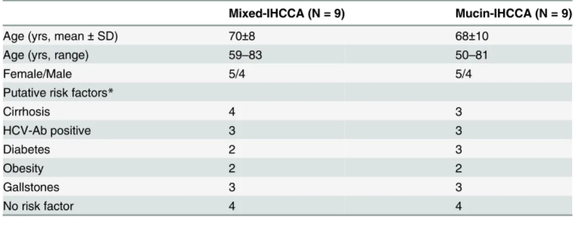
specifically: 18 patients with IH-CCA presenting as a single mass lesion within the liver. Patient characteristics were detailed inTable 1.
Human IHCCA samples were classified as mixed- or mucin-IHCCA by morphologic crite-ria and Pas staining, according to Komuta M. et al [6]. Patients characteristics CCA samples were subjected to mechanical and enzymatic dissociation with type IV collagenase (100U/ml) (Sigma Aldrich, Milan, Italy) at 37°C for 12–14 hours. Cells were plated in hormonally supple-mented medium consisting of DMEM with high glucose/DMEM:F12 mixture (1:1) (Gibco/ BRL, Life Technologies, Italia srl., Milan, Italy) supplemented with 1.8 x 10⁻⁴mol/L adenine,
5μg/ml insulin, 5μg/ml transferrin, 2 x 10⁻⁹mol/L triiodothyronine, 1.7 x 10⁻⁶mol/L
hydro-cortisone, 1.0 x 10⁻⁶mol/L human epidermal growth factor, 5.5 x 10⁻⁶mol/L epinephrine
(Sigma-Aldrich, Milan, Italy), 10% fetal bovine serum (Gibco/BRL, Life Technologies, Milan, Italy), 100 U/ml of penicillin, and 100μg/ml of streptomycin, at 37°C in a humidified
atmo-sphere of 5% CO2in air. Primary cell cultures were maintained at 37°C in a humidified atmo-sphere of 5% CO2in air. The different drugs were tested after 20–30 passages.
Characterization of Primary CCA Cell Cultures by Flow Cytometry (FC),
Immunohistochemistry/Immunofluorescence (IHC/IF) and RT-PCR
Primary IHCCA cell cultures were investigated by FC, at passages 20–30, for the expression of CSC surface markers by using the following antibodies: PE-mouse anti-human CD13 (BD Pharmigen, Milan, Italy), CD90-FITC human, CD133-APC human, CD45-PE human, EpCAM-FITC human (Miltenyi Biotec, Milan, Italy), anti-LGR5 mouse mAb PE coniugate (Origene, Unimed Scientifica, Rome, Italy). The fluorescence threshold between negative and positive cells was set on the basis of the reactivity of appropriate non-specific fluorochrome-conjugated isotypical controls. At, least 5 X 10⁵cells were analyzed using a FACS Diva software (BD).For IHC/IF, semi-confluent cultures were generated on four-chamber slides (NUNC multi-well plates, Unimed Scientific, Italy). The medium was removed, and cells were fixed in 10% buffered formalin for 10 min at room temperature. Cells were rinsed twice with PBS buffer for 2 min, blocked and incubated 1 hour with the following antibodies: Vimentin mouse monoclo-nal IgG1 (sc-32322, Santa Cruz Biotechnology),α-SMA mouse monoclonal IgG1(M0851,
Dako), E-Cadherin mouse monoclonal IgG1 (sc-21791, Santa Cruz Biotechnology), CD326/ EpCAM mouse monoclonal IgG1 (sc-59782, Santa Cruz Biotechnology), CD133/PROM1 Table 1. Patients characteristics.
Mixed-IHCCA (N = 9) Mucin-IHCCA (N = 9)
Age (yrs, mean±SD) 70±8 68±10
Age (yrs, range) 59–83 50–81
Female/Male 5/4 5/4
Putative risk factors*
Cirrhosis 4 3
HCV-Ab positive 3 3
Diabetes 2 3
Obesity 2 2
Gallstones 3 3
No risk factor 4 4
*according to ref. # 1.
mouse monoclonal IgG1 (TA309943, Origene), LGR5 rabbit polyclonal IgG (TA301323,Ori-gene), cytokeratin-19 (CK-19) sc-6278 CK-19 mouse monoclonal IgG2a (Santa Cruz Biotech-nology), Interleukin 6 (IL6) mouse monoclonal IgG2a (ab9324, Abcam) at room temperature. After rinsing twice with PBS for 2 min, cells were incubated for 40 min at room temperature with secondary biotinylated antibody (Vector laboratories,DBA Italy) rinsed twice with PBS and then incubated with Vectastain ABC reagent (Vector laboratories, DBA Italy) for 20 min. Diaminobenzidine (DAB substrate kit, Vector laboratories, DBA Italy) was used as substrate, and sections were counterstained with hematoxylin. Slides were examined by DM 2000 Light and/or Fluorescence Microscopy (Leica Microsystems, Italy) equipped with a DFC450 C Videocam (Leica Microsystems,Italy). For RT-PCR, cell cultures were extracted for total RNA by using the TRI REAGENTTM(Sigma-Aldrich, St Louis, MO, USA) and 1-bromo-3-chloro-propane in substitution of chloroform. The isolated RNA was dissolved in 55μl of RNase-free
water. RNA quality and quantity was controlled by the Experion Automated Electrophoresis System equipped with the RNA StSens Analysis Chip (Bio-Rad Laboratories, Hercules, CA, USA). The reverse transcription primed by the random hexamer (Invitrogen s.r.l., S. Giuliano Milanese, Italy) was conducted in a 20μL volume with an amount of 2.5μg of total RNA and
the M-MLV reverse transcriptase (Invitrogen s.r.l.) according the manufacturer's directions. Gene expression was determined by Real-Time PCR with a MX3000P instrument (Agilent, La J olla, CA, USA) using the averaged cycle threshold (Ct) automatically computed by the built-in software from three replicas of each sample. PCR amplifications were conducted built-into a vol-ume of 25μl, with 1.0μl of cDNA template, 12.5μl of 2x SYBR Green Brilliant QPCR Master
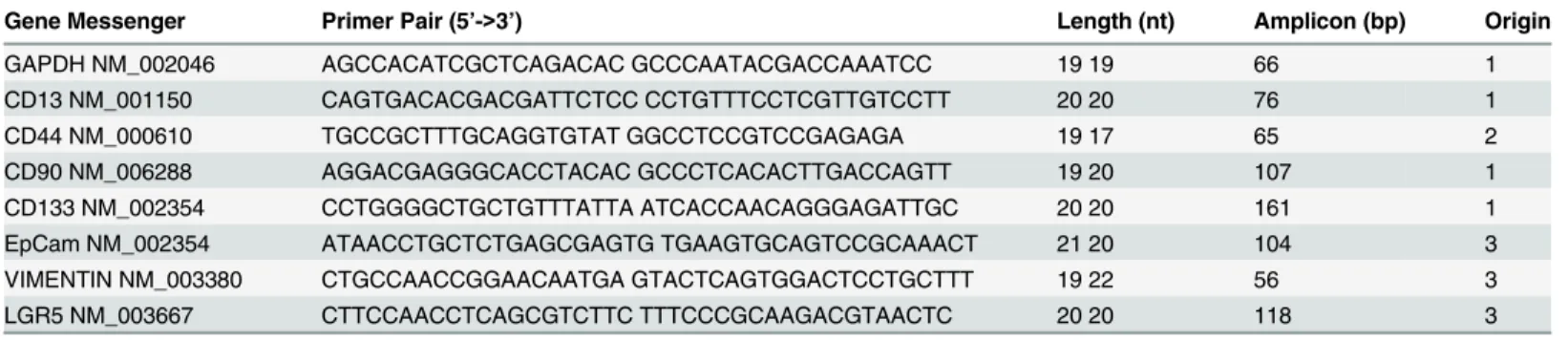
Mix (Stratagene), 3 pmoles each of upstream and downstream primer for the gene analyzed, and 0.3μl of diluted reference dye (ROX at a final concentration 30 nM). All real-time PCR
amplifications were conducted with the cycling program: 10 min at 95°C followed by 40 cycles (30 sec at 95°C, 30 sec at 58°C, 30 sec at 72°C). The fluorescence detection was performed dur-ing the extension step of each cycle. The followdur-ing genes of interest (GOI) were measured: Vimentin, CD13, CD90, CD133, EpCAM, and LGR5. All expression levels were normalized to the expression of GAPDH housekeeping gene.Table 2shows the details of primers used in the study.
In Vitro
Sensitivity to Chemotherapeutics and Molecular Targeted
Agents
Sensitivity to chemotherapeutics and molecular targeted agents was tested by evaluating cell proliferation or apoptosis in primary cell cultures exposed to increasing concentrations of dif-ferent drugs. Drugs were prepared as a stock solution in DMSO and then diluted (>1: 10,000)
in the culture medium at the desired final concentration; the same amount of DMSO was added in controls. Proliferation was evaluated by MTS assay (CellTiter 96 Aqueous One Solu-tion, PROMEGA, Milan, Italy). A total of 5x10³cells were seeded into 96-well plates in 100μL
In Vivo
Sensitivity of Human Subcutaneous Xenografts to NVP-BEZ-235
and Abraxane
Male NOD/SCID mice, 4–6 weeks old, purchased from Charles River (Italy) were maintained under standard conditions and cared according to our institutional guidelines for animal care. As previously described [5], CD13+ and CD133+ spheroids were prepared from human mucin- or mixed-IHCCA primary cultures, suspended in culture medium/Matrigel mixture (1:1 volume) and injected (approximately 10,000 cells) subcutaneously into mid-abdominal areas. We used CD13+ and CD133+ spheroids since in the previous study [5], these CSC sub-populations showed the highest tumorigenic potential in terms of xenograft generation. Tumor xenograft formation was followed by macroscopic inspection. After fifteen days, when the tumor volume was about 500 mm3, mice were treated by gavage with NVP-BEZ235 (50 mg/Kg in PBS, three times a week) and Abraxane (10mg/Kg in PBS, twice a week) for two weeks. Con-trol mice received PBS only. The health of all mice was monitored daily throughout the study. Main criteria used to assess mice health were the evaluation of body weight and consumption of food and water, other than the essentials for assessing mouse health as described by Bur-kholder et al. [7] Animal welfare was carefully ensured constantly by experienced operators every day. Every steps to avoid suffering were realized. Mice were then killed by cervical dislo-cation. The xenografts were removed after the death of the animal for histology.
Ethics Statements
The research protocol was reviewed and approved by theEthics Committee of Hospital Policli-nico Umberto I of Rome/Sapienza University of Rome(full name of the board/committee; Prot. May 2014), and was conducted according to the principles expressed in the Declaration of Hel-sinki. Subjects have been properly instructed and have indicated that they consent to partici-pate by signing the appropriate informed consent paperwork.
The experiment on animals was carried out in strict accordance with the recommendations in the Guide for the Care and Use of Laboratory Animals of the European Commission. The protocol was reviewed and approved by theEthics Committee of Hospital Policlinico Umberto I of Rome/Sapienza University of Rome(full name of the board/committee; Prot. May 2014). Animal welfare was carefully ensured constantly by experienced operators every day. Mice were then killed by cervical dislocation. All efforts were made to minimize suffering of the ani-mals along all the duration of their life and during the sacrifice.
The processing was compliant with Good Manufacturing Practice.
Table 2. Sequences of primer pairs (sense and antisense, respectively) used for amplifying the genes of interest (GOI) and the internal reference gene (GAPDH) used for their nor Primer designed by the PROBEFINDER software (https://www.roche-applied-science.com/sis/rtpcr/upl/index. jsp).
Gene Messenger Primer Pair (5’->3’) Length (nt) Amplicon (bp) Origin
GAPDH NM_002046 AGCCACATCGCTCAGACAC GCCCAATACGACCAAATCC 19 19 66 1
CD13 NM_001150 CAGTGACACGACGATTCTCC CCTGTTTCCTCGTTGTCCTT 20 20 76 1
CD44 NM_000610 TGCCGCTTTGCAGGTGTAT GGCCTCCGTCCGAGAGA 19 17 65 2
CD90 NM_006288 AGGACGAGGGCACCTACAC GCCCTCACACTTGACCAGTT 19 20 107 1
CD133 NM_002354 CCTGGGGCTGCTGTTTATTA ATCACCAACAGGGAGATTGC 20 20 161 1
EpCam NM_002354 ATAACCTGCTCTGAGCGAGTG TGAAGTGCAGTCCGCAAACT 21 20 104 3
VIMENTIN NM_003380 CTGCCAACCGGAACAATGA GTACTCAGTGGACTCCTGCTTT 19 22 56 3
LGR5 NM_003667 CTTCCAACCTCAGCGTCTTC TTTCCCGCAAGACGTAACTC 20 20 118 3
Statistical Analysis
Data are presented as arithmetic mean ± S.D. Statistical analysis was conducted using the paired or unpaired Student’st-test as appropriate or the analysis of the variance (ANOVA) when multiple comparisons were performed. A p value<0.05 was considered statistically
sig-nificant. The half maximal inhibitory concentration (IC50) values were calculated by nonlinear
regression analysis.
Results
Characterization of Primary Cultures of Human CCA
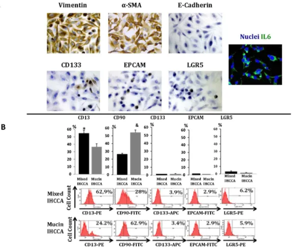
Primary cultures prepared from human mixed- and mucin-IHCCA specimens were character-ized for mesenchymal, epithelial and CSC surface markers by IH, FC and RT-PCR. As shown inFig 1A, after 20–30 passages, most cells expressed mesenchymal markers (vimentin,αSMA)
and IL6, were negative for E-Cadherin and CK-19 and rarely (<5%) positive for“epithelial”
CSC markers CD133, EpCAM, LGR5. In addition, most cells expressed the markers of EMT trait (Snail, TWIST, not shown). By FC (Fig 1B) cells positive for the mesenchymal CSC marker CD90 or for CD13, a marker of quiescent CSCs [2–4], largely predominated with respect to“epithelial”CSC markers, CD133, EpCAM and LGR5. When mixed- and mucin-IHCCCA primary cultures were compared, CD13+ cells predominated in mixed-IHCCA while the opposite was found for CD90+ cells (Fig 1B). These date were confirmed by RT-PCR (Table 3), where the expression of mRNA for vimentin and CD90 largely predominated with respect to CD133, EpCAM and LGR5, and mRNA for CD13 was more expressed in mixed-IHCCA while vimentin and the other CSC markers in mucin-mixed-IHCCA.
Effects of Chemotherapeutics on Cell Proliferation and Apoptosis
Gemcitabine and Cisplatin are the standard of care for CCA [1].Fig 2Ashows how Gemcita-bine was much more active in inhibiting cell proliferation (MTS assay) in mixed-IHCCA (IC50= 0.37 ± 0.04μM) than mucin-IHCCA primary cultures (IC50= 13.1 ± 12.8μM, p<0.01). Cisplatin, in contrast, was more active (Fig 2B) against mucin-IHCCA (IC50= 4.7 ±
1.3μM) than mixed-CCA (IC50= 13.1 ± 2.3μM, p<0.01). With respect to the two drugs
alone, the Gemcitabine-Cisplatin combination induced a higher inhibition of cell proliferation (Fig 2C) in mucin-IHCCA (IC50= 0.7 ± 0.2μM) but not in mixed-IHCCA (Gem+Cis; IC50=
14 ± 4.8,Fig 1). As shown inFig 2D, at5μM, Gemcitabine but not Cisplatin significantly
enhanced apoptosis (Caspase 3, p<0.01 vs controls) without differences between mucin- and
mixed-IHCCA primary cultures while, the combination Gemcitabine+Cisplatin does not fur-ther enhances the apoptotic effects of Gemcitabine alone.
The microtubule inhibitor, Abraxane (paclitaxel protein-bound formulation) [8], showed (Fig 2E) a significant inhibitory effect on cell proliferation in both mixed- (IC50= 0.4 ± 0.2μM)
and mucin-IHCCA (IC50= 0.1 ± 0.02μM) primary cultures, although the effect on
mucin-IHCCA was predominant (p<0.05). Abraxane (0.05μM) induced a significant increase of
apo-ptosis only in mucin- but not mixed-IHCCA (Fig 2F).
5-FU showed (Fig 2G) a slight inhibitory effect on cell proliferation in both mixed- (IC50=
9.8 ± 0.3μM) and mucin-IHCCA (IC50= 34.9 ± 5.7μM) primary cultures, although the effect
on mixed-IHCCA was predominant (p<0.05). 5-FU (20μM) induced apoptosis but only in
Effects of Molecular Targeted Agents on Cell Proliferation and Apoptosis
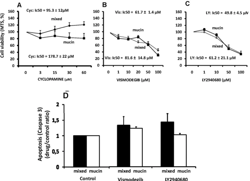
-Sonic Hedgehog (Hg) pathway inhibitors. Cyclopamine, a natural occurring alkaloid
[9], displayed no effect on cell proliferation in primary cultures of both mucin- and mixed-Fig 1. Characterization of mixed- and mucin-IHCCA primary cultures.(A) Immunohistochemical and immunofluorescence analyses of mixed- and mucin-IHCCA primary cultures for Vimentin,α-SMA, E-Cadherin and the“epithelial”cancer stem cell markers CD133+, EpCAM+, LGR5+ and for IL6. A diffuse positivity for mesenchymal markers (Vimentin,α-SMA) and IL6 was observed while E-Cadherin was virtually negative and less than 5% cells where positive for the“epithelial”cancer stem cell markers. Representative experiment of N = 12 independent staining performed in separate primary cultures. (B) Flow cytometry analyses of primary cultures of mixed- and mucin-IHCCA (20–30 passages), labeled with anti-CD13, anti-CD90, anti-EpCAM, anti-CD133, and anti-LGR5 antibodies. Bar graphs and representative plots. Cells positive for CD13 and CD90 largely predominated with respect to CD133, EpCAM and LGR5. CD13+ cells predominated in mixed-IHCCA with respect to mucin-IHCCA, while the opposite was found for CD90+ cells. Mean±SD of N = 18 independent experiments.*= p<0.05 vs mucin-IHCCA; & = p<0.01 vs mixed-IHCCA.
doi:10.1371/journal.pone.0142124.g001
Table 3. RT-PCR analysis of Vimentin and cancer stem cell surface markers.
VIM CD90 CD13 CD133 EPCAM LGR5
Mixed-IHCCA 4.3±0.36 0.49±0.002 0.05±0.0009 5.7*10−3±3.6*10−5 6.4*10−5±1.1*10−5 5.4*10−7±3.8*10−8
Mucin-IHCCA 7.2±0.4* 0.95±0.0075* 0.024±0.0011* 4.3*10−2±2.6*10−3* 7.5*10−5±1.2*10−5 5.8*10−6±7.1*10−7*
mRNA relative expression of Vimentin (VIM), CD90, CD13, CD133, EpCAM and LGR5 in primary cultures from mixed-intrahepatic (Mixed-IHCCA) or mucin-intrahepatic (Mucin-IHCCA) human cholangiocarcinoma, normalized versus the reference gene GAPDH. mRNA for CD13 was more expressed in mixed-IHCCA while vimentin, CD90, CD133 and LGR5 predominated in mucin-IHCCA. Gene expression was measured in quadruplicate. Mean±SD of N = 12 experiments.
*= p<0.05.
Fig 2. Effects of chemotherapeutics (Gemcitabine, Cisplatin, Abraxane) on proliferation and apoptosis of mucin- and mixed-CCA primary cultures.
IHCCAs (Fig 3A). In contrast, Vismodegib, a small-molecule antagonist of Smoothened [9] and LY2940680, a small-molecule antagonist of the smoothened receptor [9], showed a slight inhibitory effect on cell proliferation (Fig 3B and 3C) without differences between mucin-(IC50:Vismodecib = 61.7 ± 1.4μM, LY2940680 = 49.8 ± 4.5μM) and mixed-CCA (IC50:
Vismodecib = 81.6 ± 14.8μM, LY2940680 = 61.2 ± 21.1μM).
Apoptosis was unaffected by Vismodegib (50μM) or LY2940680 (20μM) in primary
cul-tures of mucin- and mixed-IHCCA (Fig 3D)
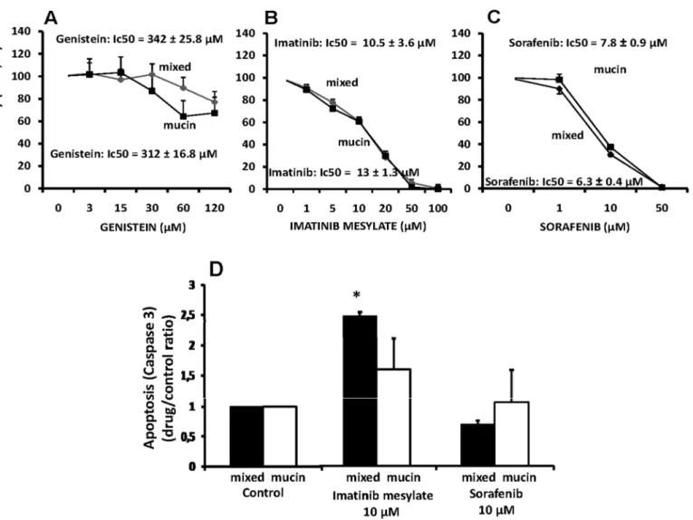
-Tyrosine kinase inhibitors. We tested the following tyrosine kinase inhibitors: Genistein
that is a broad spectrum tyrosine kinase inhibitor [10]; Imatinib Mesylate that is a c-kit tyro-sine kinase inhibitor also acting as aspecific anti-CD90+ cells [11], and; sorafenib that is a multi-targeted tyrosine kinase receptor inhibitor [12]. Primary cultures of mucin- and mixed-IHCCA were resistant to genistein (Fig 4A) but they were equally sensitive (inhibition of cell proliferation) to Imatinib Mesylate (IC50:mucin-IHCCA = 10.5 ± 3.6μM;
mixed-IHCCA = 13.0 ± 1.3μM,Fig 4B) and Sorafenib (IC50:mucin-IHCCA = 7.8 ± 0.9μM;
mixed-IHCCA = 6.3 ± 0.4μM;Fig 4C).
Apoptosis was significantly enhanced only by Imatinib Mesylate (10μM) and only in
mixed-IHCCA (Fig 4D).
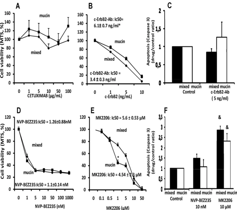
-Epidermal growth factor receptor (EGFR) antagonists. Cetuximab, a chimerical
mono-clonal EGFR IgG1 antibody, that blocks the binding of EGF or other ligands to EGFR, thus, inhibiting ligand-induced receptor phosphorylation (11), had no effect neither on mucin- nor mixed-IHCCA (Fig 5A). In contrast, the c-erbB2 blocking antibody was active against both mucin-IHCCA (IC50= 6.1 ± 0.7 ng/ml) and mixed-IHCCA (IC50= 3.4 ± 0.3 ng/ml), but with a
slight predominant effect on the latter (p<0.05,Fig 5B). Cetuximab and the c-erbB2 blocking
antibody (Fig 5C) showed no effect on cell apoptosis.
-PI3-kinase/AKT inhibitors. NVP-BEZ235, a dual PI3K and mTOR inhibitor [13],
showed a very strong inhibitory effect, at nanomolar concentration, on cell proliferation in both mucin-IHCCA (IC50= 1.1 ± 0.14 nM) and mixed-IHCCA (IC501.26 ± 0.88 nM) primary
cultures (Fig 5D). Also the allosteric AKT inhibitor, MK2206, inhibited cell proliferation (Fig 5E) in primary cultures of mucin-IHCCA (IC50= 4.5 ± 2.3μM) and mixed-CCA (IC505.6 ±
0.53μM) but at a significant lower extent in comparison with NVP-BEZ235.
As far as apoptosis is concerned, only the AKT inhibitor, MK2206, but not NVP-BEZ235, induces significant apoptotic effects, at 10μM, without differences between mucin-IHCCA and
mixed-IHCCA (Fig 5F).
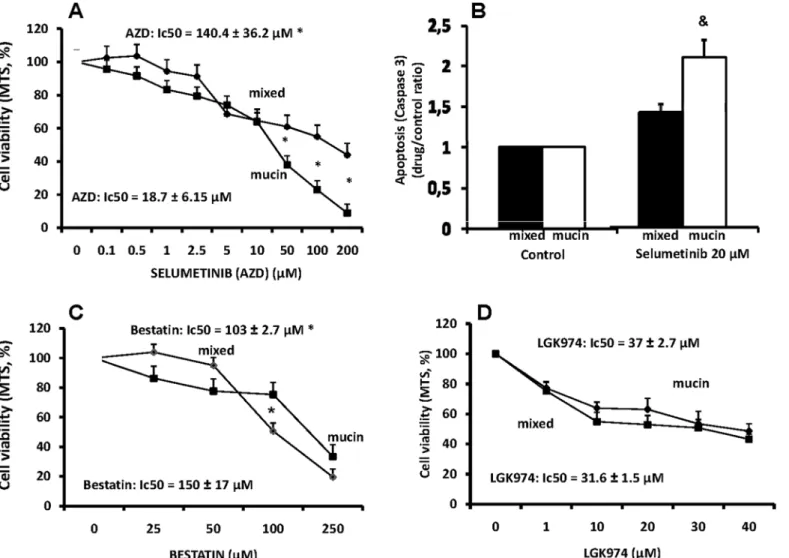
-MEK 1/2 inhibitor. The selective, ATP uncompetitive, MEK 1/2 inhibitor, Selumetinib
(AZD6244) [14] inhibits cell proliferation only at high concentrations and with a predominant effect on mucin-IHCCA (IC50= 18.7 ± 6.1μM) with respect to mixed-IHCCA (IC50140.4 ±
36.2μM, p<0.05;Fig 6A). Apoptosis was significantly enhanced by Selumetinib (20μM) only
in mucin-IHCCA (Fig 6B).
-Aminopeptidase-N inhibitor. The aminopeptidase-N inhibitor, bestatin, has been
largely tested in a large spectrum of cancer cells, as CD13 inhibitor [15]. However, in both
evaluated by measuring caspase-3 activity and expressed as ratio between drug-treated and control cells. MTS and caspase-3 were measured 72 hours after incubation with the tested drug. Gemcitabine was much more active in inhibiting cell proliferation (A) (MTS assay) in mixed-IHCCA than mucin-IHCCA primary cultures. Cisplatin (B), in contrast, was more active against mucin-IHCCA. With respect to the two drugs alone, the Gemcitabile-Cisplatin
combination induced a higher inhibition of cell proliferation (C) in mucin-IHCCA but not in mixed-IHCCA. Gemcitabine but not Cisplatin significantly enhanced Caspase-3 activity (D) without differences between mucin- and mixed-IHCCA and, the combination Gem+Cis does not further enhances the apoptotic effects of Gemcitabine alone. Abraxane showed a significant inhibitory effect on cell proliferation (E) in both mixed- and mucin-IHCCA primary cultures, although the effect on mucin-IHCCA was predominant (p<0.05). Abraxane induced a significant increase of apoptosis only in mucin-IHCCA (F). 5-FU slightly inhibited cell proliferation (G) with a more significant effect on mixed- than mucin-IHCCA (p<0.05). 5-FU induced a significant increase of apoptosis only in mucin-IHCCA (H).*p<0.05 mixed vs mucin. & = p<0.05 vs controls. Mean±SD of N = 5–7 independent experiments.
mucin- and mixed-IHCCA primary cultures, the inhibitory effect of bestatin on cell prolifera-tion was limited with an IC50of 103 ± 2.7μM for mucin-IHCCA and of 150± 17μM for
mixed-IHCCA (p<0.05,Fig 6C). No effect on apoptosis was seen at 100μM (not shown).
- Wnt signaling Inhibitor. Recent evidence suggests that enhanced Wnt signaling sustains
CCA progression and that targeting Wnt signaling could represent a potential therapeutic strategy [16]. We tested LGK974, a potent and specific small-molecule porcupine inhibitor. Porcupine is a membrane-bound O-acyltransferase that is required for palmitoylation of Wnt ligands, a necessary step in the processing of Wnt ligand secretion [17]Fig 6Dshows how LGK794 induced, already at 10μM concentration, an inhibitory effect on cell proliferation that
was similar for mixed- (IC50 = 31.6 ± 1.5μM) and mucin-IHCCA (37 ± 2.7μM, p<0.05). No
significant effect on apoptosis was seen.
Fig 3. Effect of Sonic Hedgehog (Hg) pathway inhibitors on proliferation and apoptosis of mucin and mixed CCA primary cultures.Cyclopamine (A) displayed no effect on cell proliferation in primary cultures of both mucin- and mixed-IHCCAs. In contrast, Vismodegib (B) and LY2940680 (C), showed a slight inhibitory effect on cell proliferation in both CCAs subtypes. Apoptosis was unaffected by Vismodegib or LY2940680. N experiments = 5–7.
In Vivo
Sensitivity of Human Subcutaneous Xenografts to NVP-BEZ235
and Abraxane
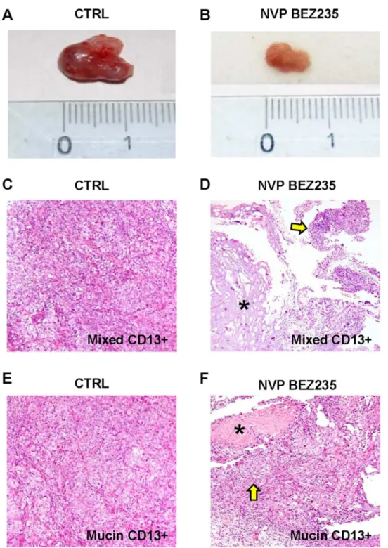
CD13+ or CD133+ spheroids prepared from primary cultures of human mucin- or mixed-IHCCA were subcutaneously injected in male NOD/SCID mice. After 2 weeks, when the tumors’volume averaged 500 mm3, mice were treated by gavage with NVP-BEZ235 or Abrax-ane dissolved in PBS, while control mice received PBS only. At the fourth week, after 2 weeks of treatment, the volume of the masses were re-evaluated. In control mice, CD133+ and CD13+ tumor xenografts increased significantly, respectively from 550 ± 50 mm3to
1400 ± 100 mm3(p<0.05), and from 590 ± 55 mm3to 1450 ± 76 (p<0.05,Fig 7A). No
differ-ences between mixed- and mucin-IHCCA were observed. In contrast, in mice treated with NVP-BEZ235 or Abraxane, the tumor volume for CD133+ spheroids remained almost stable (NVP-BEZ235: 790 ± 100 mm3for mixed-IHCCA and 690 ± 100 mm3for mucin-IHCCA; Abraxane: 730 ± 86 mm3for mixed-IHCCA and 790 ± 96 mm3for mucin-IHCCA, p<0.05 vs
controls). Similar findings were obtained when CD13+ spheroids were implanted, since the Fig 4. Effect of tyrosine kinase inhibitors on proliferation and apoptosis of mucin and mixed CCA primary cultures.Genistein showed no effect on cell proliferation (A). Imatinib mesylate (B) and Sorafenib (C) inhibited cell proliferation in mucin-IHCCA and mixed-IHCCA but only the former enhance apoptosis (D) and only in mixed-IHCCA.*p<0.05 vs controls. N experiments = 5–7.
tumor volume remained almost stable following the treatment with NVP-BEZ235 (740 ± 100 mm3for mixed-IHCCA and 650 ± 94 mm3for mucin-IHCCA,Fig 7B, p<0.05 vs
con-trols) or Abraxane (670 ± 90 mm3for mixed-IHCCA and 720 ± 85 mm3for mucin-IHCCA, p<0.05 vs controls). At the histo-pathological evaluation (H&E,Fig 7C–7F) in control mice,
tumor xenographts were composed of densely packed tumor cells (Fig 7Cmixed-IHCCA, Fig 5. Effect of Epidermal growth factor receptor (EGFR) antagonists and PI3-kinase/AKT inhibitors on proliferation and apoptosis of mucin and mixed CCA primary cultures.The EGFR antagonist, cetuximab (A), had no effect on cell proliferation neither on mucin- nor in mixed-IHCCA. In contrast, the c-erbB2 blocking antibody (B) inhibited proliferation in both mucin- and mixed-IHCCA with a predominant effect on the latter. Cetuximab and the c-erbB2 blocking antibody showed no effect on cell apoptosis (C).*p<0.05 mucin vs mixed, N experiments = 5–7. The PI3-kinase/AKT inhibitors, NVP-BEZ235 (D)
and MK2206 (E) showed strong inhibitory effect on cell proliferation in both mucin- and mixed-IHCCA primary cultures, but only MK2206 enhanced apoptosis (F) without differences between mucin-IHCCA and mixed-IHCCA.*p<0.05 mucin vs mixed, & = p<0.05 vs controls. N experiments = 5–7.
Fig 7Emucin-IHCCA) while in mice treated with Abraxane or NVP-BEZ235 (Fig 7D mixed-IHCCA,Fig 7Fmucin-IHCCA) necrotic areas were seen within the tumor mass.
Discussion
The main findings of our study, performed in primary cultures of mucin- and mixed- IHCCA subtypes, indicate that: i) among chemotherapeutics, Gemcitabine and the Gemcitabine-platin combination were more active in inhibiting cell proliferation in mixed-CCA while Cis-platin or Abraxane were more effective against mucin-CCA, where abraxane was also able to induce apoptosis. 5-FU exerted a slight inhibitory effect on cell proliferation, predominating in mixed-IHCCA, and also induced apoptosis but only in mucin-IHCCA; ii) among Hg inhibi-tors, Ciclopamine was ineffective while LY2940680 and Vismodegib showed slight effects on proliferation but not on apoptosis; iii) the tyrosine kinase inhibitor, Genistein was completely ineffective while either Imatinib Mesylate or Sorafenib showed significant inhibitory effects on Fig 6. Effect of MEK 1/2 inhibitor, Aminopeptidase-N inhibitor and Wnt inhibitor on proliferation and apoptosis of mucin and mixed CCA primary cultures.The MEK 1/2 inhibitor, selumetinib (AZD6244) (A), inhibits cell proliferation only at high concentrations and with a predominant effect on mucin-IHCCA. Apoptosis (B) was enhanced only in mucin-mucin-IHCCA.*p<0.05 mucin vs mixed, & = p<0.05 vs controls. N experiments = 5–7. The aminopeptidase-N
inhibitor, bestatin (C), showed an inhibitory effect on cell proliferation that predominated in mixed-IHCCA.*p<0.05 mucin vs mixed, N experiments = 5–7.
The Wnt inhibitor, LGK974, (D) starting from 10μM concentration inhibited proliferation of both mucin- and mixed-IHCCA, N experiments = 3–5.
Fig 7. Effect of NVP-BEZ235 on subcutaneous human tumor xenografts.In control mice (CTRL), the tumor volume of subcutaneous xenografts increased during four weeks of observation (A) while in mice treated for 2 weeks with NVP-BEZ235 (B) it remains almost stable. At the histo-pathological evaluation (Hematoxylin-Eosin), in control mice (CTRL), subcutaneous masses arisen after the implantation of CD13 + spheroids from mixed- (C) or mucin-IHCCA (E) cultures, were composed of densely packed tumor cells. Original Magnification(OM): 10x. C, E). In mice treated with NVP-BEZ235, subcutaneous xenografts arisen from CD13+ spheroids, immunosorted from mixed-IHCCA cultures (D) were characterized by extensive necrosis (asterisk) and few tumoral cells (arrow); necrosis was less extended in masses arisen after the implantation of CD13+spheroids immunosorted from mucin-IHCCAs (F, asterisk = necrosis, arrow = tumoral cells). OM: 10x (D, F).
proliferation; iv) the MEK 1/2 inhibitor, Selumetinib, was active in inhibiting proliferation only against mucin-CCA while the aminopeptidase-N inhibitor, Bestatin was more active against mixed-CCA; v) the c-erbB2 blocking antibody was more active against mixed- than mucin-IHCCA while, Cetuximab failed to show significant effects; vi) either mucin- or mixed-mucin-IHCCA showed high sensitivity to PI3-kinase inhibitors and particularly to NVP-BEZ235 that
markedly inhibited cell proliferation at nanomolar concentrations and, alsoin vivoblocked tumor progression. The main results of our study, based on experiments performed in primary cultures, indicate that mucin- and mixed-IHCCA display a different drug sensitivity. Specifi-cally, Cisplatin, Abraxane and the MEK 1/2 inhibitor, Selumetinib are more active against mucin-IHCCA while, Gemcitabine, the Gemcitabine-Cisplatin combination, 5-FU, the c-erbB2 blocking antibody and the aminopeptidase-N inhibitor, bestatin, act better against the mixed-IHCCA subtype. Remarkably, both IHCCA subtypes showed a specific sensitivity to the PI3K/AKT inhibitors and in particular to the dual PI3-kinase/mTOR inhibitor, NVP-BEZ235, that bothin vitroandin vivoexerts marked antiproliferative effect at very low concentrations.
The current standard of care for unresectable IHCCA is the Gemcitabine/Cisplatin regimen [18,19] that has been recently considered to be also cost-effective with respect to Gemcitabine alone [20]. However, this treatment is absolutely unsatisfactory since ensure only a few month improved survival [18,19] and, therefore, alternative treatments are demanding. Unfortunately, CCA is scarcely responsive to chemotherapeutics for different reasons including its desmoplas-tic nature and the high expression of drug extruding pumps [1]. In addition, we have recently realized that CCA is highly enriched with CSCs suggesting this cancer as a disease of stem cells that are typically resistant to chemotherapeutics [5]. Very few phase I/II clinical trials evaluated the effects of molecular targeted agents, alone or in combination with gemcitabile or cisplatin [4]. In all these studies no discrimination between mucin- and mixed-IHCCA subtypes has been provided. Indeed, large morphologic, biologic and clinical differences exist between the mucin- and mixed-CCA subtypes [5,6]. From a clinical point of view, the mixed-subtypes is more frequently associated with liver cirrhosis, appears macroscopically as a mass-forming with a peripheral location and progress with less lymphatic and perineural invasion and with a better prognosis with respect to the mucin-CCA subtype [5]. In the previous manuscript [6], we demonstrated the largein situdifferences in terms of CSC composition and EMT markers between the two CCA subtypes. Also primary cultures showed large differences since CD90 + cells predominated in mucin-CCA while CD13+ cells in the mixed–CCA subtype. In spite of these clinical and patho-biologic differences, no report investigated the drug-sensitivity of the two CCA subtypes and, clinically, the need of a different therapy has been so far completely neglected. We used primary cultures of mixed- and mucin-CCA to test drug sensitivity and to calculate IC50. These cultures are characterized by the predominance of cells expressing mes-enchymal markers and EMT trait. We firstly demonstrated the different sensitivity of the two CCA subtypes to chemotherapeutics since Gemcitabine and the Gemcitabine-Cisplatin combi-nation were more active against mixed-CCA while Cisplatin or Abraxane were more effective in inhibiting cell proliferation in mucin-CCA primary cultures. Abraxane is an albumin bound formulation of paclitaxel where albumin favors drug internalization and indeed, according to a recent report, this formulation showed promising results in advanced pancreatic cancer [21]. This is consistent with the present findings showing a preferential effects on mucin-IHCCA, since a number of biologic similarities exist between mucin-CCA and the adenocarcinoma of pancreatic ducts [22]. However,in vivo, in subcutaneous xenografts, Abraxane showed a simi-lar inhibitory effect on the growth of both mucin- and mixed-IHCCA.
studies suggested how the Hg signalling pathways are of relevance for CCA biology [23,24]. The expression of Hg components, for example, was associated with the progression and metastasis in IHCCA [23,24]. However, canonical Hg signaling requires cilia but CCA cells do not express cilia [25]. Nonetheless, CCA cells exhibit non-canonical Hedgehog signaling with may influence chemotaxis despite impaired cilia expression [25]. This non-canonical Hedge-hog signaling pathway appears to contribute to CCA progression, thereby, supporting a role for Hedgehog pathway inhibition in human CCA [25]. We tested ciclopamine, the clinically approved small-molecule antagonist of Smoothened, Vismodegib, and of its receptor, LY2940680. In our experiments, CCA primary cultures were completely resistant to ciclopa-mine and slightly sensitive to LY2940680 and Vismodegib in terms of inhibited cell prolifera-tion. Recently, by profiling transcriptomes in surgically resected samples, therapeutic targets for tyrosine kinase inhibitors have been identified [26]. Among the tyrosin kinase inhibitors, Genistein, a broad spectrum inhibitor [10], failed in our hands to influence cell proliferation of both CCA subtype primary cultures. In contrast, Sorafenib, a multiple kinase inhibitor [10] played antiproliferative effects without influencing apoptosis. It is noteworthy that the concen-trations demonstrated to be effective in our study correspond to the steady-state sorafenib plasma levels of 15–20μM measured in patients administered with approved dosages of this
drug [27]. In clinical trials testing Sorafenib, Dealis et al. [28] showed a controlled disease in one third of patients with advanced CCA while, Bengala et al. [29] showed an improved pro-gression free survival only in patients with better conditions. We finally tested Imatinib Mesy-late that acts not only as c-kit-R inhibitor [9] but also, as recently demonstrated [6], as aspecific blocker of CD90+ CSCs [11]. Since CD90+ cells represent a predominant CSC subpopulation in mucin-IHCCA, our result should merit investigation in clinical trials. To this regard, in a phase II study [30], the disease control rate of 26 CCA patients was 58% and this looks promising.
EGFR and/or ErbB2 are overexpressed in human and rodent CCA cells and EGFR has been implicated with CCA pathogenesis [31,32]. EGFR overexpression was found as an indepen-dent prognostic factor for survival and a risk factor for tumor recurrence after resection in intrahepatic CCA and, associated with tumor progression and invasion in extrahepatic CCA [31]. According to this literature, EGFR has been considered an attractive target for CCA ther-apy [31]. In 2010, by investigating the efficacy and safety of cetuximab in combination with gemcitabine and oxaliplatin for first-line treatment of biliary tract cancer, Gruenberger et al. [33] reported objective response in 63% of patients. Unfortunately, these promising findings have not been confirmed. In fact, in a non-comparative, open-label, randomised phase-2 trial, the addition of cetuximab to chemotherapy (cisplatin or oxaliplatin plus gemcitabine) in advanced biliary cancers, although well tolerated, failed to enhanced the activity of chemother-apeutics [34], In our experiments, cetuximab failed to influence cell proliferation or apoptosis. In contrast, the c-erbB2 blocking antibody was active against both CCA subtypes in inhibiting cell proliferation but without effects on apoptosis. To this regard, a dramatic response to Tras-tuzumab, the antibody targeting HER2 receptor molecule was described in a patient with EGFR2–positive metastatic CCA [35].
CCA cell growth and migration and significantly induced G1 arrest without apoptosis induc-tion, although increased autophagy was observed [36]. The same conclusion was raised by Ewald F. et al. [38] showing, in preclinical studies, how combined targeting of mTOR and AKT using RAD001 and MK-2206 inhibited the growth of CCA cell lines. In our experiments, NVP-BEZ235, that is now entering phase I/II clinical trials, played at nanomolar concentra-tions marked antiproliferative effects against both mucin- and mixed-CCA without enhancing apoptosis. Notably, alsoin vivo, in subcutaneous xenografts, NVP-BEZ235, blocked the pro-gression of mucin- and mixed-IHCCA. Also the pan-AKT inhibitor, MK2206, exerted antipro-liferative affects and, this compound also enhanced apoptosis. Thus our findings support future clinical trials testing PI3k/mTOR inhibitors.
Selumetinib is an allosteric inhibitor of MEK1 and MEK2 phosphorylation of ERK and of interleukin-6 secretion, the latter playng a major role in modulating CCA cell proliferation [1]. In a phase-II study, in patients with metastatic biliary tract cancer [14], 12% of patients had a confirmed objective response and, 68% of these patients experienced stable disease. In our study, Selumetinib showed inhibitory effects on proliferation and activation of apoptosis; these effects being predominant in mucin-IHCC subtype.
We also tested Bestatin, a CD13+ CSC antagonist, that showed antitumoral activity in dif-ferent cancers [15]. We found a preferential effect of Bestatin on mixed-IHCCA primary cul-tures and this is consistent with the predominance of CD13+ CSCs in mixed CCA subtypes.
Finally, we tested a Wnt signalling inhibitor, since recent evidence highlighting the role of Wnt signaling in sustaining CCA growth and progression [16]. Starting form 10μM, LGK974,
inhibited by approx. 40–50% cell proliferation without differences between Mucin- and mixed-IHCCA.
In conclusion, our findings indicate that other than biologically different, mucin and mixed-CCA subtypes are also different as far as the sensitivity to chemotherapeutics and tar-geted-agents is concerned. Future therapeutic strategies triggering IHCCA should take in con-sideration these differences and, therefore, discrimination between the two subtypes is fundamental also for therapeutic decisions.
Author Contributions
Conceived and designed the experiments: AF VC DA. Performed the experiments: AF VC MCB CN RS AML DC LN SDM AR GC. Analyzed the data: AF VC GC EG DA. Contributed reagents/materials/analysis tools: FG AMDR GLG. Wrote the paper: AF VC DA.
References
1. Razumilava N, Gores GJ. Cholangiocarcinoma. Lancet. 2014; 383: 2168–79. doi: 10.1016/S0140-6736(13)61903-0PMID:24581682
2. Yamashita T, Wang XW. Cancer stem cells in the development of liver cancer. J Clin Invest. 2013; 123: 1911–1918. doi:10.1172/JCI66024PMID:23635789
3. Magee JA, Piskounova E, Morrison SJ. Cancer stem cells: impact, heterogeneity, and uncertainty. Cancer Cell. 2012; 21: 283–296. doi:10.1016/j.ccr.2012.03.003PMID:22439924
4. Oishi N, Wang XW. Novel therapeutic Strategies for Targeting Liver Cancer Stem Cells. Int. J. Biol. Sci. 2011; 7: 517–535. PMID:21552419
5. Cardinale V, Renzi A, Carpino G, Torrice A, Bragazzi MC, Giuliante F, et al. Profiles of Cancer Stem Cell Subpopulations in Cholangiocarcinomas. Am J Pathol. 2015; 185: 1724–1739. doi:10.1016/j. ajpath.2015.02.010PMID:25892683
6. Komuta M, Govaere O, Vandecaveye V, Akiba J, Van Steenbergen W, Verslype C, et al. Histological diversity in cholangiocellular carcinoma reflects the different cholangiocyte phenotypes. Hepatology. 2012; 55: 1876–1888. doi:10.1002/hep.25595PMID:22271564
8. Green MR, Manikhas GM, Orlov S, Afanasyev B, Makhson AM, Bhar P, et al. Abraxane, a novel Cre-mophor-free, albumin-bound particle form of paclitaxel for the treatment of advanced non-small-cell lung cancer. Ann Oncol. 2006; 17: 1263–1268. PMID:16740598
9. Lin TL, Matsui W. Hedgehog pathway as a drug target: Smoothened inhibitors in development. Onco Targets Ther. 2012; 5: 47–58. doi:10.2147/OTT.S21957PMID:22500124
10. Levitzki A. Tyrosine Kinase Inhibitors: Views of Selectivity, Sensitivity, and Clinical Performance. Ann Rev Pharmacol Toxicol. 2013; 53: 161–185.
11. Yamashita T. Discrete nature of EpCAM+and CD90+cancer stem cells in human hepatocellular carci-noma. Hepatology. 2013; 57: 1484–1497. doi:10.1002/hep.26168PMID:23174907
12. Roskoski R. Pharmacol Res. ErbB/HER protein-tyrosine kinases: Structures and small molecule inhibi-tors. Pharmacol Res. 2014; 87C: 42–59.
13. Martini M, De Santis MC, Braccini L, Gulluni F, Hirsch E. PI3K/AKT signaling pathway and cancer: an updated review. Ann Med. 2014; 5: 1–12.
14. Bekaii-Saab T, Phelps MA, Li X, Saji M, Goff L, Kauh JS, et al. Multi-institutional phase II study of selu-metinib in patients with metastatic biliary cancers. J Clin Oncol. 2011; 29: 2357–2363. doi:10.1200/ JCO.2010.33.9473PMID:21519026
15. Grujic M, Renko M. Aminopeptidase inhibitors bestatin and actinonin inhibit cell proliferation of mye-loma cells predominantly by intracellularinter actions. Cancer Lett. 2002; 182: 113–119. PMID: 12048155
16. Boulter L, Guest RV, Kendall TJ, Wilson DH, Wojtacha D, Robson AJ, et al. WNT signaling drives cho-langiocarcinoma growth and can be pharmacologically inhibited. J Clin Invest. 2015; 125: 1269–1285. doi:10.1172/JCI76452PMID:25689248
17. Liu J, Pan S, Hsieh MH, Ng N, Sun F, Wang T, et al. Targeting Wnt-driven cancer through the inhibition of Porcupine by LGK974. Proc Natl Acad Sci U S A. 2013; 110: 20224–20229. doi:10.1073/pnas. 1314239110PMID:24277854
18. Valle J, Wasan H, Palmer DH, Cunningham D, Anthoney A, Maraveyas A, et al. Cisplatin plus gemcita-bine versus gemcitagemcita-bine for biliary tract cancer. N Engl J Med. 2010; 362: 1273–1281. doi:10.1056/ NEJMoa0908721PMID:20375404
19. Okusaka T, Nakachi K, Fukutomi A, Mizuno N, Ohkawa S, Funakoshi A, et al. Gemcitabine alone or in combination with cisplatin in patients with biliary tract cancer: a comparative multicentre study in Japan. Br J Cancer. 2010; 103: 469–474. doi:10.1038/sj.bjc.6605779PMID:20628385
20. Roth JA, Carlson JJ. Cost-effectiveness of gemcitabine+cisplatin vs. gemcitabine monotherapy in advanced biliary tract cancer. J Gastrointest Cancer. 2012; 43: 215–223. doi: 10.1007/s12029-010-9242-0PMID:21234709
21. Heinemann V, Haas M, Boeck S. Systemic treatment of advanced pancreatic cancer. Cancer Treat Rev. 2012; 38: 843–853. doi:10.1016/j.ctrv.2011.12.004PMID:22226241
22. Cardinale V, Carpino G, Reid L, Gaudio E, Alvaro D. Multiple cells of origin in cholangiocarcinoma underlie biological, epidemiological and clinical heterogeneity. World J Gastrointest Oncol. 2012; 4: 94–102. doi:10.4251/wjgo.v4.i5.94PMID:22645632
23. Tang L, Tan YX, Jiang BG, Pan YF, Li SX, Yang GZ, et al .The prognostic significance and therapeutic potential of hedgehog signaling in intrahepatic cholangiocellular carcinoma. Clin Cancer Res. 2013; 19: 2014–2024. doi:10.1158/1078-0432.CCR-12-0349PMID:23493353
24. El Khatib M, Kalnytska A, Palagani V, Kossatz U, Manns MP, Malek NP, et al. Inhibition of hedgehog signaling attenuates carcinogenesis in vitro and increases necrosis of cholangiocellular carcinoma. Hepatology. 2013; 57: 1035–1045. doi:10.1002/hep.26147PMID:23172661
25. Razumilava N, Gradilone SA, Smoot RL, Mertens JC, Bronk SF, Sirica AE, et al. Noncanonical Hedge-hog Signaling Contributes to Chemotaxis in Cholangiocarcinoma. J Hepatol. 2014; 60: 599–605. doi: 10.1016/j.jhep.2013.11.005PMID:24239776
26. Andersen JB, Spee B, Blechacz BR, Avital I, Komuta M, Barbour A, et al. Thorgeirsson SS.Genomic and genetic characterization of cholangiocarcinoma identifies therapeutic targets for tyrosine kinase inhibitors. Gastroenterology. 2012; 142: 1021–1031. doi:10.1053/j.gastro.2011.12.005PMID: 22178589
27. Strumberg D. Preclinical and clinical development of the oral multikinase inhibitor sorafenib in cancer treatment. Drugs Today (Barc). 2005; 41: 773–784.
28. Dealis C, Bertolini F, Malavasi N, Zironi S, Boni C, Banzi E, et al. A Phase II trial of sorafenib (Sor) in patients (Pts) with advanced cholangiocarcinoma (Cc). J Clin Oncol (Meeting Abstracts) 2008: 4590.
30. Roth A, Schleyer E, Schoppmeyer K, Kluge R, Wittekind C, Mössner J, et al. Imatinib mesylate for palli-ative second-line treatment of advanced biliary tract cancer: a bicentric phase II study. Onkologie. 2011; 34: 469–470. doi:10.1159/000331065PMID:21934349
31. Zhang Z, Oyesanya RA, Campbell DJ, Almenara JA, Dewitt JL, Sirica AE. Preclinical assessment of simultaneous targeting of epidermal growth factor receptor (ErbB1) and ErbB2 as a strategy for cholan-giocarcinoma therapy. Hepatology. 2010; 52: 975–986. doi:10.1002/hep.23773PMID:20607690 32. Nanda S. Cancer: A limited role for dual EGFR and ErbB2 inhibition in cholangiocarcinoma? Nat Rev
Gastroenterol Hepatol. 2010; 7: 591. doi:10.1038/nrgastro.2010.163PMID:21069929
33. Gruenberger B, Schueller J, Heubrandtner U, Wrba F, Tamandl D, Kaczirek K, et al. Cetuximab, gemci-tabine, and oxaliplatin in patients with unresectable advanced or metastatic biliary tract cancer: a phase 2 study. Lancet Oncol. 2010; 11: 1142–1148. doi:10.1016/S1470-2045(10)70247-3PMID:21071270 34. Malka D, Cervera P, Foulon S, Trarbach T, De La Fouchardière C, Boucher E, et al. Gemcitabine and
oxaliplatin with or without cetuximab in advanced biliary-tract cancer (BINGO): a randomised, open-label, non-comparative phase 2 trial. Lancet Oncol. 2014; 15: 819–828. doi:10.1016/S1470-2045(14) 70212-8PMID:24852116
35. Law LY. Dramatic response to trastuzumab and paclitaxel in a patient with human epidermal growth factor receptor 2-positive metastatic cholangiocarcinoma. J Clin Oncol. 2012; 30(27): e271–273. doi: 10.1200/JCO.2012.42.3061PMID:22851567
36. Yothaisong S, Dokduang H, Techasen A, Namwat N, Yongvanit P, Bhudhisawasdi V, et al. Increased activation of PI3K/AKT signaling pathway is associated with cholangiocarcinoma metastasis and PI3K/ mTOR inhibition presents a possible therapeutic strategy. Tumour Biol. 2013; 34:3637–3648. doi:10. 1007/s13277-013-0945-2PMID:23832540
37. Chen MH, Chiang KC, Cheng CT, Huang SC, Chen YY, Chen TW, et al. Antitumor activity of the combi-nation of an HSP90 inhibitor and a PI3K/mTOR dual inhibitor against cholangiocarcinoma. Oncotarget. 2014; 5: 2372–2389. PMID:24796583