DOI: 10.2298/AVB1206483R UDK 599.1.3/8.4.:611.651:616.441-008.64
EFFECTS OF INDUCED MATERNAL HYPOTHYROIDISM ON THE OVARIAN DEVELOPMENT OF OFFSPRING RATS
RADOVANOVI] ANITA, ROKSANDI] D, [IMI] MARIJA, MARKOVI] DANICA and GLEDI] D
University of Belgrade, Faculty of Veterinary Medicine, Serbia (Received 27thApril 2012)
The effects of propylthyouracil (PTU) induced hypothyroidism of rats during pregnancy and lactation on offspring ovarian development and maturation were studied. Thyroid hormones and thyroid stimulating hormone (TSH) concentrations were determined using the radioimmunoassay method in order to verify the hypothyroid status of treated mothers and their two months old pups. The ovaries of the offspring were processed for light microscopy analysis on the day of the first estrus after the 60thday of age. Histological analysis including follicle count was performed on serial sections stained with haematoxyline/eosin and on semithin sections stained with methylene blue. A significant increase of serum TSH and decrease in T3and T4 levels was observed in treated mothers compared to controls. The levels of measured hormones in the control and PTU-treated two months old rats were not significantly different. Ten percent of 60-day-old treated females did not reach estrus and they were sacrificed in diestrus. The secondary interstitial cells were the dominant structures in the ovaries. The number of healthy growing and early antral follicles was markedly decreased. Ovaries of treated rats contained relatively few antral follicles, significantly more atretic antral follicles and a decreased number of corpora lutea, compared to controls. These results indicate that lack of thyroid hormones during prenatal and early postnatal development impair ovarian development in rats.
Key words: hypothyroidism, ovarian development, rat
INTRODUCTION
dendritic branching and elongation (Lauder, 1977; Ipina et al., 1987). Even
transient undiagnosed maternal hypothyroidism can lead to measurable neurological deficits in the offspring despite the lack of neonatal hypothyroidism (Dowlinget al., 2001).
Current evidence suggests functional interrelationships between
hypothyroidism and disturbances in the development of gonads. For example, neonatal and juvenile transient hypothyroidism prolongs postnatal Sertoli cell mitogenesis (Holsberger et al., 2003) induces macroorchidism, increases the
number of Sertoli, Leydig, and germ cells (Maran, 2003) in a dose dependant
manner (Cristovao et al., 2002). Our previous results indicated that
hypothyroidism induced with very low doses of 6-propyl-2-thyouracil during pregnancy and lactation promoted changes in the Sertoli cells and resulted in disorders of germ cell differentiation control and maturation of the testicles of juvenile pups (Sto{i}-Bogdanovi} and Radovanovi}, 1992).
In addition, it was demonstrated that hypothyroidism, causes among other factors, ovarian atrophy with disturbed folliculogenesis and absence of corpora lutea (Ortegaet al., 1990; Dijkstraet al., 1996) or polycystic ovaries (Bagavandoss et al., 1998). In our previous investigations it was shown that maternal
hypothyroidism during pregnancy and lactation increased the number of atretic follicles in all stages of folliculogenesis in ovaries of 15- and 30-day-old rats, compared to control animals. Most prominent changes were in the population of antral follicles. Degenerative changes were seen in the granulosa cells, segmented oocytes and theca interstitial cells, while secondary interstitial cells were hypertrophic (Radovanovi}, 1993; Radovanovi}, 2003).
The aim of this study was to describe changes in the ovaries of pubertal rats, whose mothers were exposed to PTU during pregnancy and lactation.
MATERIAL AND METHODS
Blood samples
The blood samples of control and PTU-treated mothers and daughters on E-day were collected for the estimation of thyroid hormones and TSH. Hormone concentrations of T4 and T3 in the serum were determined using commercial radioimmunoassay (RIA) kits (INEP-Zemun). The serum TSH concentrations were determinated using rTSH RIA immunoreagents kindly supplied by Dr Parlow on behalf of the National Hormone and Pituitary Program (NHPP).
Histological analysis
The ovaries were weighed and fixed in Bouin solution and embedded in paraffin. Histological analysis including follicle count was performed on 5mm thick serial sections stained with haematoxyline/eosin.
For semithin sections, pieces of ovaries immediately after isolation were fixed in cold 4% glutaraldehyde buffered in Milloning, postfixed in 1% buffered osmic acid and embedded in araldite. Sections, 1 mm thick, were stained with methylene blue and cytological characteristics of follicular structures, interstitial and luteal cells were examined using light microscopy.
Morphometric measurements
Based on morphological and cytological criteria, healthy and atretic follicles were classified as: primary, growing, early antral and antral follicles and counted on every fifth section. Number and mean diameter of corpora lutea were measured, also.
Statistical analysis
The Student's t-test was used for evaluation of statistical significance of all the results between groups. Differences of p<0.05 were considered statistically significant.
RESULTS
Hormone concentration. Serum concentration of TSH was significantly
increased and concentration of tri-iodothyronine and thyroxine was decreased in PTU-treated mothers as compared to euthyroid mothers (Figure 1).
The levels of measured hormones in treated descendents on E-day were similar to those of corresponding controls (Figure 1). The observed differences were not statistically significant.
Body mass.The average body mass of treated pups was not significantly
changed (control: 122.43±2.26 g, treated: 115.61±3.47 g) but, while there were no respective individual variations, in the group of control animals, two subgroups formed in the group of treated 60-day-old. The body mass in one group (23.08% individuals) was lower than the lowest value of controls and equaled, on average 92.67±1.33 g***. The average mass of 15.38% was higher than the highest in the control group (145.5±2.1 g***).
Average ovarian mass was significantly reduced (p<0.05) in PTU-treated
Analysis of vaginal smears showed that 10% of treated animals did not reach estrus until the 68th day and they were sacrificed in diestrus.
Figure 1. Serum concentrations of TSH, T4and T3in control (C) and PTU-treated mothers and daughters on E-day (*- p<0.05, **- p<0.01)
Daughters E-day
Daughters E-day
Daughters E-day 1
0.9 0.8 0.7 0.6 0.5 0.4 0.3 0.2 0.1 0
TSH
(ng/mL)
T4
(nmol/L)
T3
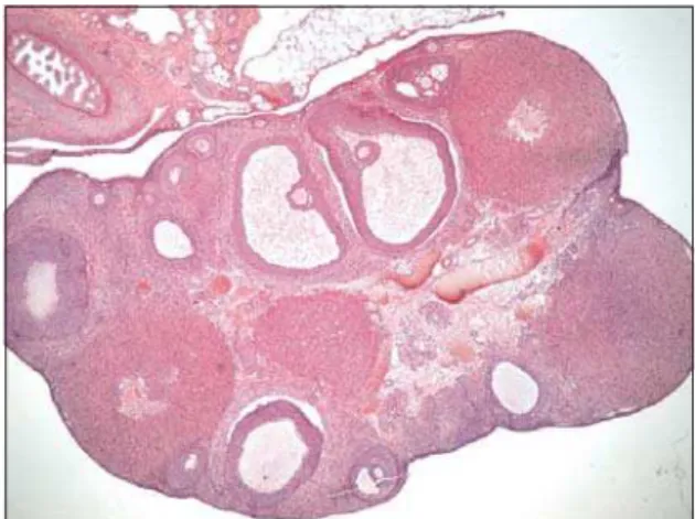
Histological examination. In the ovaries of control animals the cortex and
medulla are clearly distinguishable (Figure 2). The cortex contains follicles at different stages of development and atresia, corpora lutea and sparse interstitial cells between follicles. Interstitial cells (IC) are mostly in the medulla, forming clusters which are separated by undifferentiated stromal cells. Vesicles of different size were seen mostly at the periphery of pale cytoplasm of interstitial cells. Many lipid droplets are seen in the intercellular and perivascular spaces (Figure 3).
Ovaries of treated rats showed that the follicles are mostly in the deeper ovarian cortex, comparing to control animals. Medulla and cortical stroma contain
Figure 2. Ovary of control rat, clearly distinguishable cortex and medulla, follicles at different stages of development and atresia, corpora lutea and sparse interstitial cells between follicles, HE x 4
an unusually large amount of interstitial tissue (Figure 4). Most of the interstitial cells are large and polyhedral in shape and contain an abundance of closely packed lipid vesicles, relatively uniform in size (Figure 5).
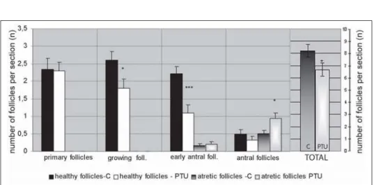
Morphometric analysis. The number of activated ovarian follicles was
significantly reduced in treated animals (p<0.05). During initial recruitment there was no appreciable difference, but the dynamic of later ovarian follicle development has been changed. The number of healthy growing (p<0.05) and early antral follicles (p<0.001) was markedly decreased in treated rats. Ovaries of PTU-treated animals contained relatively fewer antral follicles and significantly more atretic antral follicles (p<0.05) compared to controls (Figure 6).
Figure 5. Semithin section of atretic follicle and interstitial cells in ovary of treated rat, methylene blue, x 40
In the ovaries of 10% of treated animals which did not reach estrus, there were no corpora lutea. In the ovaries of the remaining 90% of treated rats, significantly fewer corpora lutea were found (p<0.001). There were no differences in the mean diameter of CL of treated animals compared to controls (670±2.91mm and 650±4.37mm, respectively).
DISCUSSION
Hypothyroidism is the most frequent endocrine disorder in the early period of development in humans and animals. However, the mechanisms, and even all the consequences on mammalian litters caused by hypothyroidism of the mother during pregnancy and lactation have not been completely understood. It was stated that the human and rat fetus is completely independent from the mother's thyroid hormones because they do not pass the placental barrier until the last third of pregnancy, in the period when the fetal thyroid gland is beginning its functional activity. The rat thyroid gland secrete thyroid hormones about day 17 p.c. At the same time thyrotropic cells in the adenohypophysis are identified for the first time (Begeotet al., 1981; Dussault and Labrie, 1975). Later examinations
revealed that thyroid hormones can be demonstrated in rat embrionic tissues using RIA 4 days after uterine implantation (Obregon et al., 1984) and their
receptors on embrionic days 13-14 (Falconeet al., 1994). This would suggest the
transplacental origin of these hormones, and potential influence of mother's thyroid hormones on fetuses. In our experiment, increased TSH and reduced serum T4 and T3 concentration, have shown a hypothyroid status in treated mothers. However, the levels of measured hormones on E-day were not significantly different between control and PTU-treated rats, but a small increase in serum T3and T4with modestly decreasing concentration of TSH was seen.
We have previously shown a significant increase of serum TSH and decrease in T3and T4level in 15-day-old treated rats under equal experimental conditions. Thirty-day-old treated rats had normal serum T3and T4with markedly elevated TSH compared to controls (Radovanovi}, 2003). Similar results were obtained by Stephan et al. (1993) who demonstrated that the
methimazole-treated pups were hypothyroid, with markedly higher TSH and lower T4
concentrations, until weaning, after which they transiently became hyperthyroid at week five, but returned to euthyroid state at week six.
It has also been noted in our present study that PTU treatment results in decreased body mass. While in control animals there were no remarkable individual variations, in the treated group, two subgroups formed animals (23.08%) whose body mass was lower from the lowest value of the control, and a subgroup with an average mass 15.38% higher than the highest in the control group. Zertashia et al. (2002) reported that body weight of offspring in the
postnatal PTU-treated group (0.1% PTU) was significantly decreased, while prenatal PTU-treatment induced a significant increase in prepubertal and pubertal rats compared to the control group. Furthermore, in our experimental model in 10% of animals ovulation did not occur until the 68th day, when they were sacrificed. These rats belong to the subgroup with the lowest body mass. All treated offspring showed reduction of ovarian mass, as a result of a decreased number of activated follicles and follicles which are able to ovulate and form corpora lutea. As a consequence of intensive follicular atresia in the previous period, secondary interstitial cells become dominant structures in the ovaries of treated animals (Radovanovi}, 1993; 2003).
The initial recruitment of follicles in the studied period is not affected in treated animals compared to controls, but dysfunction occurs in cyclic recruitment. This process starts in the secondary follicles when thetheca folliculi
is well developed, with functional receptors for gonadotrophic hormones (McGee and Huseh, 2000). These hormones, especially FSH, are important factors for the proliferation and survival of follicular somatic cells and the cyclic recruitment of antral follicles. FSH induced reduction of iRNA level for p-53 protein, which conducts cells in apoptosis and intensifies Bax protein synthesis, without changes in Bcl2 protein expression (Tilly and Tilly, 1995). Asahara et al. (2003)
demonstrated that T3 interacts synergistically with FSH to inhibit apoptosis in granulosa cells obtained from small follicles, without affecting proliferation of those cells. Oposite to FSH and T3, androgens synthesized by interstitial cells are important proapoptotic factors (Hsuehet al., 1994). In our experiment, with regard
to the respectable amount of interstitial cells, this might be the cause of an increasing number of atretic preantral and antral follicles. Another reason for the diminished number of ovulated follicles can be an increased level of mRNA for inhibin in hypothyroid rats (Tamura et al., 1998). Inhibin suppresses oestradiol
production and antagonizes activin actions and thus inhibits follicular growth and differentiation.
prenatal and early postnatal period caused an increased activation of follicles with an increased number of atretic follicles at all stages of folliculogenesis (most prominent in the population of antral follicles) compared to control animals (Radovanovi}, 2003).
According to clinical investigations, in hypothyroid women when T3 serum level is less than 80 ng/dL, no ovulation can be induced (Maruoet al.,1992). It was
shown that oocytes, cumulus and granulosa cells possess mRNA for thyroid receptors (TRa-1, TR-b-1, Trb-2 and c-erbaa-2), and that concentration of free T3 in the follicular fluid is similar to its blood concentration (Zhanget al., 1997). This
suggests that proper receptors are necessary for thyroid hormones to exert a direct influence on normal differentiation of all structures in preovulatory follicles.
In human medicine was reported that untreated thyroid disease may induce problems associated with premature ovarian failure and menopause (Buckler, 2007). Reduction in the number of follicles and corpora lutea, and hyperplasia and hypertrophy of stromal interstitial cells which is a common feature of ovarian atrophy in ageing rodents (OECD, 2009), imply premature ovarian failure in the ovaries of 60- day-old rats in our experiment.
The results presented in this paper indicate that although PTU-treated rats during prenatal and early postnatal period become euthyroid at 60 days, ovarian development was evidently impaired.
ACKNOWLEDGMENT:
This work was supported by the Ministry of Science, Republic of Serbia (grant No. 175061).
Address for correspondence: Anita Radovanovi} DVM, PhD
Department of Histology and Embryology Faculty of Veterinary Medicine
University of Belgrade 11000 Belgrade, Serbia E-mail: anitaªvet.bg.ac.rs
REFERENCES
1.Asahara S, Sato A, Aljonaid AA, Maruo T, 2003, Thyroid hormone synergizes with follicle stimulating hormone to inhibit apoptosis in porcine granulosa cells selectively from small follicles,Kobe J Med Sci, 49, 107-16.
2.Bagavandoss P, England B, Asirvatham A, Bruot BC, 1998, Transient induction of polycystic ovary-like syndrome in immature hypothyroid rats,Proc Soc Exp Biol Med, 219, 77-84.
3. Begeot M, Dupouy JP, Dubois MP, Dubois PM, 1981, Immunocytological determination of gonadotropic and thyrotropic cells in fetal rat anterior pituitary during normal development and under experimental conditions,Neuroendocrinol, 32, 285-94.
4.Buckler H, 2007, Thyroid disease and ovarian failure, Menopause Int, 13, 193.
5. Cristovao FC, Bisi H, Mendonca BB, Bianco AC, Bloise W, 2002, Severe and mild neonatal hypothyroidism mediate opposite effects on Leydig cells of rats,Thyroid, 12, 13-8.
7.Dowling ALS, Iannacone EA, Zoeller RT, 2001, Maternal Hypothyroidism Selectively Affects the Expression of Neuroendocrine-Specific Protein A Messenger Ribonucleic Acid in the Proliferative Zone of the Fetal Rat Brain Cortex,Endocrinol, 142, 390-9.
8.Dussault JH, Labrie F, 1975, Development of the hypothalamic-pituitary-thyroid axis in the neonatal life,Endocrinol, 97, 1321-4.
9.Falcone M, Takahide M, Fierro-Renoy F, Macchia E, Degroot LJ, 1994, Evaluation of the ontogeny of thyroid hormone receptor isotypes in rat brain and liver using an immunohystochemical technique,Europ J Endocrinol, 130, 97-106.
10. Fowden AL, Forhead AJ, 2004, Endocrine mechanisms of intrauterine programming, Reproduction, 127, 515-26.
11.Fowden AL, Giussani DA, Forhead AJ,2006, Intrauterine programming of physiological systems: causes and consequences,Physiology, 21, 29-37.
12.Holsberger DR, Jirawatnotai S, Kiyokawa H, Cooke PS, 2003, Thyroid hormone regulates the cell cycle inhibitor p27Kip1 in postnatal murine Sertoli cells,Endocrinology, 144, 3719-21. 13.Hsueh AJW, Billing H, Tsafriri A, 1994, Ovarian follicle atresia, Hormonally controlled apoptotic
process,Endocrine Rev, 15, 707-25.
14.Ipina SL, Ruiz-Marcos A, Escobar del Rey F, Morreale de Escobar G, 1987, Pyramidal cortical cell morphology studied by multivariate analysis, Effect of neonatal thyroidectomy, ageing and thyroxine-substitution therapy,Develop Brain Res, 37, 219-29.
15. Lauder JM, 1977, The effects of early hypo- and hyperthyroidism on the development of rat cerebellar cortex. III. Kinetics of cell proliferation in the external granular layer,Brain Res, 126, 31-51.
16.Maran RR, 2003, Thyroid hormones, their role in testicular steroidogenesis,Arch Androl49, 375-88. 17.Maruo T, Katayama K, Barnea ER, Mochizuki M, 1992, A role for thyroid hormone in the induction of
ovulation and corpus luteum function,Horm Res, 37, 12-8.
18.McGee EA, Hsueh AJW,2000, Initial and cyclic recruitment of ovarian follicles,Endocrine Rev, 21, 200-14.
19.Nicholson IL, Altman I, 1972, The effects of early hypo- and hyperthyroidism on the development of rat cerebellar cortex. I. Cell proliferation and differentiation,Brain Res, 44, 13-23.
20.Obregon MJ, Mallol J, Pastor M, Morreale de Escobar G, Escobar del Rey F, 1984, L-thyroxine and 3,5,3-triiodthyronine in rat embryos before onset of fetal thyroid function,Endocrinology, 114, 305-7.
21. OECD, 2009, Guidance Document for Histologic Evaluation of Endocrine and Reproductive Test in Rodents OECD document, ENV/JM/MONO(2009)11 No.107.
22. Ortega E, Rodriguez E, Osorio C, 1990, Activity of the hypothalamo-pituitary ovarian axis in hypothyroid rats with or without triiodothyronine replacement,Life Sci, 46, 391-5.
23. Porterfield SP, Hendrich CE, 1993, The role of thyroid hormones in prenatal and neonatal neurological development – current perspectives,Endocrinol Rev, 14, 94-106.
24. Radovanovi} A, 1993, Efekat hipotireoidizma majki i perinatalnog hipertireoidizma na jajnike juvenilnih pacova, Magistarska teza, Fakultet veterinarske medicine, Univerzitet u Beogradu. 25.Radovanovi} A, 2003, Uticaj smanjene funkcije {titaste `lezde na jajnike pacova tokom polnog
sazrevanja. Doktorska disertacija, Fakultet veterinarske medicine, Univerzitet u Beogradu. 26.Stephan DA, Menon RK, Franz B, Sperling MA, 1993, Thyroid hormone status correlates inversely
with expression of the growth hormone receptor gene in rats immediately after birth,J Dev Physiol, 19, 241-6.
27.Sto{i}-Bogdanovi} N, Radovanovi} A, 1992, The effect of hypothyroidism in pregnant and lactating dams on the testicles of juvenile pups,Acta Vet(Belgrade), 42, 99-108.
28.Tamura K, Hatsuta M, Watanabe G, Taya K, Kogo H, 1998, Inhibitory regulation of inhibin gene expression by thyroid hormone during ovarian development in immature rats, Biochem Biophys Res Commun, 242, 102-8.
30.Zahediasl S, 2010, Importance of thyroid hormones in intrauterine programming,Int J Endocrinol Metab, 8,186-7.
31.Zertashia A, Jalali S, Ahmad L, Mirza A, 2002, Effect of hypothyroidism induced by propylthiouracil on ovarian function and structure in offspring from treated mothers (rats),J Exp Zool, 293, 407-13.
32.Zhang SS, Carrillo AJ, Darling DS, 1997, Expression of multiple thyroid hormone receptor mRNAs in human oocytes, cumulus cells, and granulosa cells.Mol Hum Reprod, 3, 555–62.
UTICAJ INDUKOVANOG HIPOTIREOIDIZMA MAJKI NA RAZVOJ JAJNIKA MLADUNACA PACOVA
RADOVANOVI] ANITA, ROKSANDI] D, [IMI] MARIJA, MARKOVI] DANICA i GLEDI] D
SADR@AJ
U okviru ovog rada izu~avan je uticaj hipotireoidizma majki izazvanog propiltiouracilom (PTU) tokom graviditeta i laktacije na razvoj i sazrevanje jajnika pacova. U cilju odre|ivanja tireoidnog statusa majki i mladunaca, merena je kon-centracija tireoidnih hormona i tireostimuliraju}eg hormona (TSH) u serumu. Jaj-nici potomaka hipotireoidnih majki i kontolne grupe sprovedeni su za analizu svet-losnom mikroskopijom na dan prvog estrusa posle 60. dana starosti. Analizirani su serijski ise~ci jajnika bojeni hematoksilinom i eozinom, kao i polutanki preseci bojeni metilenskim plavim. Koncentracija T4i T3zna~ajno je smanjena u serumu tretiranih majki, dok je nivo TSH zna~ajno povi{en u odnosu na kontrole. Kod dva meseca starih kontrolnih i tretiranih `enskih potomaka nije bilo zna~ajne razlike u nivou svih odre|ivanih hormona.