J of Evolution of Med and Dent Sci/ eISSN- 2278-4802, pISSN- 2278-4748/ Vol. 4/ Issue 53/ July 02, 2015 Page 9180
OUTCOME, COMPLICATIONS AFTER CRANIOPLASTY FOLLOWING
DECOMPRESSIVE CRANIECTOMY: A PROSPECTIVE STUDY
Sathish Vandanapu1HOW TO CITE THIS ARTICLE:
Sathish Vandanapu. Outcome, Complications after Cranioplasty following Decompressive Craniectomy: A Prospective Study. Journal of Evolution of Medical and Dental Sciences 2015; Vol. 4, Issue 53, July 02;
Page: 9180-9185, DOI:10.14260/jemds/2015/1334
ABSTRACT: AIM AND OBJECTIVE: The aim and objective of this study is to evaluate the postoperative outcome, complications and cosmetic outcome after Methyl-methacrylate cranioplasty. METHODS: This study is a prospective study of two years duration. This study includes total no of 43 patients. RESULTS: The variables recorded were: age, sex, site, original diagnosis, site of defect, interval between craniectomy and cranioplasty, pre-operative symptoms, intra operative and post-operative complications, and cosmetic grading. All the skull defects were hemicranial (Involving more than two regions on one side). Intra operative dural tear occurred in two patients. Post-operative loosening of the graft seen in 4 patients and post-Post-operative subgaleal collection is seen in 4 patients. Post-operative seizures were seen in 5 patients. CONCLUSION: Methylmethacrylate cranioplasty is relatively safe, inexpensive, biologically inert and provides an excellent aesthetic reconstructive option.
KEYWORDS: Methylmethacrylate cranioplasty, Decompressive craniectomy.
INTRODUCTION: Skull defects most commonly result from trauma. Contaminated compound depressed skull fractures among civilians and penetrating head injuries among military personnel are the most frequent types. Among the possible indications for cranioplasty, the two commonly accepted ones, address issues of cerebral protection and appearance. The "Syndrome of the trephined," consisting of headaches, dizziness, intolerance, vibration and noise, irritability, fatiguability, loss of motivation and concentration, depression, and anxiety, universally accepted as an indication to reconstruct the skull.1,2
Four persuasive indications for cranioplasty,are restoration of cerebral protection, physical appearance, intracranial pressure relationships and the provision of an intact vault for the normal growth and development of cephalic structures in the young. Evidence supporting the organic basis of the syndrome of the trephined is the improvement in symptoms following cranioplasty with the reversion of intracranial pressure relationships to normal. Improvement in, electroencephalographic abnormalities, seizures, and neurological dysfunction (motor, speech) has been reported.1,3,4
Contraindications to cranioplasty include the presence of hydrocephalus, cerebral swelling, infection, a compound wound, contiguous functional paranasal sinuses (as indicated by air in a sinus on x-ray), and thin, scarred, or devitalized scalp. The timing of cranioplasty is critical for avoiding the development of infection in devitalized autografts or around alloplastic materials. It is generally accepted that cranioplastys should be delayed 3 to 6 months after compound wounds and at least 1 year after a wound infection.
J of Evolution of Med and Dent Sci/ eISSN- 2278-4802, pISSN- 2278-4748/ Vol. 4/ Issue 53/ July 02, 2015 Page 9181 post-operative complications and cosmetic grading. All the skull defects were hemicranial (Involving more than two regions on one side).
Inclusion Criteria: All traumatic intracerebral haemorrhages, hypertensive intracerebral haeorrhages.
Exclusion Criteria: Cases with cardiovascular disorder, depressed skull fractures, age less than 16 years.
Cranioplasty Procedure: The patient's head is fully shaved and prophylactic intravenous antibiotic is given prior to surgery. Old craniectomy scar is used as incision site. A scalp flap is carefully separated from the dura using sharp dissection, avoiding injury to the dura and the cranial defect is entirely exposed. Dural defects are repaired with watertight sutures or with pericranial fascia. A periosteal incision is made near the defects margin and reflected about 1-2cm away. In order to obtain good approximation of the cranioplastic plate, 3 to 5mm of outer table of defect edge is often trimmed. The cranial defect is measured intraoperatively.
Methyl methacrylate is prepared by mixing the liquid monomer (Catalyst) with the powder polymer. This mixture is constantly stirred until its consistency is doughy (Within 15 minutes). It is then place in between plastic layers and moulded according to the cranial defect with minimum 3 to 5mm thickness. The cranioplastic plate edge is trimmed with a Mayo scissors before it solidifies. Multiple angled holes are made through the outer cortical bone and acrylic plate. A nonabsorbable polyster suture material such as ETHIBONDTM 3/0 and titanium wires are inserted through the drill
holes and used to hold the cranioplasty plate in position. A subgaleal suction drain is placed and the flap closed in two layers. The subgaleal drain will be removed after 48 hours.
Cosmetic Grading: Grading system for cosmetic outcomes.5
Grade Components
A No evidence of surgery with mild prominence of temporalis
B Slight depression of temporalis fossa
C Marked depression of temporalis fossa with suggestion of zygomatic process
D Atrophy of temporalis muscle
E Atrophy with sliding/rooting of temporalis.
RESULTS AND ANALYSIS: This study includes total no of 43 patients, out of which 40 were male and 3 were female patients. 11 patients had pre-operative symptoms, out of which 3 patients had only
headache and 8 patients had syndrome of trephined . This is shown in table 1.
PREOPERATIVE SYMPTOMS No. of Patients Percentage
HEAD ACHE 3 6.97
SYNDROME OF TREPHINE 8 18.60
J of Evolution of Med and Dent Sci/ eISSN- 2278-4802, pISSN- 2278-4748/ Vol. 4/ Issue 53/ July 02, 2015 Page 9182 Out of 43 patients 38 patients comes under grade A, i. e, No evidence of surgery with mild prominence of temporalis and remaining 5 patients comes under grade B, that is Slight depression of temporalis fossa. This is shown in table no 2.
Cosmetic Grading No. of patients Percentage
Grade A 38 88.37
Grade B 5 11.63
Table 2: Post-Operative Cosmetic Results
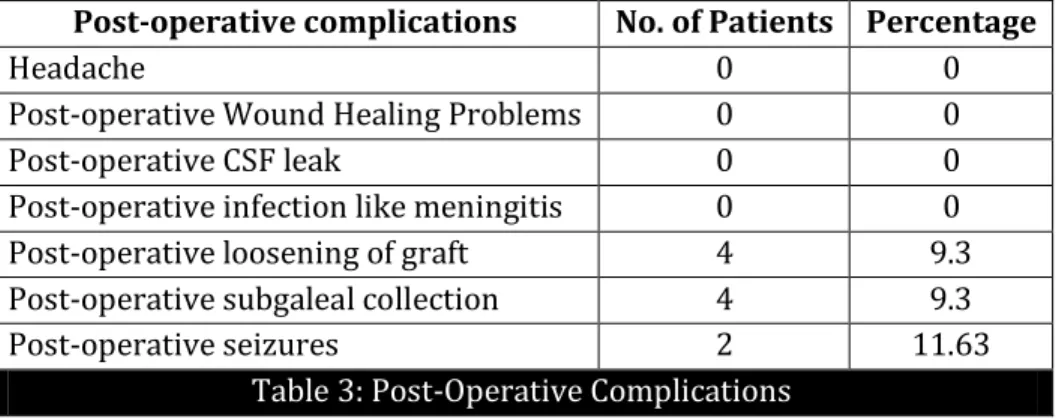
The median time interval between cranioplasty and initial surgery is 9 months. Intra operative dural tear occurred in two patients. Post-operative loosening of the graft seen in 4 patients and post-operative subgaleal collection is seen in 4 patients. Post-operative seizures were seen in 2 patients.
Post-operative complications No. of Patients Percentage
Headache 0 0
Post-operative Wound Healing Problems 0 0
Post-operative CSF leak 0 0
Post-operative infection like meningitis 0 0 Post-operative loosening of graft 4 9.3 Post-operative subgaleal collection 4 9.3
Post-operative seizures 2 11.63
Table 3: Post-Operative Complications
DISCUSSION: Common indication for decompressive craniectomies include traumatic brain injuries following road traffic accidents. In our study 39 patients underwent decompressive craniectomy following traumatic brain injury for road traffic accidents,4 patients underwent decompressive craniectomy following hypertensive gangliocapsular bleed. Meticulous cranioplasty is important for good cosmetic results, as well as long-term protection of brain from external environment.
Cerebral blood flow, brain metabolism, as well as neurological status are affected by the outside pressure in patients with skull defects.6,7,8)n our study 8 patients with Syndrome of trephined reported that they had improved after cranioplasty. Their main complaints of headache, insomnia, mental depression and local discomfort on movement had diminished. Hemiparesis improved in 3 patients. Others remained unchanged after cranioplasty.
Methyl methacrylate was first used as a cranioplastic material by Zander in 1940.9 The major
advantage of methyl methacrylate is, it is completely malleable in the initial stages of hardening, and thus can be moulded easily during surgery to fit the contour defects. It is relatively cheaper, biologically inert and does not interfere with computed tomography or magnetic resonance imagingstudies.10
The autopolymerization of methyl methacrylate during its preparation can cause thermal damage to the underlying brain due to its exothermic reaction. However, the acrylic can be irrigated with saline11 until it solidifies and can be safely put on the dura. In our study there was 4 loosening of
J of Evolution of Med and Dent Sci/ eISSN- 2278-4802, pISSN- 2278-4748/ Vol. 4/ Issue 53/ July 02, 2015 Page 9183 as a scaffold to improve the mechanical strength and cosmetic results, but this technique is more costly.
In our study 4 patients had loosening of the graft initially, when we used non absorbable suture material to hold the acrylic plate to the cortical bone. Later we used titanium wires to fix the acrylic plate to the cortical bone for preventing the loosening of the graft. After using Titanium wires to fix the acrylic plate to the cortical bone, there was no loosening of the grafts.
Complications from cranioplasty may be divided between those characteristic of the operative procedure in general and those related more to the type of implant used. Intra operative dural tear is seen in 2 patients. We closed with muscle graft before implantion of acrylic plate. The reported infection rate for methyl methacrylate is 3.8 to 12%.12 In our study there are no cases of
post operative wound healing problems, post operative CSF leak, post operative infection. The reported post-operative subgaleal collection after removal of drain is 12%13.In our study
post-operative subgaleal collection is seen in 4 patients (9.3%) and post post-operative seizures seen in 2 patients. The subgaleal collection resolved itself by applying compression dressing and post operative seizures were controlled by adequate dosage of antiepileptics.
We have adapted Raza et al5 grading system for cosmetic outcome. 4 patients had grade B
where we used non absorbable suture material to hold the acrylic plate to the cortical bone, and remaining 39 patients had grade A where we used titanium wires to fix the acrylic plate to the cortical bone.
CONCLUSION: Methylmethacrylate cranioplasty is relatively safe, inexpensive, biologically inert and provides an excellent aesthetic reconstructive option.
REFERENCES:
1. Grantham EG. Landis HP. Cranioplasty and the post-traumatic syndrome. J Neurosurg 1948; 5:19-22.
2. Walker AE. Erculei F. The late results of cranioplasty. ArchNeurol 1963; 9:105-110. 3. Rifkinson-Mann S. Cranial surgery in ancient Peru. Nero Surgery 1988; 23:411416.
4. Kent JN. Zide MF. Wound healing: bone and biomaterials. Otolaryngol Clin North Am 1984: 17:273-319.
5. Shaan M. Raza, M. D, Quoc-Anh Thai, M.D: Frontozygomatic Titanium Cranioplasty in Front osphenoteporal Pterional craniotomy. Operative neurosurgery, March 2008, volume 62, 264.
6. Yoshida K, Furuse M, Izawa A, Iizima N, Kuchiwaki H, Inao S. Dynamics of cerebral blood flow and metabolism in patients with cranioplasty as evaluated by 133Xe CT and 31P magnetic resonance spectroscopy. J Neurol Neurosurg Psychiat 1996; 61:166 – 71.
7. Richaud J, Boetto S, Guell A, Lazorthes Y. Incidence des cranioplasties sur la function neurologique et le débit sanguine cérébral. Neurochirurgie 1985;31:183-8.
8. Suzuki N, Suzuki S, Iwabuchi T. Neurological improvement after cranioplasty. Analysis by dynamic CT scan. Acta Neurochir 1993;122:49-53.
9. Abhay Sanan, Stephen J. Haines. Repairing Holes in the Head: A History of Cranioplasty. Neurosurgery 1997; 40:588.
J of Evolution of Med and Dent Sci/ eISSN- 2278-4802, pISSN- 2278-4748/ Vol. 4/ Issue 53/ July 02, 2015 Page 9184 11. Ganes AS. Cranioplasty made easier. Surg Neurol 1978; 10(4): 285-87.
12. EC Benzil, K. Thammavaram, The diagnosis of infection associated with acrylic cranioplasties, neuroradiology (1990)32;151-153.
J of Evolution of Med and Dent Sci/ eISSN- 2278-4802, pISSN- 2278-4748/ Vol. 4/ Issue 53/ July 02, 2015 Page 9185
AUTHORS:
1. Sathish Vandanapu
PARTICULARS OF CONTRIBUTORS:
1. Resident, Department of Neurosurgery, Sri Ramachandra Medical College and Research Institute, Chennai.
FINANCIAL OR OTHER
COMPETING INTERESTS: None
NAME ADDRESS EMAIL ID OF THE CORRESPONDING AUTHOR:
Dr. Sathish Vandanapu H. No.-27-9-34,
2nd Lane, Kannavari Thota,
Guntur -522004. Andhra Pradesh.
E-mail: [email protected]