Decompressive craniectomy in
massive cerebral infarction
João Paulo Mattos1, Andrei Fernandes Joaquim2,
João Paulo Cavalcante de Almeida3,
Lucas Alverne Freitas de Albuquerque4, Élton Gomes da Silva2,
Horácio Armando Marenco2, Evandro de Oliveira5
ABSTRACT
Twenty one patients were submitted to decompressive craniectomy for massive cerebral infarct. Ten patients (47.6%) presented a good outcome at the 6 months evaluation, eight had a poor outcome (38%) and three died (14.2%). There was no outcome statistical difference between surgery before and after 24 hours of ictus, dominant and non-dominant stroke groups. Patients older than 60 years and those who had a Glasgow Coma Scale (GCS)<8 in the pre-surgical exam presented worst outcome at six months (p<0.05). Decompressive craniectomy for space-occupying large hemispheric infarction increases the probability of survival. Age lower than 60 years, GCS ≥8 at pre-surgical exam and decompressive craniectomy before signs of brain herniation represent the main factors related to a better outcome. Dominant hemispheric infarction does not represent exclusion criteria.
Key words: cerebral infarction, decompressive hemicraniectomy, surgical decompression.
Craniectomia descompressiva no infarto cerebral extenso
RESUMO
Vinte e um pacientes foram submetidos a craniectomia descompressiva para o tratamento de infarto cerebral extenso. Dez pacientes (47,6%) apresentaram boa evolução em avaliação após 6 meses, 8 apresentaram evolução desfavorável (38%) e 3 faleceram (14,2%). Durante o seguimento, não se evidenciou diferença estatística na evolução entre pacientes operados antes e após 24 horas do ictus, nem entre lesões envolvendo o hemisfério dominante versus não dominante. Pacientes com mais de 60 anos e aqueles com Escala de Coma de Glasgow (ECG)<8 na avaliação pré-operatória apresentaram pior evolução após 6 meses (p<0,05). A craniectomia descompressiva para infartos hemisféricos extensos aumentam a probabilidade de sobrevivência. Idade abaixo de 60 anos e ECG ≥8 no exame pré-operatório e craniectomia descompressiva antes de sinais de herniação cerebral representam os principais fatores relacionados a uma melhor evolução clínica. Infarto hemisférico envolvendo o hemisfério dominante não representa um critério de exclusão.
Palavras-chave: infarto cerebral, hemicraniectomia descompressiva, descompressão cirúrgica.
Correspondence
Lucas Alverne Freitas de Albuquerque Rua Silva Paulet 2140 / 1402 60120-021 Fortaleza CE - Brasil E-mail: [email protected]
Received 13 May 2009
Received in final form 12 August 2009 Accepted 14 September 2009
Neurosurgery Division of Neurology and Neurosurgery Department, Campinas State University (UNICAMP), Campinas SP, Brazil: 1Assistant Neurosurgeon, Neurosurgery Department, UNICAMP; 2Resident of Neurosurgery, Neurosurgery Department, UNICAMP; 3MD, Federal University of Ceará (UFC), Fortaleza CE, Brazil; 4Medical Student, UFC; 5Professor of Neurosurgery, Neurosurgery Department, UNICAMP.
Ischemic stroke is a medical emergen-cy and the most common afection of the central nervous system (CNS). his is the second-leading cause of death worldwide and the irst cause of morbidity1. Ischemic stroke correspond to 85% of all strokes with a mortality of 10-50%. Large
“malignant” infarct and create the term malignant middle cerebral artery syndrome to describe the rapid develop-ment of fatal brain swelling6. Large space-occupying in-farction is generally secondary to an occlusion of the ca-rotid artery or the M1 segment of the middle cerebral ar-tery (MCA), including or not the anterior cerebral arar-tery (ACA) or the posterior cerebral artery (PCA). Neuroim-aging criteria varies between the authors: infarct volume on difusion-weighted magnetic resonance imaging (MRI) of more than 145 cm3; brain computed tomography (CT) ischemic changes afecting more than two-thirds of the MCA territory and including the basal ganglia; brain CT ischemic changes afecting at least two-thirds of the MCA territory with space-occupying edema; signs on CT of an infarct of at least 50% of the MCA territory, with or with-out additional infarction in the territory of the anterior or posterior cerebral artery on the same side5,7.
Many studies have suggested that decompressive sur-gery, consisting of a hemicraniectomy and duraplasty, re-duces mortality and improves outcome in patients with massive brain infarctions5,8.
We report our series of 21 patients treated with de-compressive craniectomy.
METHOD
Study design
In this retrospectively designed study, we describe the results of decompressive hemicraniectomy in 21 pa-tients with large hemispheric infarctions at UNICAMP Medical School Hospital from March 2003 to September 2007. We have considered massive cerebral infarction as brain CT ischemic changes afecting at least two-thirds of the MCA territory with space-occupying edema or both MCA and ACA or PCA infarctions leading to a minimum of 50% hemispherical volume compromised. We analyzed gender, age, Glasgow Coma Scale (GCS) on admission and pre-surgical evaluation, clinical status on pre-surgi-cal exam, time from initial symptoms to decompressive craniectomy, length of stay in the hospital and Glasgow Outcome Scale (GOS) 6 months after discharge. Data was subsequently analyzed for comparative study of patients with good to moderate outcome (GOS≥4) and patients with poor outcome (GOS≤3).
Patient selection
he decision to perform decompressive craniectomy was based on the presence of a space-occupying large
Stroke CT Yes
Trombolitic
No
Specific treatment
Yes
14-15 (<22)
Conservative treatment
Observation CT in 24h or if clinic deterioration
9-13 (>22)
No
Yes No
Decompressive hemicraniectomy +
duraplasty + ICP monitoring
Trombolitic criterea
GCS (NIHSS)
Clinical or radiological signals of malignant MCA ischemic stroke
No
GCS ≤ 8
Perfusion CT or MRI diffusion/
perfusion CT signals of poor outcome
>50y <50y
No
Poor clinical status Perfusion area>diffusion area
Yes
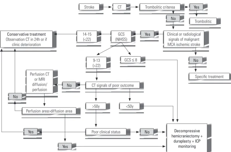
hemispheric infarction on CT scan and the clinical sta-tus of the patients. Patients with GCS >13 and no mid-line shift or basal cistern compression at initial evaluation were managed in the intensive care unit. Neurological de-terioration or development of brain herniation signs were indications to decompressive craniectomy as patients ini-tially presented with GCS ≤8 and cistern compression or midline shift at CT scan. Figure 1 shows our approach to ischemic stroke. he patients presented with GCS be-tween 9 and 13 were individually managed.
Surgical technique
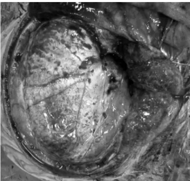
A question mark-shaped skin flap based on the ear and a wide craniotomy was performed on the afected side with partial removal of the frontal, temporal, and pa-rietal bones, so that the floor of the middle fossa could be exposed and the bone flap have a minimum of 12 cm di-ameter. he dura was opened in a “C” shape all over and 1cm distant to the border of the craniotomy. Homologous
temporal fascia was placed into the incision for volume-enlarged dural repair (Fig 2). he bone flap was placed in a subcutaneous pocket overlying the abdomen for pres-ervation until subsequent cranioplasty.
Data analysis
To make possible the comparison between the dif-ferent studies, the outcomes were classiied into 4 spe-ciic categories (Table 1): grade 1 (G1) functionally inde-pendent; grade 2 (G2) mild to moderate disability; grade 3 (G3) severely disabled; and grade 4 (G4) death8. Good outcomes were deined as functionally independent or mild to moderate disability. Poor outcomes were deined as severe disability or death. It was not necessary that the study fulill all the criteria listed, for instance, a G1 out-come could be based only on a GOS of 5, or only on a Barthel index >90, or on a mRS 0-1, not been necessary all three classiications.
Statistical analysis
All data are expressed as mean ± standard deviation (SD). Mann-Whitney U non-parametric tests, t tests and Fisher’s exact test were used for analysis of statistical evi-dence, with p<0.05 considered signiicant. Statistical soft-ware, SPSS 14.0 (SPSS Inc., Chicago, IL) was used for sta-tistical analysis.
RESULTS
A total of 21 patients (16 males and 5 females) were submitted to decompressive craniectomy during the pe-riod analyzed by the study (Table 2). he mean age was 50.09±14.29 years. On admission, the mean GCS was 12± 2.42 points (range from 6 to 14).
he mean GCS on immediate pre-surgical evaluation was 8±2.19 points. Nine patients (42.85%) presented with pupillary changes on pre-surgical evaluation; afasia oc-curred in six cases (28.5%) and hemiplegia presented in all patients.
Seventeen patients (80.95%) had “malignant” MCA infarction and 4 (19.04%) had associated ACA territo-ry infarction. he dominant hemisphere was afected in 6 cases (28.5%) and the non-dominant hemisphere in 15 cases (71.4%).
Fig 2. A right sided question mark incision. The skin lap and the temporal muscle were dissected on a separate manner to pro-vide an increase in the exposure of the middle fossa. Than a dura-plasty using aponeurotic galeal lap after decompressive craniec-tomy for brain expansion.
Table 1. Analyze of outcome.
Grade Characteristics (6) Outcome
G1 Functionally independent - BI ≥90; or mRS 0-1; or GOS 5 Good
G2 Mild to moderate disability - BI=60 to 89; or mRS 2-3; or GOS 4 Good
G3 Severely disabled - BI <60; or mRS 4-5; or GOS 2 to 3 Poor
G4 Death Poor
Time between onset of symptoms and decompressive craniectomy was less than 24 hours in 10 (47.61%), 24-48 hours in 5 (23.8%), 48-72 hours in 4 (19%) and 72-96 hours in 2 cases (9.52%). We have considered that early surgery was a decompressive craniectomy in the absence of brain herniation signs and in patients with GCS >8 despite the length of hospital stay. It was carried out in 12 cases and late surgery in other 9 patients. Length of stay in the hos-pital was of 25.76±23.3 days. Surgery improved signii-cantly the GCS of patients comparing the immediate pre-operative scores (8.0±2.19) and immediate post-pre-operative GCS (11.62±4.41), (p<0.05). Occurrence of GCS <9 in the pre-surgical evaluation was associated to a higher length of hospital stay (30.06±25.3 versus 12.0±3.46, p<0.05).
here was no statistical signiicance in the outcome between men and women, surgery before and after 24 hours of ictus, left and right side stroke groups (p>0.05). Patients older than 60 years presented worst outcome at six months (1.75±0.957 versus 3.41±0.87 points in GOS, p<0.05). he presence of brain herniation signs represent-ed a variable associatrepresent-ed to poor prognosis, but statistical signiicance was not reached in this study (2.59±1.33 ver-sus 3.5±0.67 points in GOS, in patients with and with-out pupillary changes, respectively; p=0.09). According to the Mann-Whitney test, the group of patients who had a poor GCS in the pre-surgical evaluation presented a trend toward poor prognosis, however statistical signii-cance was not demonstrated (2.88±1.14 versus 3.8±0.44
points in GOS, p=0.08). However, when GCS ≤7 is used to predict poor prognosis, we reached statistical evidence (2.72±1.27 × 3.54±0.68 points in GOS, p<0.05).
Ten patients (47.61%) presented a good outcome at the 6 months evaluation. Eight patients who survived had a poor outcome (38%). hree patients of our series died (14.2%) after the surgical procedure, secondary to the pre-sented brain lesion and hemodynamic failure.
DISCUSSION
Patients with massive space-occupying hemispheric infarction have a poor prognosis, as mass efect usually develops rapidly with occurrence of clinical deterioration in the irst 2 to 4 days5,9,10. Decompressive surgery has been studied as a way to relieve the intracranial hypertension and tissue shifts related to mass lesions. Bendszus et al. in a case report study, analyzing perfusion CT before and after decompressive craniectomy showed the value of this procedure to spare the ischemic but not infarcted area11.
Non-randomized studies suggest that late and early decompressive surgery reduces mortality and increases the number of patients with a favorable functional out-come after massive hemispheric infarction compared to the conservative treatment4,5,8,10,12. Indeed, early decom-pressive surgery with duraplasty is related with even a better outcome9,12.
Several conservative measures have been proposed to limit brain tissue shifts and reduce intracranial pressure,
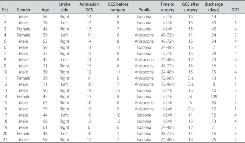
Table 2. Characteristics of 21 patients included in the study.
Pct Gender Age Stroke side Admission GCS GCS before surgery Pupils Time to surgery GCS after surgery discharge (days) GOS
1 Male 56 Right 14 8 Isocoria <24h 15 14 4
2 Male 28 Left 13 8 Isocoria <24h 15 33 3
3 Female 48 Right 13 7 Isocoria <24h 15 42 4
4 Female 59 Left 9 6 Anisocoria 48-72h 11 24 3
5 Male 53 Right 14 8 Anisocoria 48-72h 15 34 4
6 Male 56 Right 11 11 Isocoria 24-48h 15 7 4
7 Male 35 Right 15 8 Isocoria <24h 15 28 4
8 Male 42 Left 14 8 Anisocoria 24-48h 12 23 3
9 Male 21 Right 15 6 Anisocoria 48-72h 15 16 4
10 Male 30 Right 12 11 Anisocoria 24-48h 15 15 4
11 Female 39 Right 9 6 Anisocoria 72-96h Die 13 1
12 Male 71 Left 10 7 Anisocoria 72-96h Die 8 1
13 Male 56 Right 14 12 Isocoria <24h 15 10 3
14 Female 47 Right 13 8 Isocoria <24h 8 109 2
15 Male 63 Right 10 6 Anisocoria <24h 6 65 2
16 Male 79 Right 15 5 Anisocoria <24h Die 10 1
17 Male 44 Left 10 10 Isocoria <24h 11 15 4
18 Male 58 Right 13 13 Isocoria <24h 15 13 4
19 Male 61 Right 6 6 Isocoria 24-48h 12 21 3
20 Female 48 Left 10 7 Isocoria 48-72h 11 16 3
21 Male 58 Right 12 7 Isocoria 24-48h 14 25 4
including intensive care therapy, mild bed elevation, se-dation, hyperventilation, osmotic therapy, hypothermia and others. However, conservative treatment for massive brain infarction has been reported (Table 3) with a high mortality rate and poor outcome despite all those mea-sures, suggesting that they are of limited value6,13,14.
he three randomized trials, DECIMAL, DESTINY and HAMLET conirm these indings. DECIMAL and DES-TINY were interrupted because of a signiicant diference
in mortality favoring decompressive surgery and HAM-LET is still ongoing and aims to include 112 patients5,7,15,16. Several trials (Table 4) have described the effects of decompressive surgery on functional outcome after space-occupying infarction. As shown in Tables 3 and 4, the decompressive surgery increases the probability of survival from 25% to nearly 70% and the probability of a good outcome (G1 and G2) from 13% to 28%; however, the probability of surviving in a condition requiring
as-Table 3. Conservative treatment of massive cerebral infarction.
Paper Study design Patients Mean age Mortality Survivors outcome
Rieke et al.4 Open, nonrandomized, control trial 21/53 49 76% G1:0%; G2:10%; G3:14%
Holtkamp et al.2 Retrospective descriptive study 12/24 65 83% G1:0%; G2:0%; G3:17%
Kuroki et al.3 Retrospective/prospective descriptive study 7/15 79 85% G1:0%; G2:0%; G3:15%
HAMLET7 Multicenter prospective randomized open study 9/23 43 89% G1:0%; G2:11%; G3:0%
DECIMAL16 Multicenter prospective randomized open study 18/38 43 78% G1:0%; G2:22%; G3:0%
DESTINY15 Multicenter prospective randomized open study 15/32 46 53% G1:0%; G2:27%; G3:20%
Vahedi et al.5 Metanalysis of randomized trial 42/93 44 71% G1:0%; G2: 21%; G3:7%
Total – 82 51 75% G1:0%; G2:13%; G3:11%
G1 (BI ≥90; mRS 0 to 1; or GOS 5); G2 (BI 60 to 85; mRS 2 to 3; or GOS 4); G3 (BI <60; mRS 4 or 5; or GOS 2 to 3); G4 death.
Table 4. Surgical treatment of malignant middle cerebral artery infarction.
Paper Study design Pcts Mean age Treatment indication Mort Survivors outcome
Rieke et al.4 Open, nonrandomized, control trial 32/53 49 Late surgery 34% G1:0%; G2:18%; G3 47%
Holtkamp et al.2 Retrospective descriptive study 12/24 65 Late surgery 33% G1:0%; G2 + G3: 67%
Kuroki et al.3 Retrospective/prospective
descriptive study 8/15 72 Late surgery 12% G1:0%; G2:44%; G3:44%
Uhl et al.24 Multicenter retrospective
descriptive study
188 57 Late surgery 37% G1:1,6%; G2:18%; G3:49%
Woertgen et al.17 Retrospective descriptive study 48 48 Late surgery 26% G1:7%; G2:18%; G3:50%
Kilincer et al.9 Non–randomized prospective study 32 58 Late surgery 50% G1:0%; G2:0%; G3: 50%
Gupta et al.8 Metanalysis of non randomized
trials and retrospective study 138 50 12 studiesReview of 24% G1:7%; G2:35%; G3:34%
HAMLET7 Multicenter prospective
randomized open study 14/23 51 Randomized 21% G1:0%; G2:29%; G3:50%
DECIMAL16 Multicenter prospective
randomized open study 20/38 43 Randomized 25% G1:0%; G2:50%; G3:25%
DESTINY15 Multicenter prospective
randomized open study
17/32 43 Randomized 18% G1:0%; G2:48%; G3:35%
Vahedi et al.5 Metanalysis of 3 randomized trial 51/93 45 Review of 3
randomized studies
22% G1:0%; G2:43%; G3:35%
Our institution Retrospective descriptive study 21 50 Late surgery 14% G1:0%; G2:47% ;G3:38%
Total Non–randomized 435 54 31% G1:3%; G2:24% ;G3:44%
Total Randomized 51 45 22% G1:0%; G2:43%; G3:35%
Total – 486 53 30% G1:3%; G2:25% ;G3:43%
sistance and severe disability (G3) increases from 11% to 43%, probably due to the higher number of survivors. herefore, information about quality of life of survivors is essential for guiding the therapeutic decision5. In spite of previous reports in the literature difer with respect to the functional outcome and quality of life after decom-pressive surgery for space-occupying infarction5,17, even patients with aphasia may improve signiicantly18.
It is unclear which groups of patients beneit most from the procedure. Vahedi et al.5 demonstrated that sur-gery was beneicial (p<0.01) independently of age (above and below 50 years), presence of aphasia, and time to ran-domization (above and below 24 h) when compared to conservative treatment. Kuroki et al.3 describe that the decompressive surgery outcome is better than the con-servative treatment even in patient with more than 70 years old. Patients with the larger infarctions as found in the internal carotid artery (ICA) infarct were more likely to have a poorer prognosis as expected and according to Kilincer et al.9 surgery for an ICA infarction is not ben-eicial, unless exceptional cases as very young age, non-dominant hemisphere, and good clinical condition.
Identiication of patients at high risk of malignant ede-ma based on radiographic and clinical criteria might allow early hemicraniectomy, deined as a surgery performed before signs of brain stem herniation, as a mean of im-proving mortality and patient outcome6,9,10,12,19. Accord-ing to Schwab et al.10, early hemicraniectomy also reduc-es the time of critical care therapy from 13.3 to 7.4 days. Although no statistical signiicance was reached, we ob-served an important trend toward poor prognosis in the group of patients that had pupillary changes (p=0.09).
Radiographic signs such as early hypodensity of >50% of the MCA territory and/or additional vascular territo-ries (ACA or PCA)12,19-24, ICA infarct 9, midline shift ≥10 mm9,19, efacement of subarachnoid space 19,20, attenu-ation of corticomedullary diferentiattenu-ation19,20, presence of hydrocephalus19,25 may predict which patients will devel-op malignant edema or bad outcome. Infarct volume of more than 200 cm3 has 91% accuracy to predict malig-nant hemispheric infarction12 and the extent of infarct of more than two-thirds of MCA territory has a sensitivity of 93% and speciicity of 95% and they are the two most sensitive and speciic single explanatory variable for pre-diction of mortality19. On the other hand, brain edema is maximized after 24-72 hours19, so an early CT examina-tion should not be considered sensitive enough to predict the inal outcome19,20,25.
Clinical signs such as early clinical deterioration9, early nausea or vomiting22,23, and a National Institute of Health Stroke Scale (NIHSS) score ≥20 for left22,25 or ≥15 for right hemisphere infarction22, pre-operative GCS score ≤79, hypertension or heart failure, and increased
pe-ripheral white blood cell count12,22, also may predict which patients will develop malignant edema or have a poor outcome. Lam et al.19 indicate that a NIHSS >22 is dictive of high mortality. In our study, patients who pre-sented GCS <8 in the pre-surgical exam demonstrated a tendency toward poor outcome, which in our view indi-cates that the surgical approach should not be delayed un-til neurological deteriorations occurs.
Although elevated ICP was correlated with higher mortality26, ICP monitoring should not be the only pa-rameter in the determination of surgical timing as clin-ical signs of deterioration or herniation can precede the increase in ICP27.
Time from stroke to surgery has also been studied before. Non-randomized series have suggested that out-come is substantially improved if surgical treatment is ini-tiated within 24 h of stroke onset as compared with lon-ger time windows for treatment5,10,17. Schwab et al.10 pre-sented beneits of decompression before 24 hours after stroke. In a group of 31 patients, 26 (84%) had a BI >60 at follow up in their study. Gupta et al.8, however, did not show beneit to surgery<24 hours, probably due to a greater proportion of patients (64%) with signs of herni-ation before surgery in his group. Vahedi et al.5 in a sys-tematic review, conclude that the timing of surgery did not afect outcome. We did not observe diference in out-come between patients submitted to surgery before or af-ter 24 hours. We believe the reason for the no signiicant diference in timing of surgery was the poor general clin-ical status of patients who presented early to our depart-ment. In our view, timing between clinical deterioration and surgery and immediate pre-operative GCS are both more relevant factors than timing from stroke to surgery.
he age has been demonstrated to be an important predictor of outcome in decompressive hemicraniec-tomy. There are reports of poor functional outcomes and increased mortality in older patients compared to younger2,28. he cut-of point age to predict a good out-come is uncertain. Wijdicks and Diringer29 studied the natural history of 42 patients with MCA territory infarc-tion, 3 of 11 patients (28%) <45 years died, whereas 20 of 22 patients >45 years, 90.9% died. Important studies suggest that the optimal recovery occurs in patients less than 50 years8,24,28,30. However, Holtkamp et al.2 use a cut-of point cut-of 55 years and Kilincer et al.9, when selected 60 years as a cut-of point, provided one of the strongest pre-dictors of outcome. In our series, there was no statistical diference when used a cut-of age of 50 years. However, patients older than 60 years presented worst outcome at six months follow up.
quali-ty of life because of hemiplegia and aphasia28. he side of the infarct did not have prognostic relevance in our study, as demonstrated by other series8,9,17,24. In Gupta et al.8 re-view, the 27 patients who had decompression of the dom-inant hemisphere had functional outcome similar to the 111 patients who had non-dominant infarcts. In Kilincer et al.9, half of the patients had dominant hemispheric in-farction with global aphasia preoperatively, 6/7 patients in the good outcome group had a dominant hemispher-ic infarction and most of the patients showed consider-able improvements in their aphasia, a inding conirmed by other authors4,10,18,31. herefore, we believe infarction side should not be exclusion criteria for surgery. We agree with some authors that language deicits may be of small consequence in patients who are severely disabled by hemiplegia; also, non-dominant hemisphere strokes can lead to severe depressive, abulic, or neglect states that may interfere with rehabilitation eforts and are as dis-abling as aphasia8,32. On the other side, global disability scales such as the BI, mRS, and GOS may emphasize mo-bility as opposed to language dysfunction8,9.
It is not clear which patients may avoid severe disability after the procedure. A large number of patients or relatives (70%) stated that they would undergo the procedure again if faced to the same situation8. Even 79% of the patients and their family are satisied with the surgical results33.
In conclusion, decompressive craniectomy for space-occupying large hemispheric infarction increases the probability of survival that can yield good functional out-comes in some cases. Careful patient selection, made on an individual basis, and early operation may improve the functional outcome for large hemispheric infarction. In-formation about quality of life of survivors is essential for guiding such decisions because most patients require ex-tensive rehabilitative therapy and lifelong assistance.
here are limitations in our study. Although we pres-ent important data about decompressive surgery, it is a non-randomized retrospective study with results that need conirmation by larger randomized trials. Although there is not a consensus for the surgical treatment of mas-sive hemispheric infarction, we recommend: (1) in pa-tients under 60 years old; (2) in papa-tients with CT scan evidence of massive cerebral infarction with GCS ≥8; (3) decompressive craniectomy before signs of brain herni-ation if possible. Dominant hemispheric infarction does not represent an exclusion criteria.
REFERENCES
1. Murray CJ, Lopez AD. Mortality by cause for eight regions of the world: Glob-al Burden of Disease Study. Lancet 1997;349:1269-1276.
2. Holtkamp M, Buchheim K, Unterberg A, et al. Hemicraniectomy in elderly pa-tients with space occupying media infarction: improved survival but poor functional outcome J Neurol Neurosurg Psychiatry 2001;70:226-228. 3. Kuroki K, Taguchi H, Sumida M, et al. [Decompressive craniectomy for massive
infarction of middle cerebral artery territory]. No Shinkei Geka 2001;29:831-835.
4. Rieke K, Schwab S, Krieger D, et al. Decompressive surgery in space-occupy-ing hemispheric infarction: results of an open, prospective study. Crit Care Med 1995;23:1576-1587.
5. Vahedi K, Hofmeijer J, Juettler E, et al. Early decompressive surgery in malig-nant infarction of the middle cerebral artery: a pooled analysis of three ran-domised controlled trials. Lancet Neurol 2007;6:215-222.
6. Hacke W, Schwab S, Horn M, Spranger M, De Georgia M, von Kummer R. ‘Ma-lignant’ middle cerebral artery infarction: clinical course and prognostic signs. Arch Neurol 1996;53:309-315.
7. Hofmeijer J, Amelink GJ, Algra A, et al. HAMLET investigators. Hemicraniecto-my after middle cerebral artery infarction with Life-threatening Edema Trial (HAMLET): protocol for a randomised controlled trial of decompressive sur-gery in space-occupying hemispheric infarction. Trials Sep 2006;11:7-29. 8. Gupta R, Connolly ES, Mayer S, Elkind MS. Hemicraniectomy for massive middle
cerebral artery territory infarction: a systematic review. Stroke 2004; 35:539-543. 9. Kilincer C, Asil T, Utku U, et al. Factors affecting the outcome of decom-pressive craniectomy for large hemispheric infarctions: a prospective cohort study. Acta Neurochir (Wien) 2005;147:587-594.
10. Schwab S, Steiner T, Aschof A, et al. Early hemicraniectomy in patients with complete middle cerebral artery infarction. Stroke 1998;29:1888-1893. 11. Bendszus M, Mullges W, Goldbrunner R, Weigand A, Solymosi L.
Hemody-namic efects of decompressive craniotomy in MCA infarction: evaluation with perfusion CT. Eur Radiol 2003;13:1895-1898.
12. Mori K, Nakao Y, Yamamoto T, Maeda M. Early external decompressive craniectomy with duroplasty improves functional recovery in patients with massive hemispheric embolic infarction: timing and indication of decompres-sive surgery for malignant cerebral infarction. Surg Neurol 2004;62:420-430. 13. Berrouschot J, Sterker M, Bettin S, Köster J, Schneider D. Mortality of space-occupying (‘malignant’) middle cerebral artery infarction under conservative intensive care. Intensive Care Med 1998;24:620-623.
14. Hofmeijer J, van der Worp HB, Kappelle LJ. Treatment of spaceoccupying ce-rebral infarction. Crit Care Med 2003;31:617-625.
15. Jüttler E, Schwab S, Schmiedek P, et al. Decompressive Surgery for the Treat-ment of Malignant Infarction of the Middle Cerebral Artery (DESTINY): a ran-domized, controlled trial. Stroke 2007;38:2518-2525.
16. Vahedi K, Vicaut E, Mateo J, et al. Sequential-design, multicenter, random-ized, controlled trial of Early Decompressive Craniectomy in Malignant Mid-dle Cerebral Artery Infarction (DECIMAL Trial). Stroke 2007;38:2506-2517. 17. Woertgen C, Erban P, Rothoerl RD, Bein T, Horn M, Brawanski A. Quality of
life after decompressive craniectomy in patients sufering from supratento-rial brain ischemia. Acta Neurochir (Wien) 2004;146:691-695.
18. Kastrau F, Wolter M, Huber W, Block F. Recovery from aphasia after hemicraniecto-my for infarction of the speech-dominant hemisphere. Stroke 2005; 36:825-829. 19. Lam WW, Leung TW, Chu WC, Yeung DT, Wong LK, Poon WS. Early comput-ed tomography features in extensive middle cerebral artery territory infarct: prediction of survival. J Neurol Neurosurg Psychiatry 2005;76:354-357. 20. Haring HP, Dilitz E, Pallua A, et al. Attenuated corticomedullary contrast: An
early cerebral computed tomography sign indicating malignant middle ce-rebral artery infarction. A case-control study. Stroke 1999;30:1076-1082. 21. Kasner SE, Demchuk AM, Berrouschot J, et al. Predictors of fatal brain edema
in massive hemispheric ischemic stroke. Stroke 2001;32:2117-2123. 22. Krieger DW, Demchuk AM, Kasner SE, Jauss M, Hantson L. Early clinical and
radiological predictors of fatal brain swelling in ischemic stroke. Stroke 1999; 30:287-292.
23. Robertson SC, Lennarson P, Hasan DM, Traynelis VC. Clinical course and surgi-cal management of massive cerebral infarction. Neurosurgery 2004;55:55-62. 24. Uhl E, Kreth FW, Elias B, et al. Outcome and prognostic factors of hemicraniec-tomy for space occupying cerebral infarction. J Neurol Neurosurg Psychiatry 2004;75:270-274.
25. Barber PA, Demchuk AM, Zhang J, et al. Computed tomographic parame-ters predicting fatal outcome in middle cerebral artery infarction. Cerebro-vasc Dis 2003;16:230-235.
26. Frank JI. Large hemispheric infarction, deterioration, and intracranial pres-sure. Neurology 1995;45:1286-1290.
27. Schwab S, Aschof A, Spranger M, Albert F, Hacke W. The value of intracranial pressure monitoring in acute hemispheric stroke. Neurology 1996;47:393-398. 28. Carter BS, Ogilvy CS, Candia GJ, Rosas HD, Buonanno F. One-year outcome
after decompressive surgery for massive non-dominant hemispheric infarc-tion. Neurosurgery 1997;40:1168-1176.
29. Wijdicks EF, Diringer MN. Middle cerebral artery territory infarction and ear-ly brain swelling: progression and efect of age on outcome. Mayo Clin Proc 1998;73:829-836.
30. Pranesh M B, Dinesh Nayak S, Mathew V, et al. Hemicraniectomy for large middle cerebral artery territory infarction: outcome in 19 patients. J Neurol Neurosurg Psychiatry 2003;74:800-802.
31. Kalia KK, Yonas H. An aggressive approach to massive middle cerebral artery infarction. Arch Neurol 1993;50:1293-1297.
32. Walz B, Zimmermann C, Böttger S, Haberl RL. Prognosis of patients after hemicraniectomy in malignant middle cerebral artery infarction. J Neurol 2002;249:1183-1190.