International Journal of Advanced Biotechnology and Research (IJBR) ISSN 0976-2612, Online ISSN 2278–599X,
Vol-7, Special Issue-Number5-July, 2016, pp350-358 http://www.bipublication.com
Research Article
Molecular Analysis of G1691A Mutation in Factor 5 Leiden and its relation
with mtDNA common deletion in Human Recurrent Pregnancy Loss
Negin Garoosi¹, Hussein Rasi*² and Mahdi Goudarzvand³
1
Department of Biology,
Karaj branch, Islamic Azad University, Karaj, Iran
2
Department of Biology,
Karaj branch, Islamic Azad University, Karaj, Iran
3Department of Physiology and Pharmacology, School of Medicine,
Alborz University of Medical Sciences, Karaj, Iran. *Correspondence Author: [email protected]
ABSTRACT:
Background: In general, miscarriage is one of the most common complications of pregnancy and it is the
pregnancy loss before 20 weeks of gestation or birth weight below500 grams. Recurrent Pregnancy Loss is defined as two or more spontaneous miscarriages. Genetic disorders such as mutations can be involved in miscarriage. Considering the importance of this issue, in this study, G1691A mutation of coagulation factor 5 and common deletion mutation in the mitochondrial genome(4977-bp deletion in mtDNA) were investigated as factors which can influence miscarriage, especially recurrent miscarriage.
Materials and Methods: For this study 41 patients with the history of miscarriage and 48 healthy women with successful delivery were selected and completed the questionnaires which included questions such as miscarriage history, age, blood type and then Blood samples were taken. After extraction of DNA from each sample, the studied mutations were determined using PCR method. At the end, analysis of the results and assessment of other important and effective factors in them was done using Epi Info software and using chi square (X2) test.
Results: Among the patients ,there were 29.25% patients with one miscarriage, 65.85% patients with two miscarriages and 4.9% patients with three miscarriages. There was no homozygous genotype in the study of G1691A mutations in both groups, and prevalence of heterozygotes was 17% among patients and 4.17% among controls. On the other hand, frequency of 4977-bp deletion in mtDNA in patients group and control group was 68.29% and 14.58%, respectively. Analysis showed that frequency of G1691A mutations and common deletion mutation in mtDNA in patients group were higher than controls and were statistically significant . Although the opportunity to have miscarriage in GA genotype and carriers of common deletion is more than control, but there is not any correlation between these two mutations and their inheritability and also they have not affected each other. Therefore, we can use the above mutations as a molecular marker for early detection and timely treatment in patient’s likelihood of miscarriage.
Keywords: G1691A, Factor 5 Leiden, common deletion in mtDNA, recurrent miscarriage
INTRODUCTION
Miscarriage is one of the most common problems in pregnancy and includes about 15% to 25% of all diagnosed pregnancies. Recurrent pregnancy loss is also a type of miscarriage defined as two or more clinical pregnancy loss before 20 weeks of pregnancy and affects approximately 1% to 5% of women of reproductive age all over the world based on the number (1-2-3-27-28). Although various factors
remain unknown (2-10-28). Among these mentioned factors, 2% to 5% of miscarriages are due to genetic abnormalities like mutations (2). One of the important mutation is the G1691A mutation in Coagulation Factor 5 (factor 5 Leiden) which have particular importance in the creation of thrombophilia. One of the other important mutations,is the common deletion mutation in mitochondrial DNA which cause 4977-bp deletion in mtDNA and increase possibility of occurrence in mitochondria in case of mitochondrial oxidative stress (as a center for the production of free radicals of oxidative stress). G1691A point mutations in Coagulation Factor 5 known as Factor 5 Leiden.In this mutation, Factor 5 is resistance to break down and remains active for a longer time that is associated with an increased risk of thrombosis, and then aberrant blood clot or thrombus build in the capillaries of the placenta can cause confusion in the exchange of substances between mother and fetus ultimately results in miscarriage (5-9-19-21-22). Hussein Najmabadi et al (2009) examined the genetic Factor 5 Leiden which create thrombophilia, as a genetic factor for cardiovascular disease and there was no mutation of Factor 5 Leiden as homozygous and the frequency of G1691A mutation all over Iran was 2.4% in average with different rates in different ethnicities(3). Karim Shams Asanjan et al (2013) studied genetic mutation in Coagulation Factor 5 gene in the normal population of Eastern Azerbaijan province in order to be effective in prognosis of thrombotic disorders, cardiovascular disease, RSA, etc. and declared the allelic frequencies in the population (4). Oztork Nesrin et al (2014) in another study studied Coagulation Factor 5 mutation in 201 patients and found that the presence of allele A in G1691A mutation as heterozygous increases the possibility of venous thrombosis (blood clot in the vein) (5). Ashraf Moeini et al (2010) investigated thrombophilia factors association with pregnancy complications in women with polycystic ovary. The results show that there is a significant statistical difference between women with polycystic ovary syndrome and healthy women in term of pregnancy complications records but
there is no significant difference in terms of Thrombophilia factors (Factor 5 Leiden) and the relationship between these factors with pregnancy complications records between those two groups (6). Wasim Almawi et al (2002) investigated the prevalence of G1691A mutation in the Factor 5 gene and G20210A mutation in Factor 2 gene in women with recurrent pregnancy loss and it was found that the prevalence of both mutations is several times higher in patients with recurrent pregnancy loss than the control population (8). A study by Cornelia Wolf et al. (2003) investigated the recurrent pregnancy loss and its relationship with the G1691A mutation in the Factor 5 gene, G20210A mutation in Factor 2 gene and mutation in plasminogen gene inhibitors- activator, and the results showed that G1691A mutation in the Factor 5 gene was more prevalent in patients with recurrent pregnancy loss (9). common deletion mutation in the mitochondrial DNA , is important in many diseases,too.A study by Zivar Salehi et al(2013),expressed that there is a correlation between common deletion mutation in mtDNA and peptic ulcer(23).Zahiri et al(2013)studied the common deletion mutation in mtDNA in women with spontaneous miscarriage and they saw the increase risk of spontaneous miscarriage with this mutation (14).So, the current study examines the prevalence of mutations in the coagulation factor 5 (factor 5 Leiden) and the prevalence of common deletion mutation in the mitochondrial genome and If there are, the relationship between these two mutations, as well as review some important affecting factors, so that if possible to determine the prevalence and the factors affecting it help in early detection and early treatment of patients prone to be involved in abortion.
MATERIALS AND METHODS
method using Cinnagen kit (DN8115C) and were used separately to examine both mutations.
PCR reactions for G1691A mutations in Factor 5
After extraction of DNA ,PCR reaction was performed to detect G1691A mutation in Factor 5 Leiden using Cinnagen kit (PR8251C) and specific primers for this mutation according to the following protocol
Table 1. The sequence of primers used in the identification of G1691A mutations in Factor 5 Leiden
Mutation sequence of primers PCR product
FV G1691A
(C): 5'-GGA CTA CTT GAC AAT TAC TGT TCT CTT G-3' (N): 5'-GCA GAT CCC TGG ACA GAC G-3'
(M): 5'-GCA GAT CCC TGG ACA GAC A-3'
150bp
Table 2. PCR Materials required for G1691A mutations in Factor 5
Materials Quantities
Master Mix 12/5µl
Forward Primer 1µl
Reverse Primer 1µl
Distilled water 7µl
DNA 3/5µl
Total Volume 25µl
Table 3. PCR thermal cycles for G1691A mutation in Factor 5
Number of cycle Incubation
Time Temperature
Step
1 10 min
95oc
First Denaturation
10
30 sec 94oc
Denaturation
30 sec 60oc
Annealing
1min 72oc
Extension
25
30 sec 94oc
Denaturation
30 sec 55oc
Annealing
1min 72oc
Extension
1 5min
72oc Final Extension
PCR reaction for 4977 bp mutation (common deletion) in mtDNA
PCR reaction was performed to detect common deletion in mtDNA using Cinnagen kit (PR8251C) and specific primers for this mutation according to Cinnagen protocol.
Table. The sequence of primers used in the identification of common deletion in mtDNA
Table 5. PCR Materials required for common deletion mutation in mtDNA 13.0µl PCR Master Mix
2.0µl Primer Forward
2.0µl Primer Reverce
3µg DNA Template
5.0µl dH2O
Table 6. PCR thermal cycles for common deletion mutation in mtDNA
Number of cycle Incubation Time
Temperature Step
1 Cyc 10min
95˚C Pre-PCR heat step
35Cyc 30s
95˚C Denatuation
30s 60˚C
Annealing
45s 72˚C
Extension
Product PCR Primer sequences
Mutation
485bp 5′-TTC-TCC-TAG-ACC-TAA-CCT-GA-3′
MtDNA1
485bp 5′-GGA-TAT-ACT-ACA-GCG-ATG-GC-3′
MtDNA2
127bp 5′-TGT-GGT-CTT-TGG-AGT-AGA-AAC-C-3′
MtDNA3
127bp 5′- CCT-TAC-ACT-ATT-CCT-CAT-CAC-C-3′
Then, specimens were loaded on the agarose gel(1% ) and after electrophoresing, they were observed by UV Transbillimator and evaluated.
Statistical analysis
Epi Info software version 7.1.3.10 and Chi-Square test (x2) were used to analyze the data.
RESULTS
All members of two groups were investigated and then results were classified .
Patients classification based on the number of miscarriages
Frequency of miscarriage samples in patients group is presented in table 7. There were 29/25% patients with sporadic pregnancy loss, 65/85% with two and 4/9% with three pregnancy losses
Table 7. Classification of patients based on the number of miscarriage
Number of miscarriage Number of patients Percentage
Once 12 29.25%
Twice 27 65.85%
Three times 2 4.9%
Patients classification based on theage
A total of four age groups were determined. According to the survey, the lowest number of miscarriages was in the age group below 20 years with a frequency of 17.07% and the highest number of miscarriages was in patients older than 31 years with a frequency of 36.58%.
Table 8. Classification of patients based on age
Percentage No. Age group
17.07% 7 Below20
21.95% 9 21-25
24.39% 10 26-30
36.58% 15 Over 31
Patients classification based on theblood type
The results of patients classification based on the blood type are shown in Table (9). The most frequent blood type is O positive blood type (36.58%) and AB negative was not observed among individuals.
Table 9. Classification of patients based on blood type
Percentage No. blood type 17.1%
7 A+
7.3%
3 A
-9.75%
4 B+
7.3%
3 B
-4.87%
2 AB+
0%
0 AB
-36.58%
15 O+
17.1%
7 O
-Patients classification based on theconsanguinity
In the survey, there were 8 incidents of consanguineous marriages in patient groups and 2 cases in controls. Therefore, statistical analysis shows a significant difference in two groups of patients and controls in terms of marriage and parental consanguinity has increased miscarriage rate.
Table 10. Classification of patients based on consanguinity marriage
Percentage No. Consanguinity marriage
19.5% 8 Patients
4.16% 2 Non- patients
(X2=5.22, P<0.05)
Patients classification based on the family history of miscarriage
Table 11. Classification of patients based on the family history of miscarriage
Percentage No.
the family history of miscarriage
24.3% 10
Patients
8.33%
4
Non- patients (X2=4.30, P<0.05)
The frequency of G1691A mutation in coagulation factor 5 in the groups studied
The frequency of G1691A mutation in coagulation Factor 5 heterozygous was 17% in the patients group and 4.17% for the control group. homozygous mutation was not observed in the studied populations.
Table 12. The G1691A mutation frequency in the coagulation Factor 5 in sample groups Control Patient
Genotype
Percentage No.
Percentage No.
95.83% 46
82.93%
34
GG
4.17% 2
17.07%
7
GA
0 0
0
0
AA
Analysis of the frequency of GG and GA genotype in two groups of patients and controls were statistically significant (p≤0.05) and the likelihood of miscarriage for genotypes GA is about three times higher than the control group.
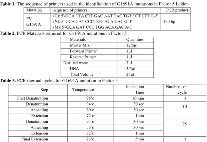
Figure 1. The results of electrophoresis of PCR products for the detection of G1691A mutation in coagulation
factor 5
In the figure, Lane M- 100 bp is molecular marker, lanes 1, 2 are for negative control specimens of normal and mutant type, lanes 3,4, 5 are normal type specimens and lanes 6-7 -8 are mutants specimens.
Frequency of patients with G1691A mutationin different age ranges
In this classification, the maximum heterozygous patients was for the Group over 31 years with a frequency of 60% but statistical analysis shows that there is no significant relationship between G1691A mutations and age.
Table 13. Frequency of patients with G1691A mutation in different age ranges Percentage No. Age group
20% 1 Below20
20% 1 21-25
0 0 26-30
60% 3 Over 31
Prevalence and statistical analysis of common deletion in mtDNA in the groups studied
the frequency of common deletion in mtDNA in both patients and controls was significant (p <0.01) and the risk of miscarriage for patients with this mutation are more than control group.
Table 14. The frequency of common deletion in mtDNA
Controls(No., percentage ) patients(No., percentage )
the presence or absence of common
remove mutations absence of mutation presence of mutation absence of mutation presence of
mutation
41 (85.42%) 7(14.58%) 13 (31.71%) 28 (68.29%)
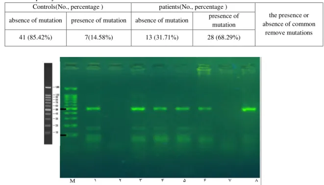
Figure 2. Electrophoresis of PCR products for the detection of common deletion in mtDNA
According to the figure, the bold bands close to 500 bp are 485 bp normal types and band close to 100 bp is common mutation band size 127 bp. Lane M, molecular marker sized 100 bp, lanes 1, 3, 4, 5 and 6 have common deletion in mtDNA with 127 bp band and normal type of 485 bp. Specimen No. 8 has no common deletionbut has normal band, but specimens No. 2 and 7are negative control samples where the DNA is replaced by distilled water.
Frequency of patients with common deletion in mtDNA in different age ranges
In this classification the most frequent age group which has the mutation, was over 30 years with the frequency of 50%. Statistical analysis (p <0.01) showed that common deletion in mtDNA occurrence and increasing age were significant, so that as age increases, the likelihood of common deletion in mtDNA increases.
Table 15. Frequency of patients with common deletion in mtDNA in different age ranges
Patients (No. and
percentage) Age group
1 (3.5%) Below20
6 (21.5%) 21-25
7 (25%) 26-30
14 (50%) Over 31
The relationship between factor V Leiden and common deletion in mtDNA
The statistical analysis shows that there is no significant correlation between the frequency of G1691A mutation in coagulation factor 5 and common deletion mutation in mitochondria genome. This means that both mutations produce no effect on each other, but independently effective on miscarriage.
DISCUSSION AND CONCLUSION
5's G1691A mutation causes a defect in coagulation pathway causing a misplaced blood clot or thrombus within the blood and since fetal health have direct contact with the maternal circulation any factor disruptive in this respect is detrimental to the fetus(20-21). There many researches in regard to G1691A mutation and Factor 5 Leiden. There are many studies that have introduced the mutation as a risk factor for recurrent miscarriage. While there are other reports that rejected the relationship between the mutations and recurrent miscarriage. In fact, the differences in the studies could be related to differences in the number of cases and also differences in inclusion criteria. The current study found significant relationship between G1691A mutations in Factor 5 (Factor 5 Leiden) and recurrent miscarriage in both groups of patients and control indicates approximately 3 times higher risk of miscarriage at the presence of this mutation. Rai et al (2001) studied women with RPL and found no increase in G1691A mutation compared to the control group (13). In another study by Rai et al (2006) three common mutations in thrombophilia factor 5 Leiden include mutations (G1691A) prothrombin (G20210A), and MATHFR (C677T) reviewed and confirmed that both patient group and the control group show the sme frequebcy of mutations but simaltaneous mutations in the patient group that increases the likelihood of recurrent pregnancy loss (11). On the other hand the results of the current study show (17%) prevalence of genotype heterozygous GA in the patients group and (4.7%) in the control group that is less compared to another study conducted by Behjati et al (2006) in an Iranian population with the 20% frequency of mutations in the patient population (13). In the study conducted by Behjati mutation was absent in the control group and generally in both studies G1691A mutation prevalence was higher in the patient group with significant difference between G1691A mutations and recurrent pregnancy loss in the two groups(7). These results are consistent with that of Wolf et al (2003) which showed a 10% frequency of factor 5 Leiden in patients with recurrent miscarriage and 19% frequency of factor 5 Leidenin infertility
with the results of Zahiri et al (2013) with the subject of “study of common deletion mutation in the mitochondrial genome of women with spontaneous miscarriage”, which showed the 30% and 6.66% prevalence of common deletion mutation in patients and controlls,respectively, with a relationship between the increased risk of spontaneous miscarriage.(14) A study by Sumitha et al (2015) at the age group of 21 to 45 years revealed that oxidative stress and damage to nuclear DNA and mitochondrial DNA in patients with recurrent pregnancy loss increases with age that is consistent with our results indicating the prevalence of mutaions with increased age (18). Generally, it can be concluded that the frequency of coagulation Factor 5 G1691A mutation and mitochondrial common deletion mutation were statistically significant and of in both groups of patients and controls with the several times higher risk of miscarriage in GA genotypes and carriers with a chance for common deletion mutation in mitochondrial DNA. However, there are no significant relationship between miscarriage and inheritance in terms of both mutations. But given the high incidence of it in RPL, the two above-mentioned mutations can be used as a molecular marker for early detection of individuals prone to miscarriage and timely treatment.
REFERENCES
1. Williams Obstetrics (Cunningham, Kenneth Leveno , Gill Strip, Huth , Westermann) 2010
2. Kashanyan M., Akbarian A. , Shbandvst Sh., “The effects of one occurrence of spontaneous miscarriage pregnancy on the next pregnancy”, Iran University of Medical Sciences, 2004, 463 Vol. XI, No. 41
3. Najmb Abadi H., Imanian H., Hadavi V., Kariminejad A., Rostami M., Abrokaniz K., “genetic study of factor 5 Leiden polymorphisms as genetic factor of cardiovascular disease in Iran population”, genetic the third millennium, winter, 2009,7 (4): 1844-1847
4. Shams Asanjan K., Pourakbar A., Farsh Dosti Haqi M., Karimiyan Fathi N.,
“Coagulation Factor 5 gene G1691A mutation in the normal population of East Azerbaijan province, Urmia Medical Journal, June 2013,24 (3): 219- 225
5. Ozturk ErÇelen N., Öztürk B., Cömert H.,
Diken M., Gültomruk M., Coşkun H., Akat
A.,Allelic Frequencies of Mutations in Blood Coagulation Factor Genes(Factor V,Factor II)and Methylenetetrahydrofolate Reductase (MTHFR)in 201 Turkish Patients with Venous Thrombosis Complications; J Mol Genet Med,2014;8:1
6. Moeini A., Sarrafiun F., Ziyaee S., Faghihzadeh S., “The relationship between thrombophilia factors and pregnancy complications in Pcos women”, bimonthly Journal of Shahed University, July 2010, Issue 87
7. Behjati R., Modarressi M. ,Jeddi- Tehrani M., Dokoohaki P., Ghasemi J.,Zarnani AH., Thrombophilic mutations in Iranian patient with infertility and recurrent spontaneous abortion. Ann Hematol 2006, 85 (4): 268-71 8. Almawi W.,Finan R.,Tamim H.,Ameen
G.,Sharida H.,Rashid M.,Prevalence of factor V G1691A(Factor V-Leiden)and Prothrombin G20210A Gene Mutations in a Reccurent Miscarriage Population,American Journal of Hematology ,2002 ; 71:300-305. 9. Wolf C., Haubelt H., Ulrich Pauer H.,
Hinney B., Krome-Cesar C., Legler T., Hellstern P., Emons G., Zoll B., Köhler M., Recurrent pregnancy loss and Relation to FV Leiden,FII G20210A and Polymorphisms of Plasminogen Activator and Plasminogen Activator Inhibitor,Pathophysiol Haemost Thromb,2003; 33:134-137.
10.Hizem S., Almawi W.,Finan R.,Tamim H.,Mahjoub T. ,Mtiraoui N.,Nsiri B. ,Association between adverse pregnancy outcomes and maternal factor V G1691A(Leiden)and Prothrombin G20210A genotypes in women with a history of recurrent idiopathic miscarriages,American Journal of Hematology,2005;80:12-19 11.Rai R., Jivraj S., Underwood J., Regan L.,
12.Glueck C., Gogenini S., Munjal J., Tracy T., Pranikoff J., Wang P., Factor V Leiden mutation:a treatable etiology for sporadic and recurrent pregnancy loss ,Fertil Steril 2008 Feb;89:410-416.
13.Rai R., Shlabak A.,Cohen H.,Backos M.,Holmes Z., Marriott K., Regan L., Factor V Leiden and acquired activated protein C resistance among 1000 woman with recurrent miscarriage, HumReprod 2001;16(5): 961-5 14.Zahiri Sarvari, Z, Askafi Sabet, Salehi Z.,
Ghanami., Gashti N., “To study of bp- 4977 deletion in mitochondrial DNA in women with spontaneous miscarriage”, Journal of Gilan University of Medical Sciences, January 2013, Issue twenty-second, No. 88, p. 1-6
15.Azem F.,Many A.,Ben Ami I.,Yovel I., Amit A., Lessing J.,Increased rates of thrombophilia in woman with repeated IVF failures .Hum Reprod 2004;19(2): 368-70. 16.Gluek C. ,Pranikoff J.,Aregawi D.,Haque
M.,Zhu B.,Tracy T., Wang P., The factor V Leiden mutation , high factor VIII, and high plasminogen activator inhibitor activity: etiologies for sporadic miscarriage, Metabolism ,2005 : 54(10 )1345-9
17.Glueck C., Wang P.,Bornovali S., Goldenberg N., Sieve L., Polysystic ovary syndrome,The G1691A factor V Leiden mutation , and plasminogen activator inhibitor activity: association with recurrent pregnancy loss, Metabolism 2003;52 (12) 1627-32
18.Sumitha Prabhu P.,Aneesh P.,Jiju J .,Reshma P.,Dinesh Roy D., Evidence of increased oxidative sress and DNA damages in women with recurrent miscarriages,International Journal of Scientific and Engineering Research,2015May;6(5)
19.Zoossmann-Diskin A.,Gazit B.,Peleg L.,Shohat M.,Turner D.,Thrombophilic polymorphisms in Israel,Blood Cells Mol Dis 2008;41:230-3
20.Silver Robert M., Fetal Death, Obstetrics and Gynecology,2007Jan;109(1)
21.Stief Thomas W., Single oxygen inactivates fibrinogen, factor V, factor VIII, factorX and
platelet aggregation of human blood, Thrombosis research 2000;97(6):473-480 22.Zammiti W., Miraoui N.,Mrcier E. ,Abboud
N., Saidi S.,Mahjuob T., Almawi W., Gris J., Association of factor V gene polymorphism ( Leiden ,Cambridge,Hong Kong and HR2 haplotype) with recurrent idiopathic pregnancy lossin Tunisia.Thromb Haemost,2006;95(4):612-7
23.Salehi Z,Haghighi A,Fakhrie Asl S,Aminian K,Investigate prevalence of 4977bp deletion in mitochondrial DNA in patients with peptic ulcer, Birjand Medical Journal, 2013,21 (1): 48-55
24.Teede H., Deeks A., Moran L.,Polycystic ovary syndrome:a complex condition with psychological,reproductive and metabolic manifestations that impacts on health across the lifespan, BMCMed 2010;8:41
25.Thayer R., Wittock R., Parr R., Zullo S., Birch-Machin M., A maternal line study investigating the 4977-bp mitochondrial DNA deletion, Experimental Gerontology 38, 2003; 567-571
26.Malini S., Chaithra P., Kumar C., An Overview of Genetic and Molecular Factors Responsible for Recurrent Pregnancy Loss,Int Hum Genet,2011;11(4):217-225 27.M.Abou-Gabal K., Fayoumi H.,
El-Shabrawy E., Amani H., Roshdi A.,Prevalence and Causes of Recurrent Abortion among Women in Beni-Suef Governorate,Life Science Journal2013;10(3) 28.Vanniarajan A., Govindaraj P., Carlus
S., Aruna M., Aruna P., Kumar A., Jayakar R., Lionel A., Gupta S., Rao L., Gupta N.,Chakravarthy B., Deenadayal M., Selvaraj K., Andal S., Reddy B., Singh L., Thangaraj K.., Mitochondrial DNA variations associated with recurrent pregnancy loss
among Indian
women,Mitochondrion,2011;10.1016.